Insulinoma diagnosis and characterization with intratumoral heterogeneity employing [18F]FB(ePEG12)12-exendin-4 PET/MRI
Hayao Yoshida, Takaaki Murakami, Kanae Kawai Miyake, Yoichi Shimizu, Koji Itagaki, Kentaro Sakaki, Daisuke Otani, Hiroyuki Fujimoto, Daisuke Yabe, Nobuya Inagaki, Yuji Nakamoto

Abstract
Genes, proteins, chemicals, diseases, species, mutations and cell lines named across the full text — each resolved to its canonical identifier and authoritative record.
Click any figure to enlarge with its caption.
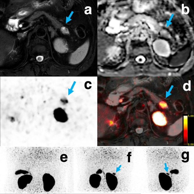 Figure 1
- —http://dx.doi.org/10.13039/501100001691Japan Society for the Promotion of Science
- —Moriya Scholarship Foundation
- —http://dx.doi.org/10.13039/501100007664Manpei Suzuki Diabetes Foundation
- —http://dx.doi.org/10.13039/100007434Suzuken Memorial Foundation
- —http://dx.doi.org/10.13039/100008735Japan Foundation of Applied Enzymology
- —Terumo Health Management Research Foundation
- —Advanced Science, Technology & Management Research Institute of KYOTO
- —The Japan Health Foundation
Peer Reviews
No public reviews on file for this paper yet. If you reviewed it on a platform where reviews are public (OpenReview, ICLR, NeurIPS, ICML), you can paste yours below so the community can read it here.
Videos
No videos yet. Explain this paper in a talk, walkthrough, or lecture? Add one.
Taxonomy
TopicsNeuroendocrine Tumor Research Advances · Neuroblastoma Research and Treatments · Lung Cancer Research Studies
Insulinomas are rare, typically small pancreatic neuroendocrine tumors that often elude detection on widely used imaging methods like computed tomography (CT), magnetic resonance imaging (MRI), and somatostatin receptor (SSTR)-based positron emission tomography (PET), owing to their small size and low levels of SSTR expression [1]. Technically demanding procedures like endoscopic ultrasound and selective arterial calcium stimulation testing can improve sensitivity, but their invasive nature may preclude clinical use [2]. Conversely, because most benign insulinomas display increased levels of glucagon-like peptide-1 receptors (GLP-1R), we developed a PEGylated exendin‑4 probe labeled with [^18^F]FB(ePEG12)12‑exendin‑4 (^18^F-exendin-4)—that enables both qualitative and quantitative diagnosis [3–5].
A 76‑year‑old woman with symptomatic hypoglycemia (plasma glucose 37 mg/dL; C‑peptide 1.87 ng/mL) underwent integrated ^18^F-exendin-4 PET/MRI at the expiratory phase. Figure shows matched panels: (a) fat-saturated T2-weighted MRI and (b) apparent-diffusion-coefficient (ADC) map depict a tumor with a hyperintense cystic component; (c) respiratory-gated PET slice and (d) the fused PET/MRI, (plus (e)–(g), maximum intensity projection for orientation) demonstrates intense uptake confined to the solid, low-ADC core while the cystic component shows negligible activity. After enucleation, hypoglycemia resolved completely and histology confirmed insulinoma.
Owing to its high soft-tissue contrast and motion-corrected spatial resolution, integrated PET/MRI not only delineated the intratumoral characteristics but also clearly distinguished the lesion from adjacent renal physiologic activity—an advantage over sequential PET/CT, which is prone to misregistration and false negatives near the kidney [6, 7]. This case highlights the diagnostic value of GLP-1R-targeted PET/MRI for precise insulinoma characterization and localization.
The reference list from the paper itself. Each links out to its DOI / PubMed record.
- 1Murakami T, Yoshida H, Sakaki K, et al. Qualitative and quantitative analyses of noninvasive diagnosis of insulinoma using [18F]FB(e PEG 12) 12-Exendin-4 PET/CT. J Clin Endocrinol Metab. Published online May 20, 2025:dgaf 253. 10.1210/clinem/dgaf 25310.1210/clinem/dgaf 25340391925 · doi ↗ · pubmed ↗