The Salivary Tumor That Lost Its Way: A Case Report of Sinonasal Pleomorphic Adenoma
Áine O'Brien, Eoin F Cleere, Thomas Crotty, Mohammad Habibullah Khan

TL;DR
A rare case of nasal pleomorphic adenoma, a salivary gland tumor, was diagnosed through histology and RNA sequencing in a 46-year-old man.
Contribution
This case report highlights the rare occurrence of pleomorphic adenoma in the nasal cavity and its diagnosis using RNA sequencing.
Findings
Histology showed biphasic ductal epithelial-like and myoepithelial architecture consistent with pleomorphic adenoma.
RNA sequencing detected an NCALD::PLAG1 fusion, confirming a salivary cell origin.
The case emphasizes the importance of considering pleomorphic adenoma in nasal obstruction or mass differential diagnosis.
Abstract
Pleomorphic adenoma (PA) is a benign tumour that most commonly arises in the salivary glands but can occur in other sites. Occurrence in the nasal cavity is rare, arising from minor salivary tissue. A 46-year-old man with primary ciliary dyskinesia and type 2 diabetes presented with bilateral nasal obstruction and chronic rhinosinusitis. Examination showed bilateral nasal polyposis, with the right one being more obstructive. Computed tomography (CT) demonstrated bilateral diffuse pansinus mucosal thickening with polypoidal transformation. The patient underwent bilateral endoscopic sinus surgery. Nasal polyps were sent for a routine histological examination. Histology revealed sheet-like fascicular growth of uniform ovoid-spindled cells with biphasic ductal epithelial-like and myoepithelial architecture, in keeping with PA. Targeted RNA sequencing detected an NCALD::PLAG1 fusion,…
Genes, proteins, chemicals, diseases, species, mutations and cell lines named across the full text — each resolved to its canonical identifier and authoritative record.
Click any figure to enlarge with its caption.
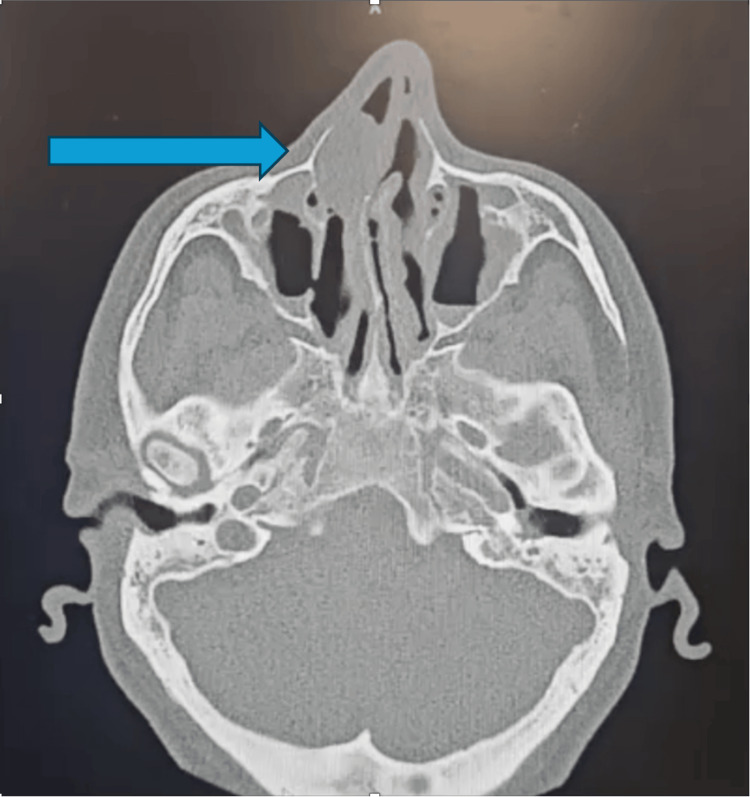 Figure 1
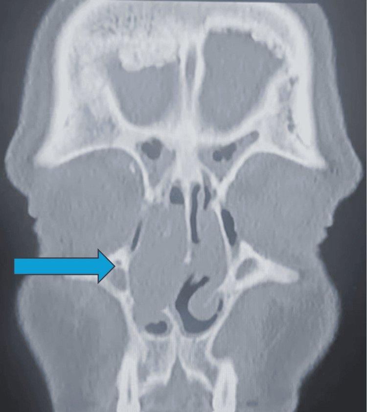 Figure 2
Peer Reviews
No public reviews on file for this paper yet. If you reviewed it on a platform where reviews are public (OpenReview, ICLR, NeurIPS, ICML), you can paste yours below so the community can read it here.
Videos
No videos yet. Explain this paper in a talk, walkthrough, or lecture? Add one.
Taxonomy
TopicsSalivary Gland Tumors Diagnosis and Treatment · Head and Neck Surgical Oncology · Ear and Head Tumors
Introduction
Pleomorphic adenoma (PA) is the most common benign salivary gland neoplasm, accounting for approximately 75% of all salivary gland tumours [1]. It most frequently arises in the parotid gland (approximately 84%), followed by the submandibular gland (8%) and minor salivary glands (6-7%) [2]. These tumours typically present between the third and sixth decades of life and demonstrate a female predominance. Clinically, they manifest as slow-growing, painless, well-circumscribed masses. Diagnosis may be suspected by radiological or cytological investigation and is confirmed on histopathological examination [3].
Although PA predominantly arises within the major salivary glands, it may also develop in ectopic or minor salivary gland sites, including the soft palate, larynx, epiglottis, and sinonasal tract [4]. Sinonasal PA is rare and poses unique diagnostic challenges. In contrast to their major salivary gland counterparts, sinonasal tumours frequently exhibit hypercellularity and a relative paucity of myxoid stroma, potentially leading to misdiagnosis as a malignant neoplasm. Furthermore, while PAs are benign, incomplete excision carries a risk of recurrence and malignant transformation to carcinoma ex-PA [5].
We report a case of hypercellular sinonasal PA arising in the setting of chronic rhinosinusitis, with molecular confirmation of an uncommon NCALD::PLAG1 gene fusion, highlighting the diagnostic challenges and the value of adjunct molecular testing.
Case presentation
A 46-year-old man with a background of primary ciliary dyskinesia, situs inversus with dextrocardia, and type 2 diabetes mellitus presented to the otolaryngology outpatient clinic. He presented with progressive right-sided nasal obstruction and longstanding symptoms of chronic rhinosinusitis, including nasal congestion and facial pressure, ongoing for three years. There was no history of significant epistaxis, weight loss, or facial numbness.
Flexible nasoendoscopy demonstrated a right-sided obstructive nasal polypoidal mass with diffusely inflamed and erythematous mucosa bilaterally. The lesion appeared polypoid and was clinically suspected to represent an inflammatory nasal polyp in the context of chronic rhinosinusitis. Computed tomography (CT) of the paranasal sinuses revealed bilateral diffuse mucosal thickening with polypoid changes and a dominant right-sided nasal cavity lesion causing obstruction (Figures 1, 2). There was no evidence of bony erosion or destructive change. Given the patient’s symptom burden, unilateral obstructive mass, and radiologic findings, he underwent endoscopic sinus surgery and excision of the mass. The mass was grossly excised in its entirety endoscopically and submitted for histopathological evaluation. Intraoperatively, the lesion appeared similar to an inflammatory polyp, without necrosis, ulceration, or obvious invasion. Other differentials included an anterochoanal polyp and an inverted papilloma.
CT sinuses: dominant right-sided nasal cavity lesion causing obstruction on a background ofbilateral diffuse mucosal thickening with polypoid changes
CT sinuses: dominant right-sided nasal cavity lesion causing obstruction on a background of bilateral diffuse mucosal thickening with polypoid changes
Histological examination of the right nostril polyp demonstrated a cellular proliferation with sheet-like and focally fascicular architecture, composed of uniform ovoid to spindled cells within a fibromyxoid stroma and focal adipocytic metaplasia. No high-grade features, tumour necrosis, or lymphovascular invasion were identified.
Immunohistochemical studies were performed to confirm the suspected diagnosis, considering the unusual site of the suspected pathology. Immunohistochemistry showed diffuse MNF116 and SOX10 positivity, with AE1/AE3 and CK7 highlighting epithelial elements, and S100 and SMA confirming myoepithelial differentiation. The tumour cells were negative for beta-catenin (wild-type), INSM1, synaptophysin, TTF-1, CD31, CD34, STAT6, and desmin. These findings supported a diagnosis of PA; however, given the relative stromal paucity and diagnostic uncertainty, molecular testing was pursued.
Targeted RNA sequencing identified an NCALD::PLAG1 gene fusion. Rearrangements involving PLAG1 are well-recognised oncogenic drivers in PA and provided molecular confirmation of the diagnosis [6]. Due to the discordant findings, the specimen was also sent to a world-leading expert on salivary tumours, who concurred with the diagnosis of PA. Postoperatively, no tumour was evident on clinical examination. There is no evidence for follow-up imaging to monitor recurrence of PA when the initial lesion was excised in its entirety. At the most recent follow-up, 12 months post-surgery, the patient remained well with no evidence of recurrence. He will remain under close clinical surveillance for at least six years, as this is the median length of time of PA recurrence.
Discussion
PA of the sinonasal tract is rare, with most cases arising from minor salivary glands of the nasal septum. Less commonly, lesions originate from the lateral nasal wall or paranasal sinuses. Patients typically present with unilateral nasal obstruction, congestion, epistaxis, hyposmia, or facial pressure [7]. Because these symptoms overlap significantly with inflammatory sinonasal disease, lesions may initially be presumed to represent nasal polyps, as in this case.
Histologically, PA is characterised by three components: epithelial ductal structures, myoepithelial cells, and a mesenchymal-like stroma that may be myxoid, chondroid, or myxochondroid [8]. However, sinonasal PA frequently demonstrates reduced stromal elements and increased cellularity compared with tumours of the major salivary glands [9]. This hypercellular pattern can mimic other spindle cell or epithelial neoplasms of the sinonasal tract, including adenoid cystic carcinoma, myoepithelioma, basal cell adenoma, and low-grade adenocarcinoma [10]. Recognition of the biphasic architecture and confirmation with immunohistochemistry are therefore critical. Molecular alterations provide additional diagnostic support. PLAG1 (pleomorphic adenoma gene 1) is a proto-oncogene encoding a zinc finger transcription factor. In PA, chromosomal rearrangements involving PLAG1 lead to promoter swapping and overexpression.
PLAG1 fusions are among the most common molecular alterations in PA across anatomical sites [6]. Numerous fusion partners have been described; however, NCALD is an uncommon partner, and the true prevalence of this rearrangement remains uncertain, as molecular testing is not routinely performed in all cases. In diagnostically challenging lesions, in particular those with unusual morphology, molecular analysis can provide valuable confirmation to ensure adequate treatment and clinical follow-up.
Complete surgical excision with negative margins remains the definitive management for sinonasal PA. Enucleation or incomplete excision is associated with recurrence. Although PA is benign, there is a recognised risk of malignant transformation to carcinoma ex-PA, estimated at approximately 1.5% within five years and increasing with prolonged duration [3]. Data specific to sinonasal lesions are limited, but PA is associated with a potential risk of recurrence, with reported mean or median times to recurrence of approximately 6 years, and recurrences documented even decades after initial excision [11, 12]. In the sinonasal tract, complete excision with wide margins may be challenging to achieve endoscopically due to anatomical constraints. Identification of this entity on histopathology may prompt consideration of further formal resection in selected cases and, at a minimum, long-term clinical surveillance compared to nasal polyps, which do not require long-term follow-up. Accurate pathological diagnosis is therefore critical in guiding appropriate follow-up to detect potential recurrences or malignant transformations.
Conclusions
Sinonasal PA is a rare but important differential diagnosis in patients presenting with unilateral nasal obstruction. Hypercellularity and stromal paucity may create significant diagnostic uncertainty and mimic malignant neoplasms. Recognition of characteristic biphasic morphology, supported by immunohistochemistry and molecular confirmation of PLAG1 rearrangements, was essential for accurate diagnosis. Complete surgical excision with long-term follow-up is recommended to detect recurrence early and monitor for malignant transformation.
The reference list from the paper itself. Each links out to its DOI / PubMed record.
- 1Tracking pleomorphic adenoma incidence trends over 47 years: a population-based study Otolaryngol Head Neck Surg Rourk KS Daher GS Schwartz JR 65165917320254032318110.1002/ohn.1292 · doi ↗ · pubmed ↗
- 2Benign parotid tumors Otolaryngol Clin North Am Zhan KY Khaja SF Flack AB Day TA 3273424920162704058410.1016/j.otc.2015.10.005 · doi ↗ · pubmed ↗
- 3Salivary neoplasms: overview of a 35-year experience with 2,807 patients Head Neck Surg Spiro RH 17718481986374485010.1002/hed.2890080309 · doi ↗ · pubmed ↗
- 4Pleomorphic adenomas of the salivary glands: retrospective multicentric study of 130 cases with emphasis on histopathological features Eur Arch Otorhinolaryngol Lopes ML Barroso KM HenriquesÁCG Dos Santos JN Martins MD de Souza LB 54355127420172752057010.1007/s 00405-016-4253-5 · doi ↗ · pubmed ↗
- 5Malignant transformation of salivary gland pleomorphic adenoma: proof of principle J Pathol Clin Res Valstar MH Mast H Ten Hove I 432437720213439032010.1002/cjp 2.216PMC 8363925 · doi ↗ · pubmed ↗
- 6Clinicopathological effect of PLAG 1 fusion genes in pleomorphic adenoma and carcinoma ex pleomorphic adenoma with special emphasis on histological features Histopathology Asahina M Saito T Hayashi T Fukumura Y Mitani K Yao T 5145257420193030705510.1111/his.13759 · doi ↗ · pubmed ↗
- 7Pleomorphic adenoma of the nasal cavity Cureus Basharat R Bjorling A Samara G 016202410.7759/cureus.65969 PMC 1136560539221300 · doi ↗ · pubmed ↗
- 8Molecular Pathology of Salivary Gland Neoplasms: Diagnostic, Prognostic, and Predictive Perspective Adv Anat Pathol Toper MH Sarioglu S 81932820213340540010.1097/PAP.0000000000000291 · doi ↗ · pubmed ↗