Intracranial Metastasis of a Primary Mediastinal Seminoma Mimicking a Convexity Meningioma: A Case Report
Rikiya Kameno, Shinjitsu Nishimura, Asuhito Takemura, Sumito Okuyama, Keiichi Kubota, Junko Matsuyama, Tadao Matsushima, Hideo Sakuma, Yuya Yoshida, Sadayoshi Watanabe

TL;DR
A young man with a brain tumor initially thought to be a meningioma was found to have a rare germ cell tumor that had spread from the chest, highlighting the importance of considering this diagnosis in similar cases.
Contribution
This case report highlights the diagnostic challenge of convexity germ cell tumors mimicking meningiomas and emphasizes the need for tumor marker evaluation in young males.
Findings
A convexity germ cell tumor was misdiagnosed as a meningioma due to similar imaging features.
The tumor exhibited dural invasion, skull destruction, and high proliferative activity (Ki-67).
Systemic evaluation revealed a primary mediastinal seminoma with intracranial metastasis.
Abstract
Convexity germ cell tumors are exceedingly rare and may closely mimic meningiomas, particularly in young men. This case report presents a rare convexity germ cell tumor characterized by dural invasion and skull destruction. An 18-year-old man presented with headache and nausea. Neuroimaging demonstrated a right frontoparietal extra-axial mass accompanied by adjacent skull hyperostosis and significant cerebral edema. Based on these radiological findings, a convexity meningioma was strongly suspected, and a craniotomy was performed for tumor resection. Unexpectedly, histopathological examination revealed a germ cell tumor with diffuse infiltration of the dura and bone, characterized by a high Ki-67 labeling index and immunoreactivity for placental alkaline phosphatase. Subsequent systemic evaluation identified a mass in the anterior mediastinum, leading to a final diagnosis of…
Genes, proteins, chemicals, diseases, species, mutations and cell lines named across the full text — each resolved to its canonical identifier and authoritative record.
Click any figure to enlarge with its caption.
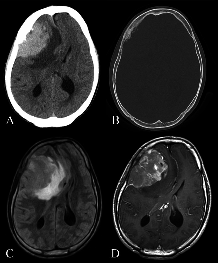 Figure 1
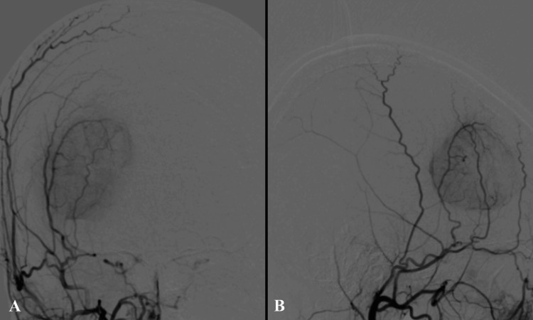 Figure 2
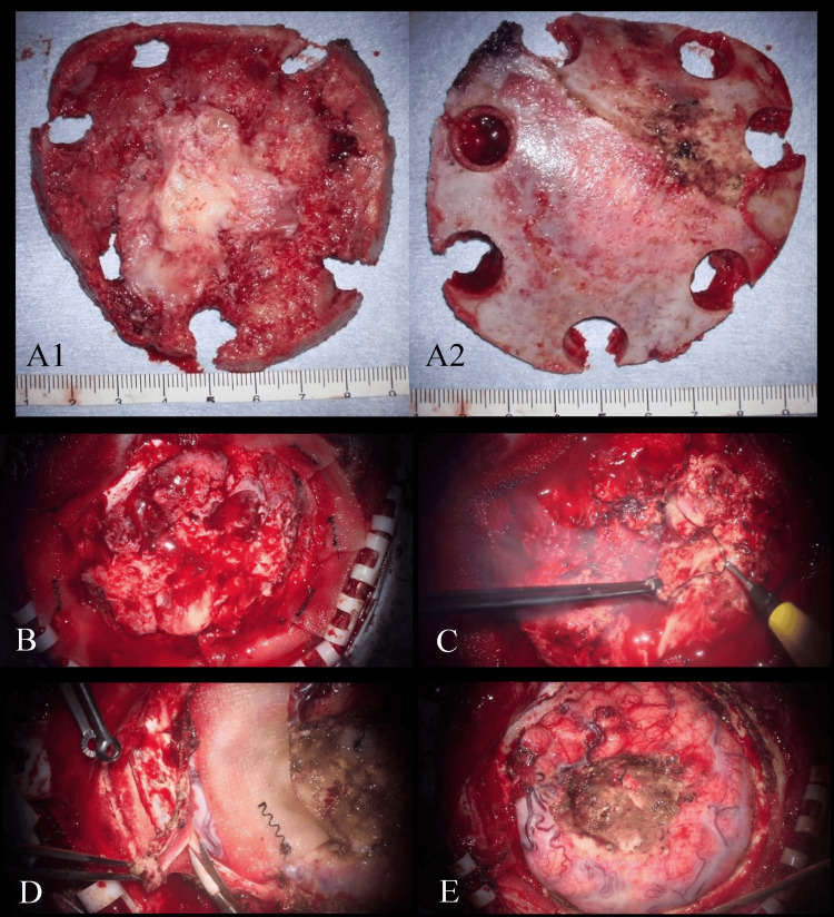 Figure 3
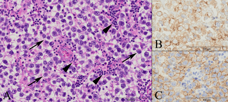 Figure 4
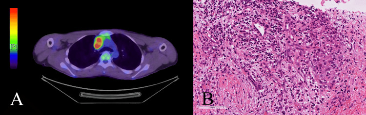 Figure 5
Peer Reviews
No public reviews on file for this paper yet. If you reviewed it on a platform where reviews are public (OpenReview, ICLR, NeurIPS, ICML), you can paste yours below so the community can read it here.
Videos
No videos yet. Explain this paper in a talk, walkthrough, or lecture? Add one.
Taxonomy
TopicsGlioma Diagnosis and Treatment · Meningioma and schwannoma management · Testicular diseases and treatments