Gliomap-GAN: A conditional generative adversarial network to visualize glioblastoma’s cell density from contrast-enhanced magnetic resonance imaging
Manabu Kinoshita, Keisuke Miyake, Wataru Ide, Hideyuki Arita, Kayako Isohashi, Jun Hatazawa, Haruhiko Kishima

TL;DR
This paper introduces Gliomap-GAN, an AI tool that creates images resembling 11C-methionine PET scans from MRI data to visualize glioblastoma cell density.
Contribution
The novel contribution is a GAN-based method to generate glioblastoma cell density maps from contrast-enhanced MRI.
Findings
Gliomaps visually resembled original 11C-methionine PET images with a tumor-to-normal tissue residual error of 0.07 ± 0.04.
The Sørensen-Dice coefficient between Gliomap and PET lesion predictions was 0.88 ± 0.07 at a threshold of 1.5.
Gliomap values correlated significantly with tumor cell density (P = 0.02).
Abstract
11C-methionine positron emission tomography is one of the most reliable imaging modalities for glioblastoma visualization. This investigation aimed to generate an 11C-methionine positron emission tomography-like image, “Gliomap,” from contrast-enhanced magnetic resonance imaging via a conditional Generative Adversarial Network (Gliomap-GAN). Eighty-one newly diagnosed glioblastoma patients with preoperative contrast-enhanced magnetic resonance imaging and 11C-methionine positron emission tomography were retrospectively collected. T1-weighted, T2-weighted, and Gd-enhanced T1-weighted images were co-registered and intensity normalized, followed by the creation of a contrast-enhancement subtraction map. They were used as source data to train Gliomap-GAN, targeting the corresponding 11C-methionine positron emission tomography image. The training dataset comprised 2459 images augmented to…
Genes, proteins, chemicals, diseases, species, mutations and cell lines named across the full text — each resolved to its canonical identifier and authoritative record.
Click any figure to enlarge with its caption.
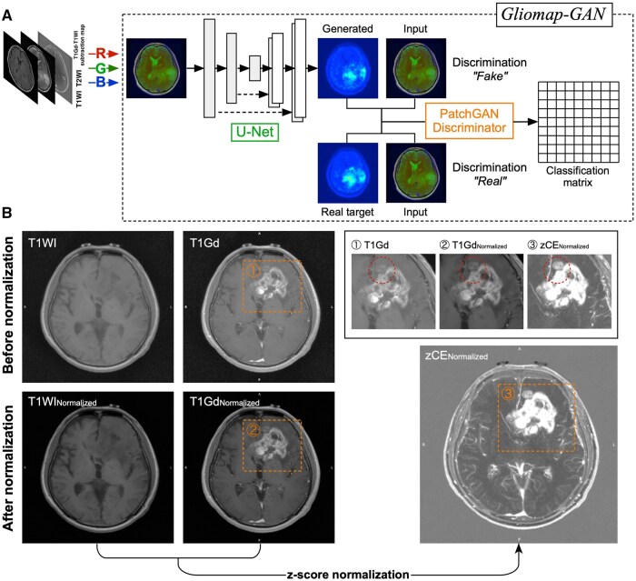 Figure 1
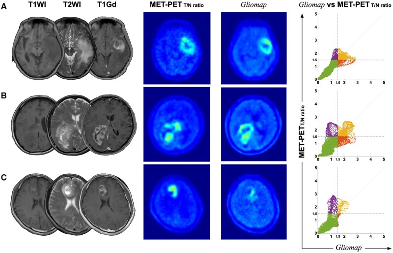 Figure 2
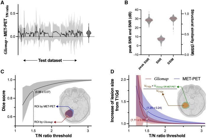 Figure 3
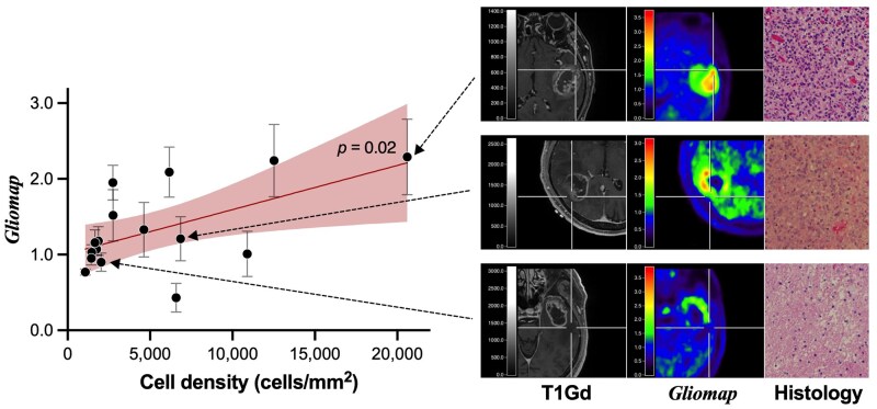 Figure 4
Peer Reviews
No public reviews on file for this paper yet. If you reviewed it on a platform where reviews are public (OpenReview, ICLR, NeurIPS, ICML), you can paste yours below so the community can read it here.
Videos
No videos yet. Explain this paper in a talk, walkthrough, or lecture? Add one.
Taxonomy
TopicsCell Image Analysis Techniques · Radiomics and Machine Learning in Medical Imaging · AI in cancer detection