Particle‐Based Detection of Surface Chemistry via Optical Microscopy—Integrating Microfluidics, Light‐Induced Activity of Colloids and Data Science
Fabian Rohne, Daniela Vasquez Muñoz, Isabel Meier, Anne Nitschke, Florian Schmitt, Nino Lomadze, Martin Reifarth, Andreas Taubert, Svetlana Santer, Marek Bekir

TL;DR
This paper introduces a new microscopy-based method to measure the surface area and porosity of microparticles using minimal sample amounts and standard lab equipment.
Contribution
The novel integration of microfluidics, optical microscopy, and data science enables per-particle surface area analysis without drying or bulk preparation.
Findings
The method achieves a relative precision of approximately 9% when validated on SiO2 microparticles.
It allows high-resolution analysis of particle-to-particle heterogeneity by summing individual particle measurements.
The workflow is rapid and uses a self-generated reference data library for surface area calculation.
Abstract
We present particle‐resolved methods for determining the porosity and surface area of microparticles, based on single‐particle trajectory analysis conducted via optical video microscopy integrated with microfluidics and LED illumination. This technique introduces a unique combination of analytical advantages that address key limitations of conventional methods, such as BET nitrogen adsorption. Notably, (1) the method operates with extremely low analyte quantities on the order of micrograms or less and in the form of dilute aqueous dispersions, eliminating the need for drying or bulk sample preparation; (2) surface area quantification is performed on a per‐particle basis, with total surface area determined by summing up all individual particle measurements, enabling high‐resolution analysis of particle‐to‐particle heterogeneity; and (3) the entire workflow from sample preparation to data…
Genes, proteins, chemicals, diseases, species, mutations and cell lines named across the full text — each resolved to its canonical identifier and authoritative record.
Click any figure to enlarge with its caption.
 Figure 1
 Figure 2
 Figure 3
 Figure 4
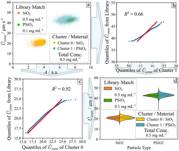 Figure 5
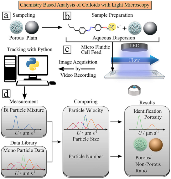 Figure 6
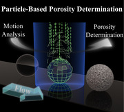 Figure 7
Peer Reviews
No public reviews on file for this paper yet. If you reviewed it on a platform where reviews are public (OpenReview, ICLR, NeurIPS, ICML), you can paste yours below so the community can read it here.
Videos
No videos yet. Explain this paper in a talk, walkthrough, or lecture? Add one.
Taxonomy
TopicsAdvanced Biosensing Techniques and Applications · Mesoporous Materials and Catalysis · Force Microscopy Techniques and Applications