Distinct macular structural and microvascular alterations differentiate neuromyelitis optica spectrum disorder from myelin oligodendrocyte glycoprotein antibody–associated disease in optic neuritis
Mi Zhang, Zhongzhong Liu, Yunfei Li, Yanli Li, Ruili Ma, Qingli Lu, Pei Liu, Yan liu, Qiaoqiao Chang, Yan Wang, Chensheng Song, Yan Huo, Lanping Rao, Shundao Cao, Ning Wang, Guo Li, Fanyan Wu, Tong Liu, Linna Peng, Yunlong Hao, Zijing Cao, Xuemei Lin, Xiaolai Zhou, Songdi Wu

TL;DR
This study finds that NMOSD and MOGAD cause distinct changes in eye structure and blood flow, which can help differentiate these conditions in patients with optic neuritis.
Contribution
The study identifies unique microvascular and structural differences between NMOSD and MOGAD in optic neuritis, offering potential biomarkers for differentiation.
Findings
NMOSD-ON eyes showed greater reduction in choriocapillaris blood flow area compared to MOG-ON eyes.
Both NMOSD and MOGAD caused significant reductions in macular microvascular and structural parameters compared to healthy controls.
Microvascular and structural parameters in NMOSD-ON were inversely correlated with visual acuity and disability scores.
Abstract
Neuromyelitis optica spectrum disorder (NMOSD) and myelin oligodendrocyte glycoprotein antibody–associated disease (MOGAD) are among the leading causes of optic neuritis. This study aimed to examine differences in macular retinal structure and microvascular characteristics between affected and unaffected eyes in individuals with NMOSD and MOGAD. This cross-sectional study enrolled both eyes of patients diagnosed with optic neuritis (ON)secondary to NMOSD (22 patients: 36 NMOSD-ON eyes and 8 NMOSD-NON eyes), MOGAD (23 patients: 34 MOG-ON eyes and 12 MOG-NON eyes), and 20 age- and sex-matched healthy controls (HCs, 40 eyes) recruited from the First Affiliated Hospital of Northwest University (Xi’an No.1 Hospital) between February 2023 and January 2025. Microvascular density (MVD), vascular density (VD), blood flow area (BFA), and macular ganglion cell–inner plexiform layer (GCIPL)…
Genes, proteins, chemicals, diseases, species, mutations and cell lines named across the full text — each resolved to its canonical identifier and authoritative record.
Click any figure to enlarge with its caption.
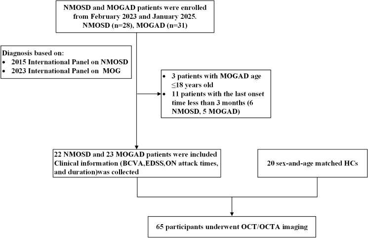 Figure 1
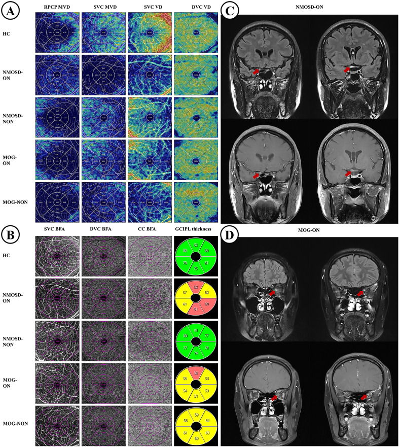 Figure 2
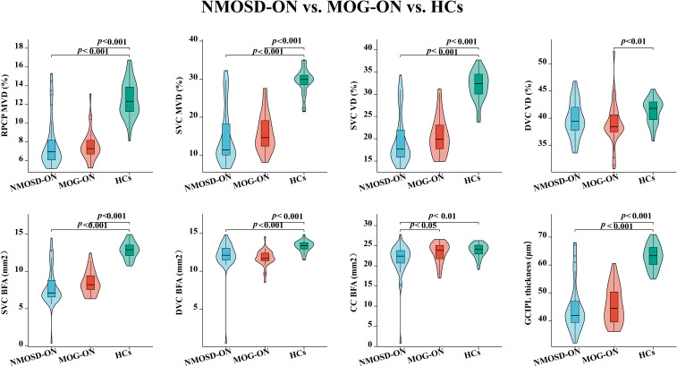 Figure 3
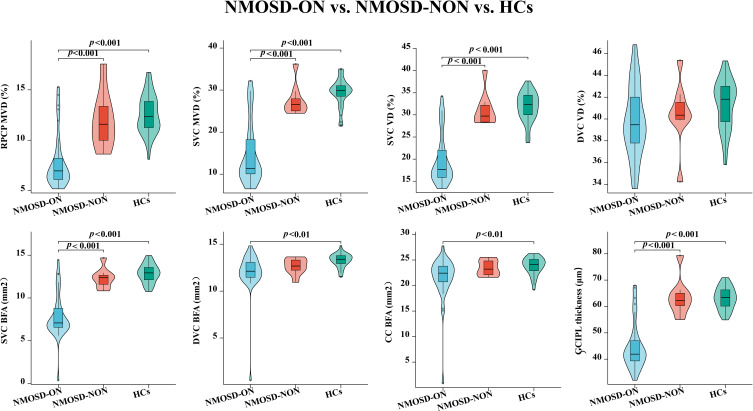 Figure 4
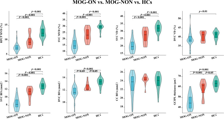 Figure 5
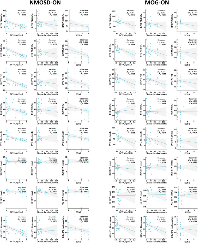 Figure 6
Peer Reviews
No public reviews on file for this paper yet. If you reviewed it on a platform where reviews are public (OpenReview, ICLR, NeurIPS, ICML), you can paste yours below so the community can read it here.
Videos
No videos yet. Explain this paper in a talk, walkthrough, or lecture? Add one.
Taxonomy
TopicsMultiple Sclerosis Research Studies · Ophthalmology and Eye Disorders · Peripheral Neuropathies and Disorders