A Single-Cell Atlas of Pan-Cancer Liver Metastasis Reveals Dynamic Cellular Programs Driving Metastatic Progression and Immune Modulation
Xinyu Tong, Haoyu Chao, Chenlu Zhang, Zhuojin Li, Quan Han, Lishan Wang, Nuo Wu, Ruidong Chen, Jian Gao, Shu Zhang, Lei Xu, Ming Chen, Hui Zhao, Lei Wang, Dijun Chen

TL;DR
This study creates a detailed map of liver metastases across various cancers, revealing how different cell types and interactions drive cancer spread and immune evasion.
Contribution
The paper introduces a pan-cancer single-cell atlas of liver metastases and identifies four dynamic cellular programs linked to metastatic progression and immune modulation.
Findings
Four cellular programs were identified that reflect transitions from immune-active to immunosuppressive tumor environments.
Natural killer cells and macrophages are associated with early immune surveillance, while regulatory T cells dominate immunosuppressive states.
The study highlights tumor-intrinsic adaptations and stromal remodeling as key drivers of metastatic niche formation.
Abstract
Liver metastasis remains a major challenge in cancer treatment, yet its cellular and molecular landscape remains poorly defined at the pan-cancer level. Here, we construct a single-cell transcriptomic atlas of liver metastases across multiple cancer types by analyzing 100 single-cell RNA sequencing samples, profiling over 460,000 cells, and identifying 121 distinct cellular subtypes. We define 4 representative cellular programs (CPs) associated with liver metastasis, revealing how cellular composition and intercellular interactions within the tumor microenvironment drive metastatic progression and immune modulation. These CPs recapitulate a dynamic transition from immunoactive states, marked by natural-killer-cell-mediated immune surveillance and macrophage-driven angiogenesis, to immunosuppressive environments dominated by regulatory T cell infiltration and immune exclusion. The shift…
Genes, proteins, chemicals, diseases, species, mutations and cell lines named across the full text — each resolved to its canonical identifier and authoritative record.
Click any figure to enlarge with its caption.
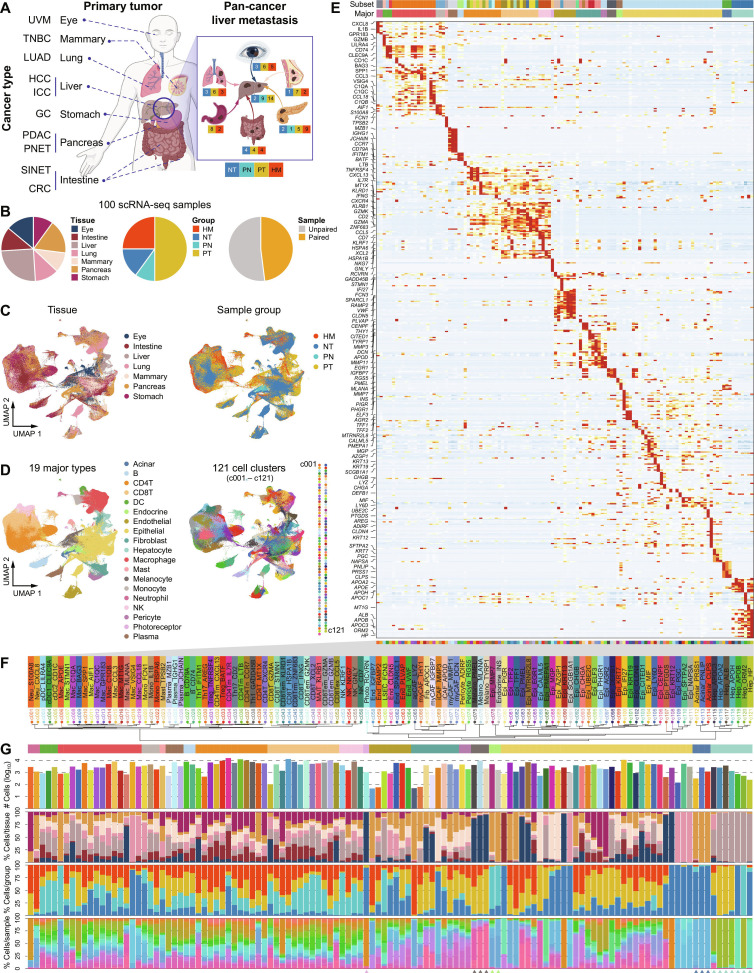 Figure 1
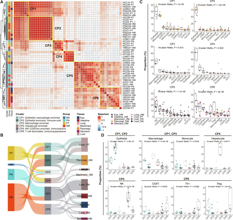 Figure 2
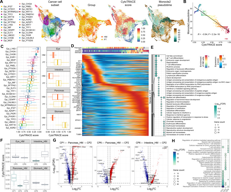 Figure 3
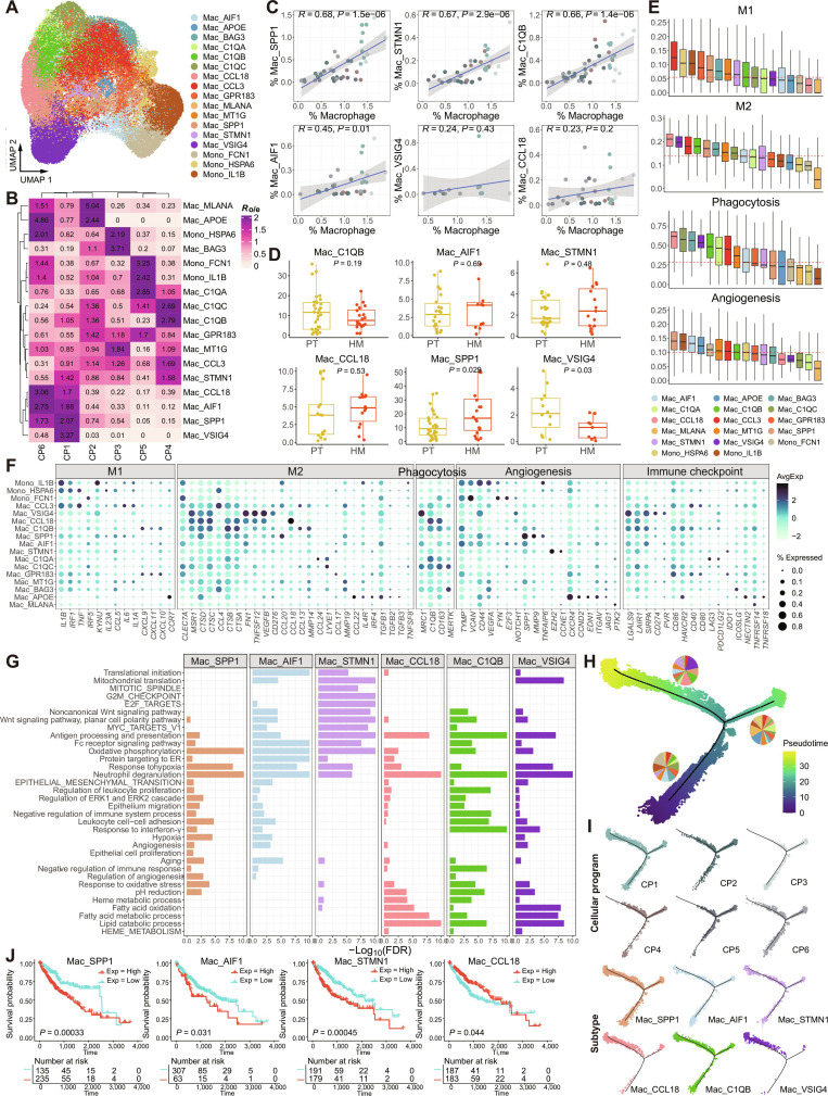 Figure 4
 Figure 5
 Figure 6
 Figure 7
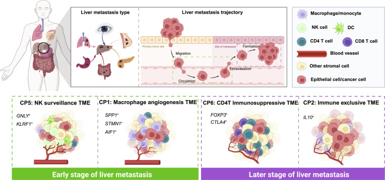 Figure 8
Peer Reviews
No public reviews on file for this paper yet. If you reviewed it on a platform where reviews are public (OpenReview, ICLR, NeurIPS, ICML), you can paste yours below so the community can read it here.
Videos
No videos yet. Explain this paper in a talk, walkthrough, or lecture? Add one.
Taxonomy
TopicsSingle-cell and spatial transcriptomics · Immune cells in cancer · Cancer Immunotherapy and Biomarkers