Intracellular fluid accumulation underlies brain volume increases in early Alzheimer’s disease
Michalis Kassinopoulos, Paula Montesinos, Carles Falcon, Jordi Huguet, Carolina Minguillon, Karine Fauria, Gwendlyn Kollmorgen, Clara Quijano-Rubio, José Luis Molinuevo, Oriol Grau-Rivera, Henrik Zetterberg, Kaj Blennow, Marc Suárez-Calvet, Javier Sanchez-Gonzalez

TL;DR
This study finds that brain volume increases in early Alzheimer’s disease may be due to changes in water diffusion linked to neuroinflammation, not just amyloid buildup.
Contribution
The study introduces a three-compartment diffusion MRI model to show that neuroinflammation and microstructural changes, not amyloid pathology, drive brain volume increases in early Alzheimer’s.
Findings
Diffusion parameters strongly correlate with neuroinflammation and neurodegeneration markers, not amyloid pathology.
Grey matter volume increases in amyloid-related regions are negatively linked to slow diffusion coefficient and perfusion fraction.
Intracellular water diffusion changes may reflect glial remodelling or cellular complexity in amyloid-positive individuals.
Abstract
In the preclinical stages of Alzheimer’s disease, increased brain volume has been associated with amyloid-beta pathology, particularly in regions that undergo volume reductions as the disease progresses. Glial reactivity and water diffusion alterations have been linked to such macroscopic volumetric changes. Brain volume reductions have also been reported following amyloid-beta removal with anti-amyloid therapies with beneficial clinical effects, but it remains unclear whether these changes result from resolving amyloid-triggered neuroinflammation or neurodegeneration. Intravoxel incoherent motion modelling based on multi-shell diffusion-weighted imaging may provide a better understanding of the processes underlying these paradoxical changes. This study used intravoxel incoherent motion diffusion MRI to examine how alterations in cerebral water pools contribute to increased brain volume…
Genes, proteins, chemicals, diseases, species, mutations and cell lines named across the full text — each resolved to its canonical identifier and authoritative record.
Click any figure to enlarge with its caption.
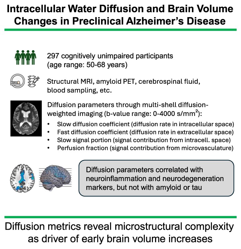 Figure 1
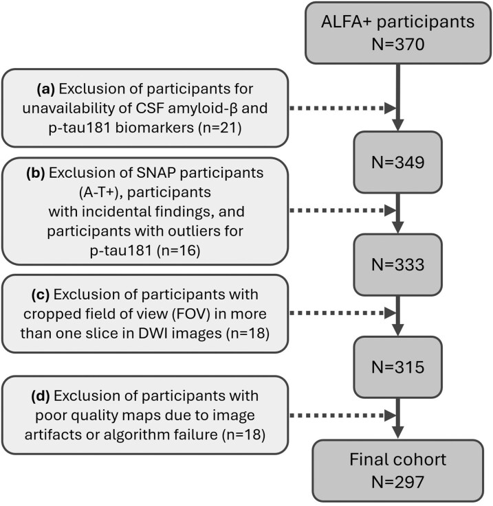 Figure 2
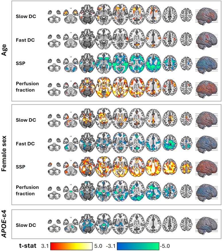 Figure 3
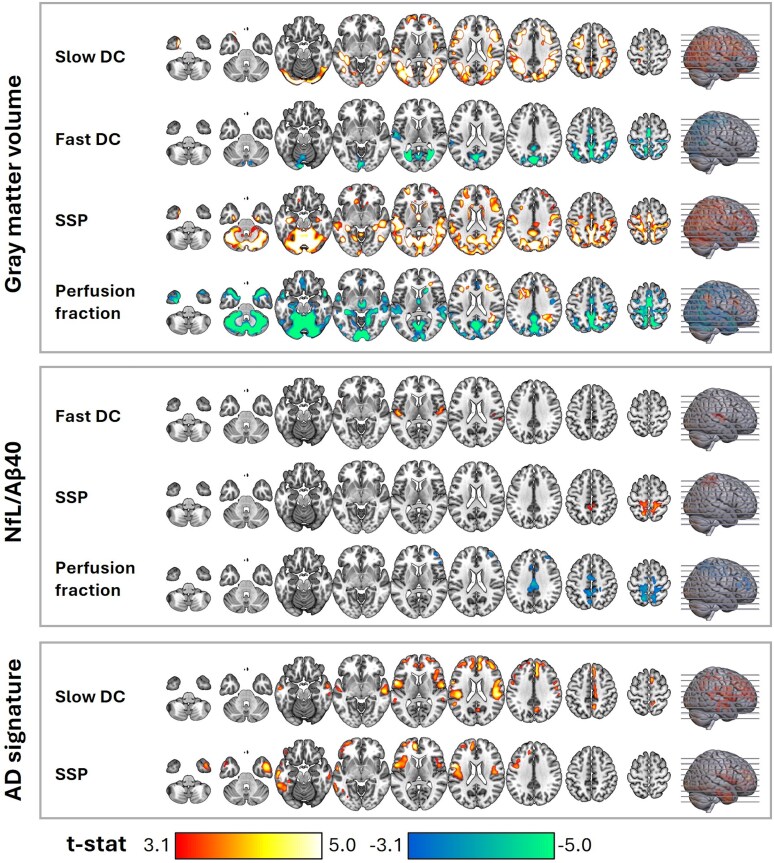 Figure 4
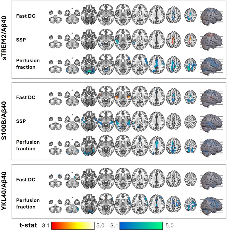 Figure 5
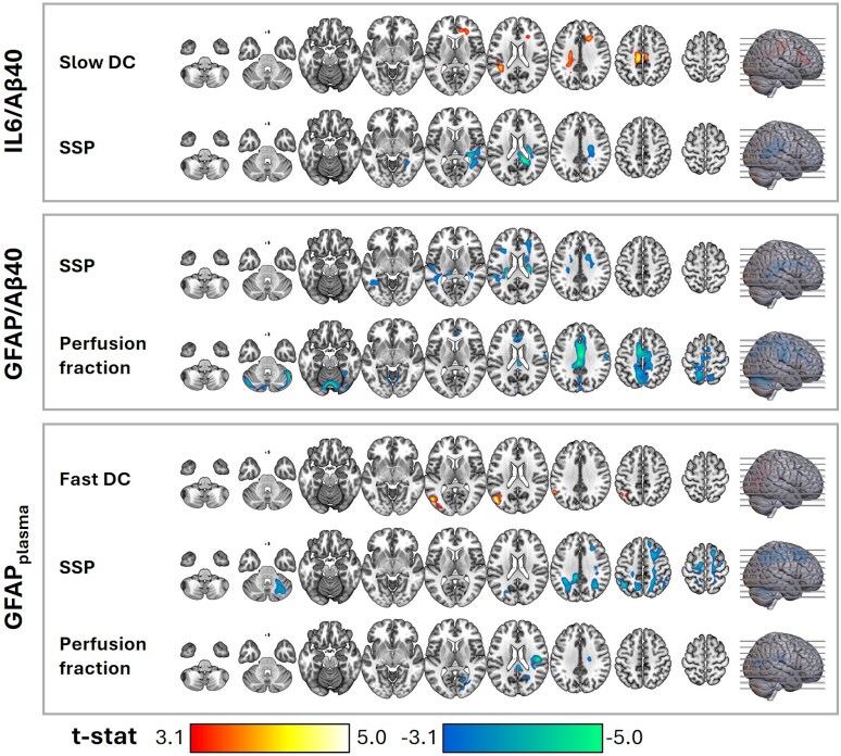 Figure 6
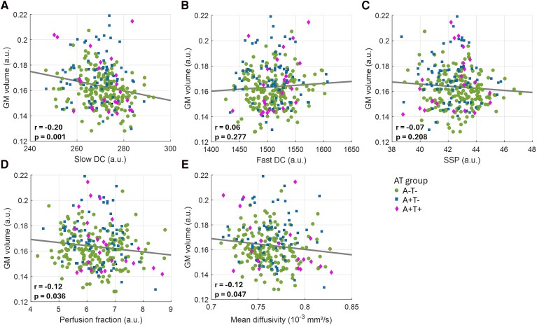 Figure 7
Peer Reviews
No public reviews on file for this paper yet. If you reviewed it on a platform where reviews are public (OpenReview, ICLR, NeurIPS, ICML), you can paste yours below so the community can read it here.
Videos
No videos yet. Explain this paper in a talk, walkthrough, or lecture? Add one.
Taxonomy
TopicsAdvanced Neuroimaging Techniques and Applications · Barrier Structure and Function Studies · Neuroinflammation and Neurodegeneration Mechanisms