Microstructural variation of hippocampal substructures across childhood and adolescence quantified with high-gradient diffusion MRI
Bradley G. Karat, Sila Genc, Erika P. Raven, Marco Palombo, Ali R. Khan, Derek K. Jones

TL;DR
This study uses advanced MRI to track changes in the hippocampus during childhood and adolescence, revealing age-related differences in brain microstructure.
Contribution
The study introduces high-gradient diffusion MRI and SANDI modeling to quantify hippocampal microstructural development in children and adolescents.
Findings
Hippocampal volume, gyrification, and thickness remain stable during childhood and adolescence.
Age-related changes in MR-derived neurite and soma parameters are correlated with adult microstructure markers like myelin and iron content.
Distinct age-related profiles of hippocampal substructures are revealed using high-gradient diffusion MRI.
Abstract
The hippocampus plays a crucial role in cognition, yet its microstructural development during childhood and adolescence remains poorly understood. Here, we investigate age-related differences in hippocampal microstructure using diffusion MRI with ultra-strong gradients (300 mT/m) in a cohort of 88 participants aged 8–19 years. Surface-based hippocampal modelling was combined with established microstructural approaches, and a more advanced biophysical model (Soma and Neurite Density Imaging: SANDI) suited for studying cortical microstructure. Hippocampal volume, gyrification, and thickness remained stable across this developmental window, however we observed significant differences across age related to MR-derived neurite and soma parameters. Diffusion-derived changes across age were found to be correlated with adult microstructure maps related to myelin and iron content, synaptic…
Genes, proteins, chemicals, diseases, species, mutations and cell lines named across the full text — each resolved to its canonical identifier and authoritative record.
Click any figure to enlarge with its caption.
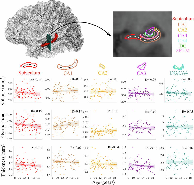 Figure 1
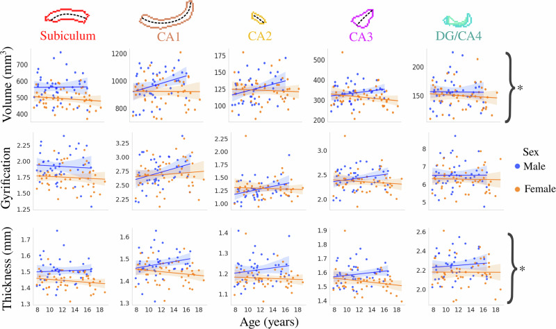 Figure 2
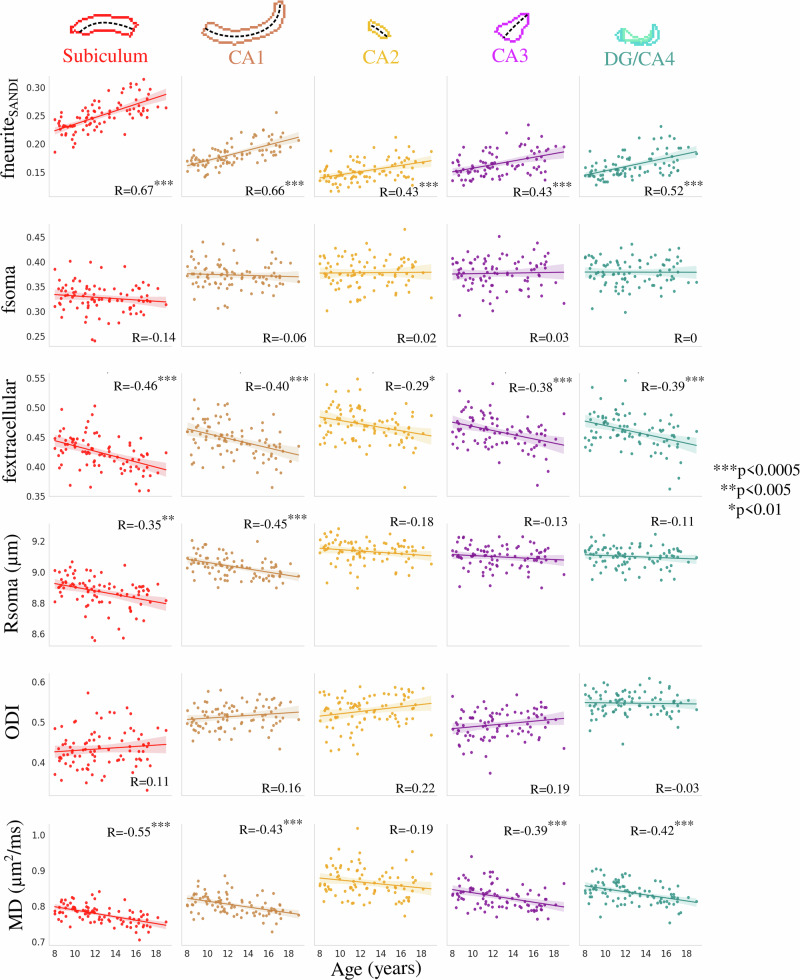 Figure 3
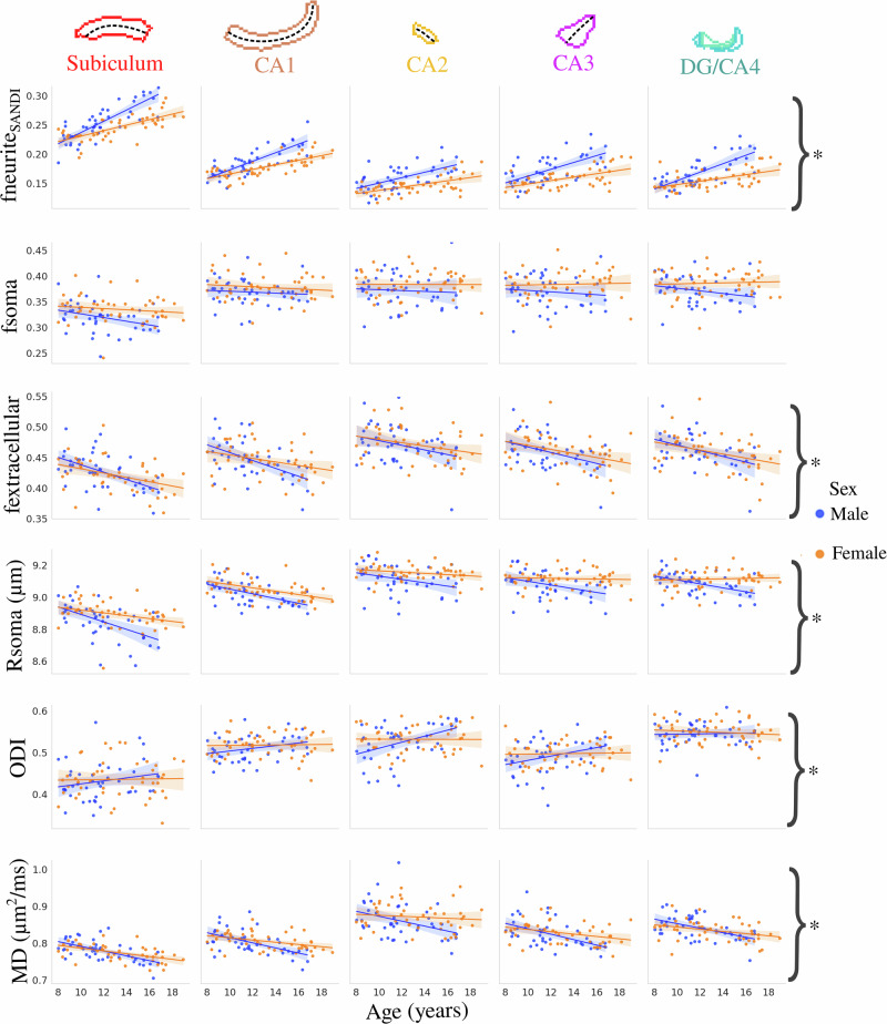 Figure 4
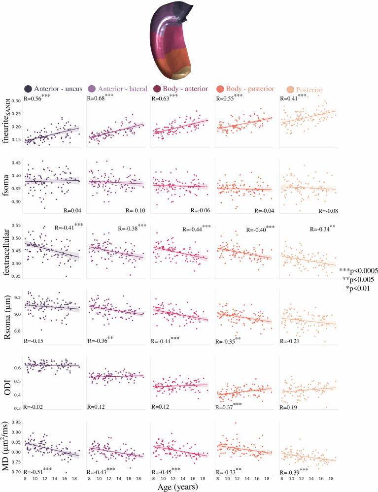 Figure 5
 Figure 6
 Figure 7
 Figure 8
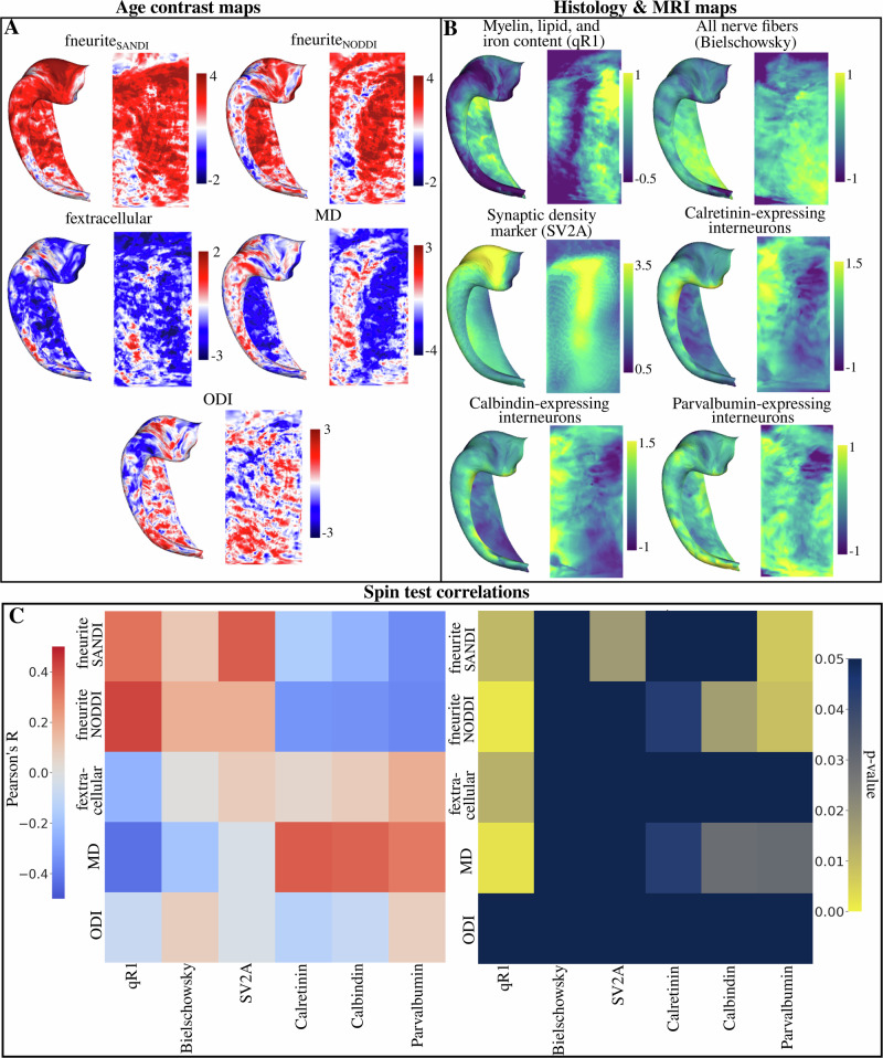 Figure 9
Peer Reviews
No public reviews on file for this paper yet. If you reviewed it on a platform where reviews are public (OpenReview, ICLR, NeurIPS, ICML), you can paste yours below so the community can read it here.
Videos
No videos yet. Explain this paper in a talk, walkthrough, or lecture? Add one.
Taxonomy
TopicsAdvanced Neuroimaging Techniques and Applications · Functional Brain Connectivity Studies · Neurogenesis and neuroplasticity mechanisms