Standardized methodology for assessing the presence, variants and area of the interthalamic adhesion using anatomical MRI (SNAP-IA): multicentric validation on 565 healthy individuals and multiple neurological disorders
Julie P. Vidal, Gonzalo Forno, Michael Hornberger, Meritxell Bach Cuadra, Lola Danet, Vinod J. Kumar, Patrice Péran, Thomas Tourdias, Emmanuel J. Barbeau

TL;DR
Researchers developed a standardized MRI method to assess the interthalamic adhesion, finding differences in its presence and size across age, gender, and neurological conditions.
Contribution
A reproducible MRI protocol (SNAP-IA) for consistent identification and quantification of the interthalamic adhesion across populations and MRI sequences.
Findings
The interthalamic adhesion was absent in 22.8% of healthy controls and showed gender and age-related differences in presence and area.
Neurodevelopmental and neuropsychiatric patients had significantly lower IA presence and smaller IA areas compared to controls.
SNAP-IA demonstrated high reproducibility (mean Dice ≈ 0.92) and average identification time of 35 seconds across MRI datasets.
Abstract
The interthalamic adhesion (IA) connects both thalami. Emerging research suggests it may support thalamo-cortical connectivity and could be involved in neurodevelopmental and neuropsychiatric conditions. However, inconsistent MRI evaluation hinders progress on this subject. We developed SNAP-IA, a standardized anatomical imaging protocol for consistent IA identification and quantification. This work leveraged the expertise from seven research teams (Toulouse, Santiago, Southampton, Lausanne, Tübingen, and Bordeaux). SNAP-IA includes three steps: (1) determination of IA presence/absence on T1-weighted MRI; (2) classification of IA variants (simple, broad, double, bilobar, and filiform); (3) segmentation-based area assessment. It was tested on 500 controls (20–69 yo) and patients (stroke, schizophrenia, bipolar disorder, and ADHD) with 0.6–1 mm isotropic T1-weighted MRI (3T to 9.4T).…
Genes, proteins, chemicals, diseases, species, mutations and cell lines named across the full text — each resolved to its canonical identifier and authoritative record.
Click any figure to enlarge with its caption.
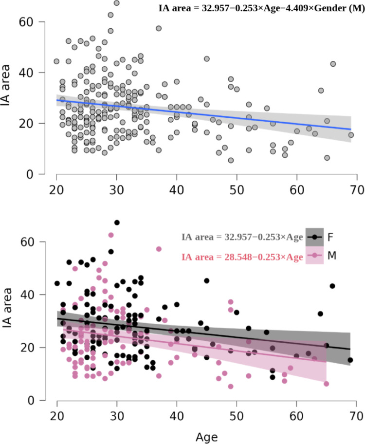 Figure 10
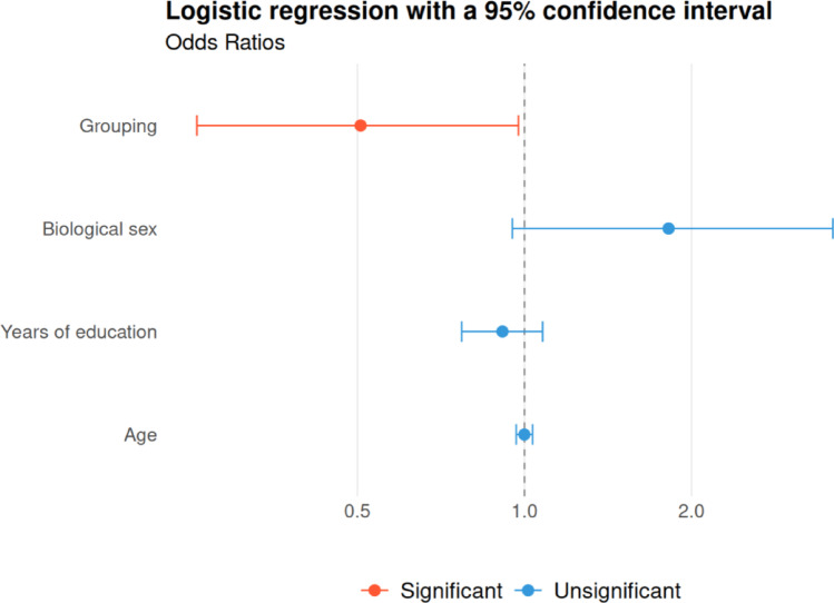 Figure 11
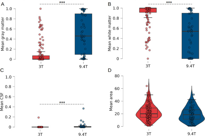 Figure 12
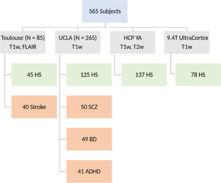 Figure 1
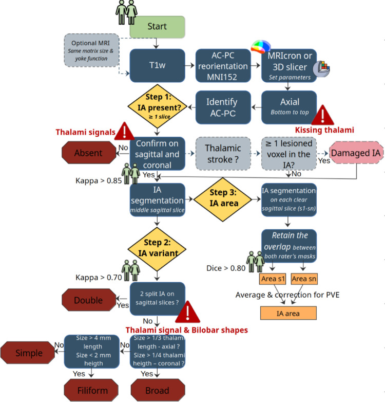 Figure 2
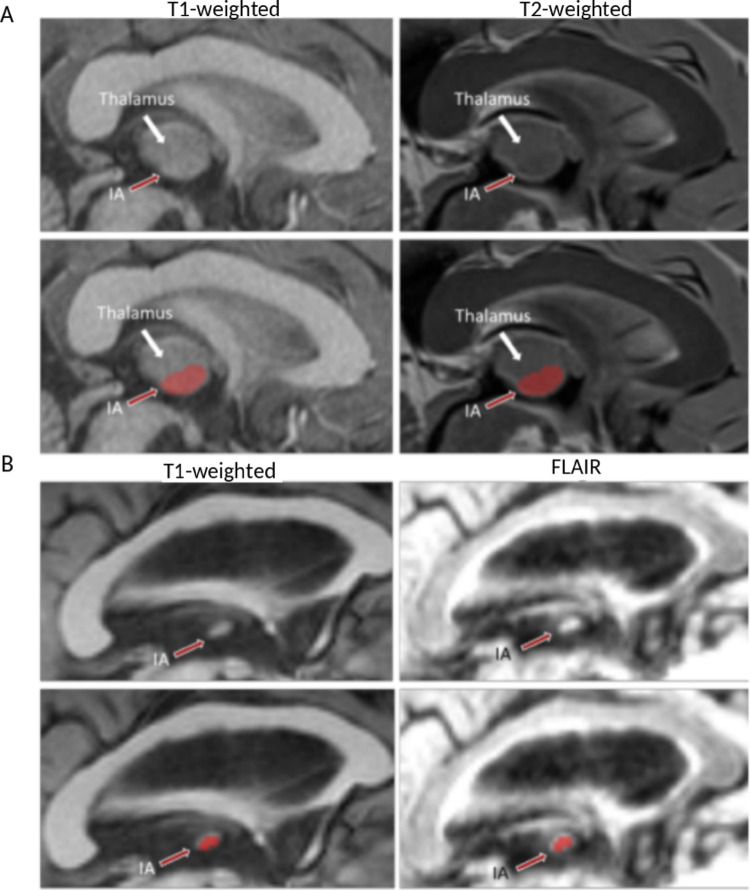 Figure 3
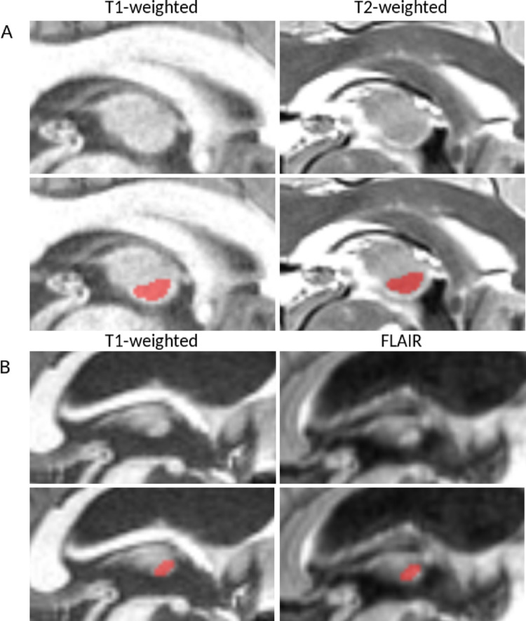 Figure 4
 Figure 5
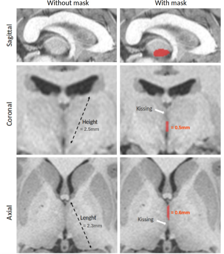 Figure 6
 Figure 7
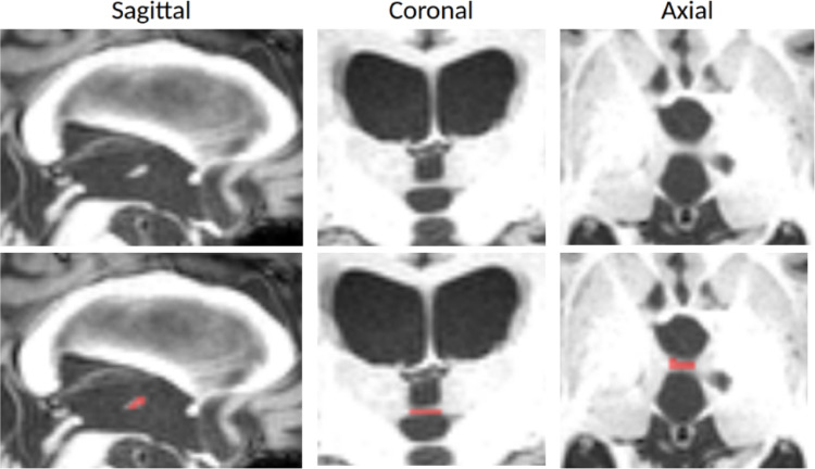 Figure 8
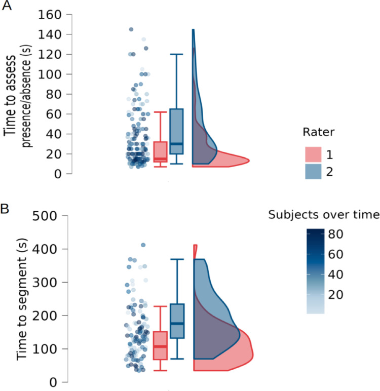 Figure 9
Peer Reviews
No public reviews on file for this paper yet. If you reviewed it on a platform where reviews are public (OpenReview, ICLR, NeurIPS, ICML), you can paste yours below so the community can read it here.
Videos
No videos yet. Explain this paper in a talk, walkthrough, or lecture? Add one.
Taxonomy
TopicsNeurological disorders and treatments · Epilepsy research and treatment · Advanced Neuroimaging Techniques and Applications