Electromechanical wave imaging vs electrocardiographic imaging: a direct comparison of non-invasive ventricular activation mapping modalities
Johanna B. Tonko, Melina Tourni, Aikaterini Afentouli, Joseph Hansen-Shearer, Biao Huang, Mengxing Tang, Anthony Chow, Elisa Konofagou, Pier D. Lambiase

TL;DR
This study compares two non-invasive methods for mapping the origin of heart arrhythmias, finding that both have strengths and weaknesses in identifying the correct location.
Contribution
The paper provides a direct comparison of EWI and ECGI for non-invasive ventricular activation mapping, highlighting their specific advantages and limitations.
Findings
ECGI correctly identified the anatomical site of origin in 77.8% of cases, while EWI did so in 80%.
EWI accurately determined transmural sites in 77.1% of cases, which ECGI could not reliably do.
ECGI excels in mapping multifocal or infrequent arrhythmias due to its panoramic single-beat mapping capability.
Abstract
Precise non-invasive identification of the site of origin (SoO) of ventricular arrhythmias (VA) could inform ablation strategies. To compare spatial accuracy of ultrasound-based electromechanical wave imaging (EWI) and ECG imaging (ECGI) to estimate the anatomical and axial (endo- vs epicardial) SoO of focal VA or pace maps employing contact mapping as gold standard. Patients awaiting a catheter ablation procedure underwent preprocedural EWI and ECGI to non-invasively map the SoO of VE/VT or RV and LV pacing sites. A commercial CT-ECGI system was used to reconstruct epicardial activation maps. Unipolar EGM morphology and slew rate were employed to estimate axial SoO. EWI was performed using high frame rate (2000fps) transthoracic echocardiography with simultaneous ECG. Contact mapping and pacing sites were used as gold standard to define SoOs. Thirty-three patients with 36 maps in…
Genes, proteins, chemicals, diseases, species, mutations and cell lines named across the full text — each resolved to its canonical identifier and authoritative record.
Click any figure to enlarge with its caption.
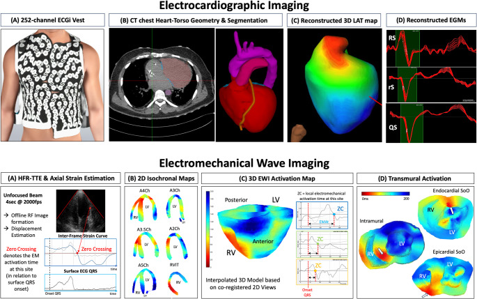 Figure 1
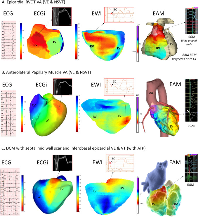 Figure 2
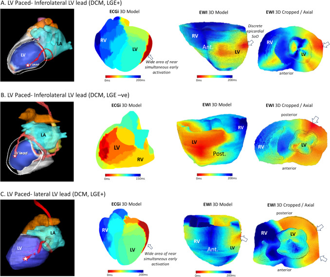 Figure 3
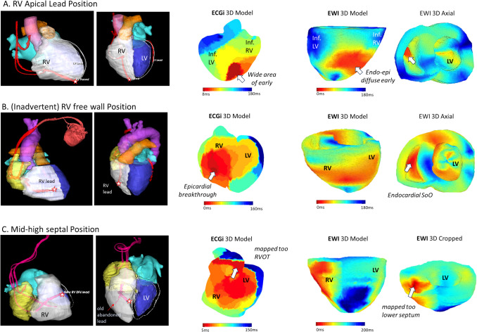 Figure 4
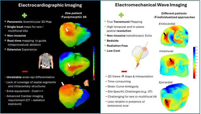 Figure 5
Peer Reviews
No public reviews on file for this paper yet. If you reviewed it on a platform where reviews are public (OpenReview, ICLR, NeurIPS, ICML), you can paste yours below so the community can read it here.
Videos
No videos yet. Explain this paper in a talk, walkthrough, or lecture? Add one.
Taxonomy
TopicsCardiac electrophysiology and arrhythmias · Cardiac Arrhythmias and Treatments · ECG Monitoring and Analysis