Prognostic impact of radiotherapy timing in WHO grade 2 and 3 meningiomas utilizing an integrated molecular-morphologic classification
Claire Delbridge, Helen X. Hou, Thomas Hielscher, Benedikt Wiestler, Chiara Negwer, Lena Schenck, Jan Peeken, Christian Diehl, Kai Borm, Sandro Krieg, Kaywan A. Aftahy, Sophia M. Leiss, Friederike Schmidt-Graf, Meike Mitsdörffer, Igor Yakushev, Andreas Von Deimling, Jens Gempt

TL;DR
This study shows that combining molecular and morphological data improves prediction of meningioma outcomes and helps determine the best timing for radiotherapy.
Contribution
The study introduces an integrated molecular-morphological score that enhances risk prediction for meningioma patients beyond traditional WHO classification.
Findings
MF-malignant meningiomas had a 0% local failure-free survival after 5 years, while MF-benign had 77%.
Copy Number Variations correlated significantly with local failure-free survival in patients receiving radiotherapy.
The IntS model showed distinct survival disparities across risk categories, emphasizing the value of combined molecular and morphological data.
Abstract
Meningiomas (MNGs) occur in different histopathological subtypes. The WHO grading system classifies a subset as grade 2 and 3, indicating a more aggressive course. Recent advances in risk stratification introduces an integrated molecular-morphological score (IntS), offering improved risk prediction over the traditional WHO classification. This study aims to evaluate the prognostic utility of IntS in the context of the timing of adjuvant radiotherapy (RT). This retrospective study analyzed 55 patients with histologically diagnosed WHO grade 2 and 3 MNG treated with adjuvant RT. Molecular analyses using Illumina 450k Human BeadChip and Illumina 850k EPIC stratified patients into 3 risk groups (low, intermediate and high) using an integrated model that combines WHO grading, Copy Number Variations (CNVs), and Methylation Families (MF). After 5 years a local failure-free survival (LFFS)…
Genes, proteins, chemicals, diseases, species, mutations and cell lines named across the full text — each resolved to its canonical identifier and authoritative record.
Click any figure to enlarge with its caption.
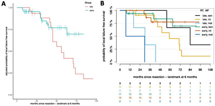 Figure 1
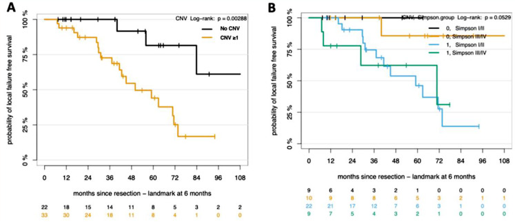 Figure 2
 Figure 3
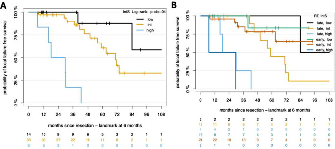 Figure 4
Peer Reviews
No public reviews on file for this paper yet. If you reviewed it on a platform where reviews are public (OpenReview, ICLR, NeurIPS, ICML), you can paste yours below so the community can read it here.
Videos
No videos yet. Explain this paper in a talk, walkthrough, or lecture? Add one.
Taxonomy
TopicsMeningioma and schwannoma management · Brain Metastases and Treatment · Neurofibromatosis and Schwannoma Cases