Acute Esophageal necrosis associated with diabetic ketoacidosis and alcohol intoxication
Arpit Shastri, K B Naveen, Arka De

Abstract
Genes, proteins, chemicals, diseases, species, mutations and cell lines named across the full text — each resolved to its canonical identifier and authoritative record.
Click any figure to enlarge with its caption.
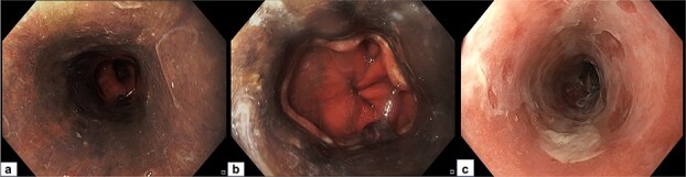 Figure 1
Peer Reviews
No public reviews on file for this paper yet. If you reviewed it on a platform where reviews are public (OpenReview, ICLR, NeurIPS, ICML), you can paste yours below so the community can read it here.
Videos
No videos yet. Explain this paper in a talk, walkthrough, or lecture? Add one.
Taxonomy
TopicsPotassium and Related Disorders · Gastroesophageal reflux and treatments · Neurological and metabolic disorders
Case
A 52-year-old male presented with complaints of coffee-brown vomitus and melena for two days. There was no associated history of jaundice or abdominal distention. The patient had reported binge drinking few hours prior to his presenting complaints. Initial investigations revealed anemia (Hb: 10.2 g/L), deranged liver function parameters (total bilirubin: 3.8 mg/dL, aspartate aminotransaminase: 154 IU/L, alanine aminotransaminase: 60 IU/L) and high blood glucose levels (454 mg/dL) with detectable urinary ketones bodies and metabolic acidosis (pH:7.2). Upper gastrointestinal endoscopy revealed erosions and diffuse circumferential and necrotic blackening of entire esophageal mucosa which terminated abruptly at gastroesophageal junction. The gastric and duodenal mucosa were spared (Fig. 1a and b). Esophageal mucosal biopsies showed loss of surface epithelium with extensive necrosis in the lamina propria, thereby confirming the diagnosis of acute esophageal necrosis (AEN). The patient was managed with intravenous fluids, insulin, antibiotics and proton pump inhibitors (PPIs). After resolution of ketoacidosis and establishment of oral intake, the patient was discharged on oral pantoprazole and subcutaneous insulin. Repeat endoscopy after 4 weeks revealed significant healing of esophageal mucosa (Fig. 1c).
a: Diffuse circumferential blackening of entire esophageal mucosa. b: Distal esophagus showing sparing of gastroesophageal junction. c: Healed esophageal mucosa after 4 weeks.
AEN or ‘black esophagus’ is a rare entity with a reported incidence of 0.008% to 0.2% [1]. Patchy or diffuse circumferential ‘black’ discoloration of esophageal mucosa which often terminates at gastroesophageal junction, with sparing of gastric mucosa is a characteristic feature. This is likely due to the increased susceptibility of ischemic esophageal mucosa to acid exposure. Risk factors include alcohol binging, diabetic ketoacidosis, connective tissue disorders, malignancy and fungal infections. Management is primarily conservative focusing on timely resuscitation, intravenous hydration, and PPIs. Ryle’s tube insertion is usually avoided for risk of perforation. As such, prompt and appropriate treatment of the underlying medical condition is the central pillar of management of acute esophageal necrosis [2, 3]. Mortality is uncommon and is usually due to the severity of underlying illness rather than esophageal complications like perforation [3].
Guarantor
Arka De.
The reference list from the paper itself. Each links out to its DOI / PubMed record.
- 1Grudell AB, Mueller PS, Viggiano TR. Black esophagus: report of six cases and review of the literature, 1963-2003. Dis Esophagus 2006;19:105–10. 10.1111/j.1442-2050.2006.00549.x 16643179 · doi ↗ · pubmed ↗
- 2Gurvits GE . Black esophagus: acute esophageal necrosis syndrome. World J Gastroenterol 2010;16:3219–25. 10.3748/wjg.v 16.i 26.321920614476 PMC 2900712 · doi ↗ · pubmed ↗
- 3Sharma V, De A, Lamoria S. et al. Black esophagus: acute esophageal necrosis due to alcohol intoxication. Trop Gastroenterol 2017;38:47–8. 10.7869/tg.391 · doi ↗