A comprehensive framework for the interpretation of TTN missense variants
Maria Francesca Di Feo, Martin Rees, Victoria Lillback, Ay Lin Kho, Angelina Meybatova, Mark Holt, Heinz Jungbluth, Francesco Muntoni, Giovanni Baranello, Anna Sarkozy, Chiara Fiorillo, Serena Baratto, Claudio Bruno, Monica Traverso, Michele Iacomino, Marina Pedemonte

TL;DR
This paper presents a new framework to interpret TTN missense variants in genetic diagnostics, combining clinical, computational, and functional data.
Contribution
A novel framework integrating clinical, computational, and functional data to classify TTN missense variants in myopathy.
Findings
30 patients with TTNtv/missense combinations showed heterogeneous myopathic phenotypes.
Proline substitutions in β-sheets of Ig domains caused impaired folding and aggregation.
Two variants, p.(Gln7023Pro) and p.(Arg25480Pro), meet ACMG criteria as likely pathogenic.
Abstract
Missense variants in TTN pose a major challenge in genetic diagnostics due to their high frequency in the general population, the large size of the gene, and the complex multidomain architecture of the titin protein. While the contribution of truncating variants (TTNtv) to titinopathies is well established, the role of rare TTN missense variants remains poorly defined. Advances in computational prediction and functional testing offer new tools to assess their potential pathogenicity, which however are currently not fully utilized for clinical application. We analyzed an international cohort of unsolved myopathy cases selected based on the presence of a rare missense variant in trans with a TTNtv. Clinical data were collected from neuromuscular centers worldwide. In silico predictions were generated using AlphaMissense and complemented by minor allele frequency (MAF) and exon usage…
Genes, proteins, chemicals, diseases, species, mutations and cell lines named across the full text — each resolved to its canonical identifier and authoritative record.
Click any figure to enlarge with its caption.
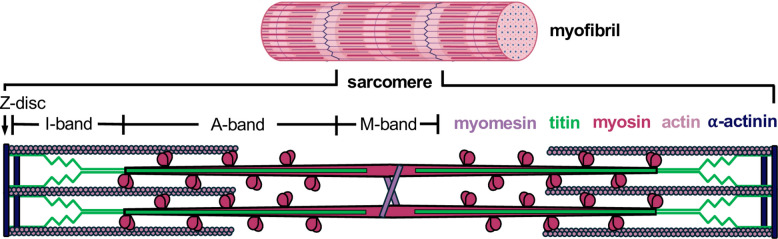 Figure 1
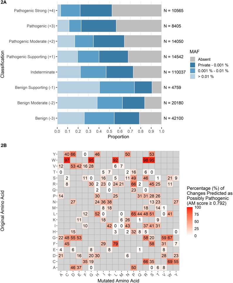 Figure 2
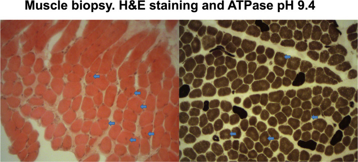 Figure 3
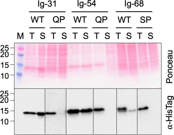 Figure 4
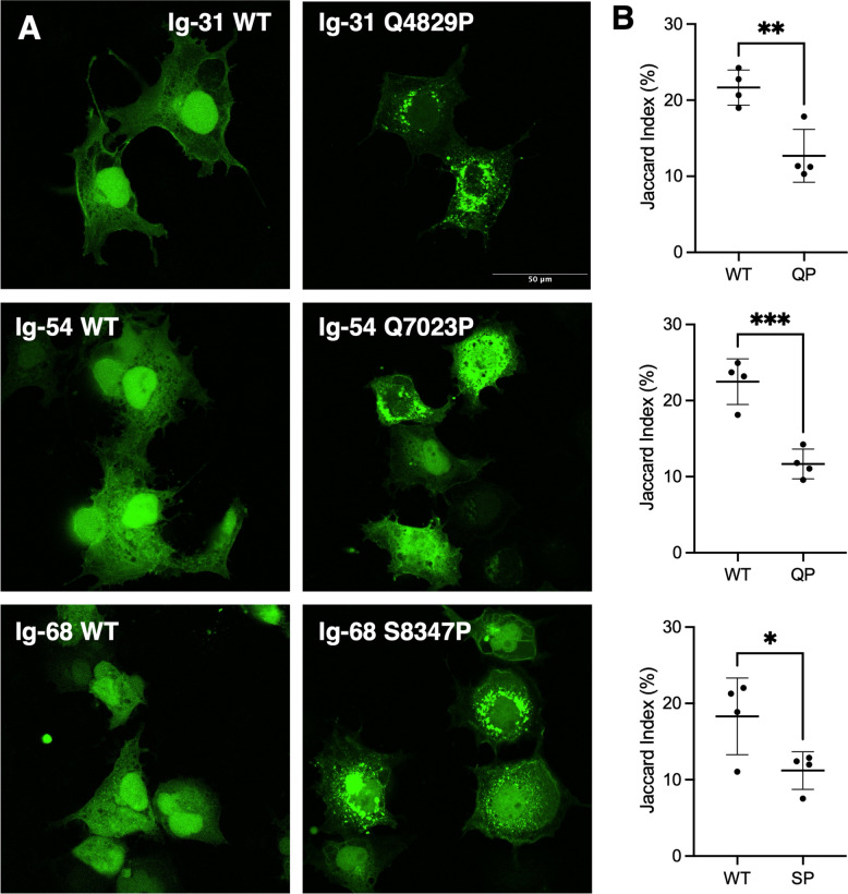 Figure 5
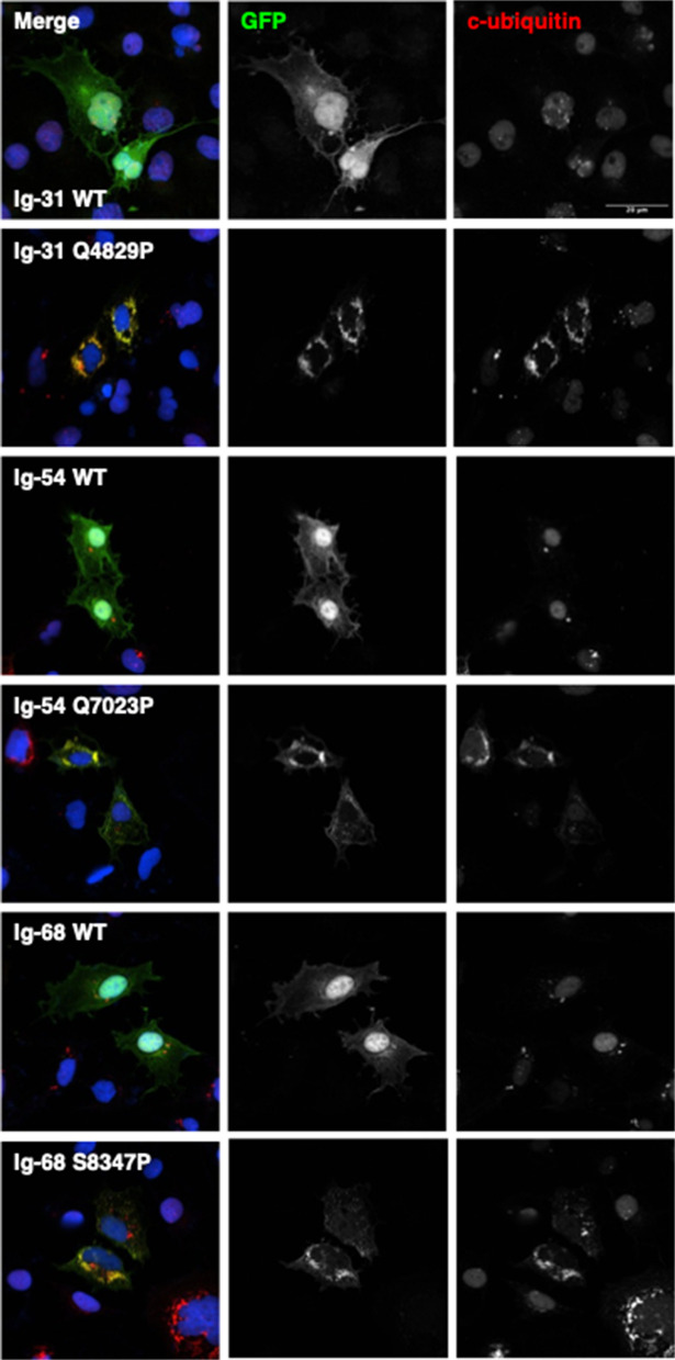 Figure 6
- —University of Helsinki (including Helsinki University Central Hospital)
Peer Reviews
No public reviews on file for this paper yet. If you reviewed it on a platform where reviews are public (OpenReview, ICLR, NeurIPS, ICML), you can paste yours below so the community can read it here.
Videos
No videos yet. Explain this paper in a talk, walkthrough, or lecture? Add one.
Taxonomy
TopicsCardiomyopathy and Myosin Studies · Genomics and Rare Diseases · Neurogenetic and Muscular Disorders Research
Background
TTN is a 294-kb gene of 364 exons that encodes the protein titin, the largest protein in the human body and among the largest known in nature [1]. Titin is expressed in striated muscle and spans half a sarcomere, the basic contractile unit of muscle, from the Z-disk to the M-band (Fig. 1). The protein plays roles in muscle development, structure, and stabilization and is composed of 132 fibronectin III-like (Fn3) domains, up to 169 immunoglobulin-like (Ig) domains, a single kinase domain, and unstructured regions [2, 3]. Each region of titin has specific features and functions: the Z-disk-anchored region links titin to the actin-based thin filament via alpha-actinin; the I-band, including the segmental duplication region of TTNspanning from exon 173 to exon 199, plays a crucial role in muscle stiffness and elasticity; the A-band region helps assemble and regulate the length of the myosin-based thick filaments whilst, finally, the M-band region contributes to sarcomere structural integrity and myofibril formation while interacting with several ligands [4]. The length and composition of TTNregions vary between isoforms, which are differentially expressed depending on the stage of development and type of muscle [5, 6]. The A-band is the most constitutively expressed region, with minimal exon usage variation reported across major transcripts. In contrast, other regions contain exons with variable percent spliced-in (PSI) values [6, 7].Fig. 1. Schematic representation of the sarcomere, illustrating its main structural regions (Z-disk, I-band, A-band, M-band) and key structural proteins
Pathogenic TTN variants cause a wide range of skeletal myopathies, cardiomyopathies or combinations of both, varying by their mode of inheritance (autosomal dominant, autosomal recessive, or digenic, the latter involving a TTNvariant in combination with a mutation in an X-linked gene), age of onset, muscle involvement, severity, and rate of progression [8–12]. Pathogenic missense variants in two specific exons, 344 and 364, cause hereditary myopathy with early respiratory failure (HMERF) and tibial muscular dystrophy (TMD or Udd myopathy), respectively, with the variants shown to render their domain insoluble or unfolded when expressed in bacteria, or to reduce the thermal stability of the domain [10, 13, 14].
Heterozygous TTNtruncating variants (TTNtv) in exons with high PSI values—representing the proportion of transcripts in which the exon is included—in transcripts expressed in the heart are observe in approximately 20% of all dilated cardiomyopathy (DCM) cases and are thought to contribute to disease by causing haploinsufficiency and/or resulting in a poison peptide [15–17].
Notably, monoallelic TTNtv are enriched in neuromuscular cohorts compared with the general population [18–21]. However, in most cases they are unlikely to fully explain the phenotype, suggesting that additional hits are often required (e.g. missense variants in trans) [22]. Indeed, most of TTN-related skeletal myopathies identified to date are linked to biallelic TTNtv [23]. Population studies have found that up to 1% of the general population carries a TTNtv, and up to 20% carry a rare missense variant with a minor allele frequency (MAF) < 1% [24, 25]. Recently, there has been increased focus on the investigation of rare missense variants throughout TTN; with the exception of pathogenic dominant variants in exons 344 and 364, however, only a few studies have addressed this issue, and fewer than a dozen missense variants linked to disease reported — mainly in sporadic cases — and with limited supporting evidence for their pathogenicity according to the criteria established by the American College of Medical Genetics and Genomics (ACMG) [22, 26, 27].
Clinical interpretation of TTNmissense variants is complex, given the large size of the gene, the frequency of the variants, and the default application of the BP1 benign “supporting” criterion (“missense variant in a gene for which primarily truncating variants are known to cause disease”) [27].
Our study represents an international effort to collect unsolved cases with a suspected recessive titinopathy to gather further evidence on rare TTN missense variants which are more likely to affect the protein function or structure. We hypothesize that missense changes in TTN may represent a major but currently frequently overlooked contributor to inherited titinopathies.
Methods
In silico prediction and variants assessment
We collected all the available predictions for missense variants in the TTNgene generated by AlphaMissense, without pre-filtering for additional criteria. AlphaMissense is the state-of-the-art artificial intelligence model developed to assess the pathogenic potential of missense variants by integrating sequence context, evolutionary conservation, and other biological features [28]. AlphaMissense provides precomputed pathogenicity predictions for all possible single amino acid substitutions in the human proteome**,and the complete dataset is openly accessible [28]. As a cut-off for AlphaMissense predictions—which range from 0 to 1, with higher scores indicating a greater likelihood of pathogenicity—we applied the thresholds proposed in the updated ClinGen recommendations for PP3/BP4 variant classification criteria [29]. In this recent work, Bergquist and colleagues assessed the validity of the most recent in silico tools to support a deleterious effect on the gene or gene product [29]. After posterior probability-based calibration, AlphaMissense reached the Strong level of evidence for pathogenicity (PP3) and at least the Moderate level for benignity (BP4), although at score thresholds more stringent than those originally recommended by its developers. We therefore applied the following recommended thresholds: ≤ 0.070 for “Benign” BP3 (−3 points); [0.071, 0.099] for “Benign Moderate” BP3 (−2 points); [0.100, 0.169] for “Benign Supporting” (- 1 point); [0.170, 0.791] for “Indeterminate”; [0.792—0.905] for “Pathogenic Supporting” PP3 (+ 1 point); [0.906—0.971] for “Pathogenic Moderate” PP3 (+ 2 points); [0.972, 0.989] for “Pathogenic” PP3 (+ 3 points); ≥ 0.990 for “Pathogenic Strong” (+ 4 points). Together with AlphaMissense predictions, we reported available data on Minor Allele Frequency (MAF) for each TTN missense variants from the gnomAD database (Additional File 1) [30]. For each variant, whether TTNtv or missense, the PSI of the corresponding exon was assessed. PSI values—derived from our recent study (open-access database: http://psivis.it.helsinki.fi:3838/TTN_PSIVIS/)—are—are) reported in Tables 1 and 2 [7].Table 1. Clinical details of patients carrying missense variants to proline (MAF < 0.010, AlphaMissense score ≥ 0.792) that have been functionally assessed in this study, in trans with a TTNtvOnsetFamily and Patient IDCountry of diagnosis[Patient/family origin]Missense variant, position and skeletal muscle PSITruncating variant, position and skeletal muscle PSIAge at last examination, sexShort clinical report(including cardiac findings)^***^Congenital onsetF1-P1, F1-P2Italy (Romania)c.14486A>Cp.(Gln4829Pro) exon 51, proximal I-bandPSI: 97% → 94%c.24729C > A p.(Cys8243Ter) exon 86 proximal I-bandPSI: 94% → 86%F1-P1, M,6 years (deceased) F1-P2, F7 yearsCongenital myopathy, scapular winging, myopathic facies;Pectus excavatum, scoliosis, underweight;Respiratory insufficiency Congenital onsetF2-P1Germanyc.55351C > T p.(Arg18451Ter) exon 287A-bandPSI:94% → 87%9 years, FCongenital myopathy, contractures at birth, scapular winging, myopathic facies;Clubfeet detected prenatally, scoliosis;Respiratory insufficiency Congenital onsetF3-P1Italyc.68575_68576dup p.(Ile22861SerfsTer14) exon 324A-bandPSI:93% → 86%12 years, FCongenital myopathy; Scoliosis;Respiratory insufficiency Congenital onsetF4-P1Russiac.61876C > T p.(Arg20626Ter) exon 305A-bandPSI:96% → 91%11 years, FCongenital myopathy, waddling gait, difficulty climbing stairs, myopathic facies;Scoliosis, underweight; Respiratory insufficiency;*No cardiac signs (only mitral valve prolapse);No family history of cardiac diseases Congenital onsetF5-P1Greecec.26173_26174delAC p.(Thr8725LeufsTer19) exon 91 proximal I-bandPSI:95% → 85%Pediatric age, MCongenital myopathy, hypotonia, difficulty climbing stairs, difficulty running Congenital onsetF6-P1US (Hungary)(BOS1693-1)^a^c.32092C > T p.(Arg10698Ter) exon 126I-bandPSI:89% → 70%17 years, FCongenital myopathy, classified as multiminicore myopathy, hypotonia, scoliosis s/p surgical repair, difficulty climbing stairs, cannot runAdult onsetF7-P1Francec.14486A > C p.(Gln4829Pro) exon 51, proximal I-bandPSI: 97% → 94%c.42472dup p.(Thr14158AsnfsTer7) exon 232A-bandPSI:97% → 91%Adult age, MNoncongenital distal myopathyAdult onset F8-P1F8-P2Swedenc.26028del p.(Trp8676CysfsTer16) exon 91I-bandPSI:95% → 85%F8-P1, 71 years (deceased), F F8-P2, 80 years, MNoncongenital myopathy, proximal weakness, facial involvementCongenital onsetF9-P1Hungaryc.25039 T > C p.(Ser8347Pro) exon 87, proximal I-bandPSI:94% → 86%c.76109_76110delTA, p.(Ile25370ArgfsTer6) exon 327A-bandPSI:97% → 93%14 years, MCongenital myopathy, contractures at birth, “frog-like” position; scoliosis, pectus excavatum; Mild facial dysmorphisms (mandibular prognathism, maxillary hypoplasia);Dysphagia at 2 months (then solved);Bi-PAP home ventilation supportPrenatal and postnatal skeletal muscle PSI are indicated for each variant’s exon, with the arrow indicating the prenatal-to-postnatal PSI change^^Cardiac phenotypes and family history were negative unless otherwise indicated^a^Internal laboratory ID for US cases; not further referenced in the manuscriptTable 2Clinical details of patients carrying missense variants (MAF < 0.010, AlphaMissense score ≥ 0.792), not functionally assessed in this study, in trans with a TTNtvOnsetFamily and Patient IDCountry of diagnosis[Patient/family origin]Missense variant, position and skeletal muscle PSITruncating variant, position and skeletal muscle PSIAge at last examination, sexShort clinical report (including cardiac findings)**Congenital-neonatal onsetF10-P1Italyc.88658 T > G p.(Leu29553Arg) exon 333PSI: 93% → 84%c.36285C > G p.(His12095Gln) exon 170 *(PSI decreasing from prenatal to postnatal)*SpliceAI delta score 0.89PSI: 57% → 10%6 years, MCongenital myopathy, arthrogryposis, myopathic facies; Micrognathia; improved (now walking with aids) Congenital-neonatal onsetF11-P1US (BOS1118-1)^a^c.23035 T > G p.(Tyr7679Asp) exon 80PSI: 96% → 85%c.47399_47402dupp.(Ser15802AspfsTer14) exon 254PSI: 97% → 94%17 years, MCongenital myopathy, classified as multiminicore myopathy, mildly delayed motor milestones, cannot run, proximal and distal weakness, mild facial weakness;*No cardiac signs;**Family history of arrhythmia/conduction disease in both paternal and maternal grandmothers (TTNtv on the paternal side; missense on the maternal side)*Congenital-neonatal onset F12-P1US (BOS1380-1)^a^c.41124 T > Gp.(Cys13708Trp)exon 226PSI: 97% → 94%c.29245C > Tp.(Gln9749Ter)exon 103PSI: 97% → 95%13 yrs, FCongenital myopathy, classified as centronuclear myopathy, unable to climb stairs;Bilateral femur fractures at birth, scoliosis s/p surgical repair;Respiratory insufficiency at birth;Dilated cardiomyopathy; Father with heart disease (deceased, unspecified), carrying the TTNtv Congenital-neonatal onsetF13-P1US (BOS1223-1)^a^c.84523 T > C p.(Trp28175Arg) exon 327PSI: 97% → 93%c.57215delp.(Gly19072GlufsTer12) exon 294PSI: 93% → 87%17 years, MCongenital myopathy with increased internal nuclei and myofibrillar disarray, gross motor delay, ambulant at age 17;Dilated cardiomyopathy (onset at 4 months of age);Maternal grandmother with tachycardia(TTNtv om the paternal side; missense on the maternal side) Congenital-neonatal onsetF14-P1US (BOS0878-1)^a^c.85302 T > A p.(Asn28434Lys) exon 327c.83281G > C p.(Val27761Leu) exon 327PSI: 97% → 93%c.103360delp.(Glu34454AsnfsTer3) exon 359PSI: 94% → 94%14 months (deceased), MCongenital myopathy, unspecific pattern, severe hypotonia, delayed motor milestones;Respiratory insufficiency; Required g-tube Congenital-neonatal onsetF15-P1F15-P2US (BOS0246-3)^a^c.99518G > A p.(Cys33173Tyr)exon 356PSI: 96% → 94%c.68885_68888dup p.(Ile22964TyrfsTer8) exon 325PSI: 92% → 87%F15-P18 year, M F15-P24 year, FCongenital myopathy, core myopathy Congenital-neonatal onsetF16-P1US (BOS0129-1)^a^c.29800 T > C p.(Trp9934Arg) exon 106PSI: 97% → 95%c.91669C > Tp.(Arg30557Ter) exon 338PSI: 91% → 83%14 years, MCongenital myopathy, classified as multiminicore myopathy, unable to climb stairs;Scoliosis s/p surgical repair; Respiratory insufficiency; Feeding difficulties from birth;*Dilated cardiomyopathy;No family history of cardiac diseases Congenital-neonatal onsetF17-P1Canadac.102200C > A p.(Ala34067Asp) exon 359PSI: 94% → 94%c.37246del p.(Ser12416ArgfsTer531) exon 181 (PSI decreasing from prenatal to postnatal)PSI: 7% → 1%1 year, FCongenital myopathy, arthrogryposis, slightly improved;Respiratory insufficiency Congenital-neonatal onsetF18-P1Russiac.14588G > A p.(Gly4863Glu) exon 51PSI: 58% → 39%(PSI decreasing from prenatal to postnatal)*c.67_81delinsCTCCAGTGGTAGTAC p.(Ala23_Ala27delinsLeuGlnTrpTer) exon 2PSI: 100% → 100%14 years, FCongenital myopathy with motor delay (acquired independent walking at the age of 3 yrs), proximal weakness, waddling gait, reduced leg reflexes, myopathic faces; Scoliosis, feet deformity;*Cardiac arrhythmia;No family history of cardiac diseases Congenital-neonatal onsetF19-P1F19-P2UKc.98171 T > C p.(Leu32724Pro) exon 353PSI: 95% → 91%c.37543 + 1G > Tintron 184 *(low PSI both in pre and postnatal muscles)SpliceAI delta score 0.99PSI: 21% → 22%19 years, M and 12 years, FNeonatal-onset myopathy, hypotonia, stable course, independent walking acquired Congenital-neonatal onsetF20-P1UKc.76439G > Cp.(Arg25480Pro) exon 327PSI: 97% → 93%c.27435G > Ap.(Trp9145Ter) exon 96PSI: 98% → 94%13 years, MReported onset at 9 months, multiple contractures, hypotonia, stable course Congenital-neonatal onsetF21-P1UK(Thailand)c.53348 T > C p.(Leu17783Pro) exon 279PSI: 97% → 92%c.39109G > T p.(Glu13037Ter) exon 203 *(PSI decreasing from prenatal to postnatal)*PSI: 35% → 21%8 years, MCongenital myopathy, hypotonia, dysphagia, *improved (now walking independently)*Childhood onsetF22-P1Italyc.100237 T > C p.(Trp33413Arg) exon 358PSI: 90% → 89%c.7856-1G > A intron 33SpliceAI delta score 1 (RNA-seq performed on muscle biopsy, splice anomalies confirmed)PSI: 99% → 96%14 years, MChildhood onset myopathy, steppage, distal > proximal weakness;No cardiac signs;Family history of cardiac disease is uncertain; father referred with suspected Takotsubo, carrying the splicing variant Childhood onsetF23-P1Estoniac.106919 T > C p.(Leu35640Pro) exon 361PSI: 94% → 94%c.91476 T > A p.(Tyr30492Ter) exon 337PSI: 92% → 84%16 yrs, FChildhood onset myopathy, post-exercise stiffness, fatigue in lower limbs during walking; biopsy showing dystrophic changes with fibrosis Childhood onsetF24-P1Chinac.25006 T > C p.(Cys8336Arg) exon 87PSI: 94% → 86%c.33938dup p.(Glu11314ArgfsTer24) exon 146(PSI decreasing from prenatal to postnatal)*PSI: 67% → 46%21 years, MChildhood onset myopathy, distal lower limbs weakness, distal contractures;Restrictive respiratory impairment;*Multifocal atrial premature beats and ventricular premature contractions;The younger brother was found to have a small ventricular septal defect at birth—no genetic testing performed Childhood onsetF25-P1Swedenc.99400 T > Ap.(Trp33134Arg) exon 356PSI: 96% → 94% c.54142 T > C p.(Ser18048Pro) exon 281PSI: 97% → 91%c.83416C > T p.(Arg27806Ter) exon 327PSI: 97% → 93%14 years, FChildhood onset myopathy, proximal lower limb weakness starting around age 3 and slow progression, myopathic facies from birth, walking acquired at 16 months (upper range of normal motor development)Late adult onsetF26-P1UKc.72146 T > C p.(Leu24049Pro) exon 327PSI: 97% → 93%c.62251G > T (p.Glu20751Ter) exon 305PSI: 96% → 91%75 years, FLate axial myopathy, mild limb-girdle with proximal lower limb involvementTachycardia;No family history of cardiac diseases^^Cardiac phenotypes and family history were negative unless otherwise indicated^a^Internal laboratory ID for US cases; not further referenced in the manuscript
Data collection and study cohort
We collected from several international neuromuscular centers TTNdata from unsolved myopathy cases carrying a heterozygous likely pathogenic/pathogenic TTNtv, in compound heterozygosity with a rare missense variant. Only cases with missense variants showing a MAF < 0.010 and an AlphaMissense score ≥ 0.792 were included in this study, for a total of 30 patients from 26 unrelated families [31]. All these patients had undergone exome/genome sequencing in their reference hospitals or as part of a resequencing project for unsolved cases, and did not carry other candidate variants with a compatible mode of inheritance (Likely Pathogenic/Pathogenic according to ACMG criteria). CNV analysis was also performed using standard computational tools integrated into the respective diagnostic pipelines. In addition, RNA sequencing (RNA-seq) was performed in two cases, and long-read sequencing in another. We excluded from the current study patients with missense variants in exons 344 and 364, as pathogenic missense variants in these exons have already been associated with specific phenotypes [10, 13]. All variants were reassessed using SpliceAI and Varsome; no evidence of splicing effects was found for variants classified as missense [32, 33]. In case of splicing variants found in trans with a missense variant, SpliceAI scores have been specified in Tables 1 and 2; all reported scores are above 0.8 [34]. For each potential amino acid substitution, we assessed key parameters including: the pathogenicity score from AlphaMissense, the recurrence of the variant across unrelated families, and the availability of functional evidence. Detailed phenotypic description and age at onset and at last examination were also collected. Biopsies, MRI reports, and clinical photographs were collected where possible as part of routine diagnostic evaluation (Fig. S1-S3: Additional File 2; Additional data and materials available upon reasonable request).
Bioinformatics
Titin domain boundaries were based on those from TITINdb [35, 36]. Alphafold-predicted structures for the Ig-31 and Ig-54 domains were retrieved from TTITINdb, and the Ig-68 domain was from the experiential structure 3B43; all were visualized using PyMol (Schrödinger Inc.) [37]. The surface accessible area of each wild type (WT) residue of interest mutated in patients was calculated using POPSCOMP data retrieved from TITINdb [35].
Cloning, protein expression and cell fractionation
Sequences encoding titin domains Ig-31, Ig-54 and Ig-68 (whose structures are shown in Fig. S4: Additional File 2) were amplified from a human skeletal muscle cDNA library by polymerase chain reaction and inserted into a modified pET-14 vector encoding an N-terminal His_6_-tag. Once vectors encoding the WT sequences were obtained, mutations encoding the missense variants p.(Gln4829Pro), p.(Gln7023Pro) and p.(Ser8347Pro) were induced by mutagenesis PCR. From these vectors, the sequences were amplified and inserted into pCMV_GFPC2 vectors. All vector sequences were confirmed by Sanger sequencing. The pET vectors were transformed into BL21-Star (Thermo Fisher) and cultured overnight in LB broth supplemented with carbenicillin (100ug/mL) at 37 °C. 80ul overnight culture was used to inoculate 4 mL MagicMedia (Thermo Fisher) which was cultured at 37 °C for 8 h and then 18 °C for 48 h. Cultures were harvested by centrifugation and stored at −80 °C.
For western blot solubility analysis, the cell pellet was resuspended in 20 mM HEPES pH7.5, 300 mM NaCl and 15 mM imidazole, supplemented with cOmplete protease inhibitor (Roche) and then lysed with B-Per (Thermo Fisher) supplemented with lysozyme (0.1 mg/mL) and Benzonase (40U/mL, Merck) for 30 min at room temperature. 20ul of this sample was mixed with 20ul SDS-PAGE loading buffer for the “total” fraction, and then the lysed cells were separated into soluble and insoluble fractions by centrifugation of 1000 g RCF at 4 °C for 30 min. 20ul of the supernatant was then mixed with 20ul SDS-PAGE loading buffer for the “soluble” fraction. The “total” and “soluble” samples were then incubated at 98 °C for 10 min.
Western blotting of protein fragments
10ul of each sample were loaded onto a precast 4–20% polyacrylamide gel (BioRad) and electrophoresis was run until the dye front reached the bottom of the gel. Following electrophoresis, proteins were transferred to nitrocellulose membrane (GE), stained with Ponceau S and imaged. The membrane was then blocked in 5% milk in low-salt buffer (LSB) for 1 h and incubated in primary antibody (anti-His tag, Novagen, 1:2000 dilution) overnight at 4 °C. After washing in LSB, the membrane was incubated in secondary antibody (HRP-tagged anti-mouse, DAKO, 1:1000 dilution) for 1 h and then washed again. HRP activity was measured following incubation with 2 ml Clarity ECL substrate (BioRad) using a ChemDoc (BioRad).
COS-7 transfection, fixation, and immunostaining
COS-7 cells were prepared using standard protocols and plated at a cell density of 7.5 × 10^4 cells per well of a 24-well plate (Azenta). Vectors encoding GFP-tagged WT Ig-31, Ig-54, and Ig-68 and their variants were transfected using Escort IV (Sigma, 1.7ul per well, added to 0.3ug DNA) according to the manufacturer’s instructions. Following incubation for 24 h at 37 °C, 5% CO_2_, cells were fixed by washing in PBS, incubated with 4% PFA for 10 min, and finally washed again in PBS. A subset of cells were permeabilised with 10 mg/ml digitonin (30 min) before being blocked with 5% normal goat serum in antibody dilution buffer (1% bovine serum albumin, 1 mM Tris pH 7.5, 15.5 mM NaCl, 0.2 mM EGTA, 0.2 mM MgCl_2_, 30 min) and incubated with a primary antibody against conjugated ubiquitin (1:100, FK2, 04–263, Merck, overnight at 4 °C) followed by Cy3-conjugated anti-mouse IgG secondary antibody (1:100 Jackson Immunoresearch, 1 h at RT), and DAPI.
Cell imaging
Widefield images for fluorescence intensity quantification: Fixed cells expressing GFP-tagged titin domains were imaged using a 10 × objective on a fluorescence widefield microscope (Leica). Confocal images were obtained using a 60 × oil immersion objective on a Nikon AXR inverted confocal microscope (Nikon Imaging Centre, King’s College London).
Image analysis
Cell expression patterns in widefield fluorescence microscope images were quantified by measuring the Jaccard Index of nuclear overlap with the whole cell mask using two different filters to segment the images into binary masks. Nuclei were identified by their size and roundness. Image segmentation and analysis were performed using a custom script written in Wolfram Mathematica 13.3 (Champaign, Il).
Statistics
Statistical differences between Jaccard Index values from the analysed images for WT-variant domain pairs were calculated using the Student’s t-test.
Results
Overall AlphaMissense predictions on titin
Of the 224,638 possible TTN single nucleotide variants resulting in missense mutations listed in Additional File 3, a total of 47,562 are predicted by AlphaMissense as possibly pathogenic (AlphaMissense score ≥ 0.792, corresponding to PP3 evidence with at least “Supporting” strength), as outlined in Fig. 2A and Fig. S7 (Additional File 2). This analysis relies on predictions generated for the canonical UniProt isoform (Q8WZ42-1), encompassing all exons present in the major adult cardiac and skeletal muscle isoforms, but excluding exons in the meta transcript. Of the variants scored as possibly pathogenic, 44,476 (93.5%) are absent from gnomAD. Only 95 out of 224,638 missense variants have a MAF > 1%, and only 4 of them have an AlphaMissense score ≥ 0.792. An additional 207 variants fall within the 0.1–1% MAF range, of which 21 have AlphaMissense scores ≥ 0.792. As illustrated in Fig. 2A, more than 80% of amino-acid substitutions with AlphaMissense scores ≥ 0.792 occur at population frequencies ≤ 0.01%. These data indicate that allele frequency alone has limited discriminatory value for TTN missense variants, as the vast majority are rare in population datasets.Fig. 2. AlphaMissense predictions for TTN missense variants. A Distribution of AlphaMissense prediction scores across Minor Allele Frequency (MAF) categories for TTN missense variants. Variants are grouped by predicted pathogenicity strength, from “Benign” (− 3) to “Pathogenic Strong” (+ 4). B Heatmap showing the proportion of amino acid substitutions in titin predicted as possibly pathogenic (Pathogenic Supporting or stronger evidence, AlphaMissense score ≥ 0.792), with enrichment for specific changes
Our analysis also identified the amino acid substitutions most frequently predicted as pathogenic by AlphaMissense, across all possible changes in TTN. The most frequently damaging change is from tryptophan: over 92% of all amino acid changes from tryptophan to another amino acid are predicted to be possibly pathogenic. Titin contains 466 tryptophan residues, from which 3,262 possible missense changes are predicted. Regarding other amino acids, the proportion of damaging changes depends on the specific combinations; e.g. changes to proline, cysteine, arginine, and asparagine are also frequently predicted to be pathogenic, but with greater variability according to the original amino acid (Fig. 2B).
Molecular and clinical findings
We selected 30 unsolved myopathy patients from 26 different families with both a TTNtv and a rare missense variant (MAF < 0.010, AlphaMissense score ≥ 0.792) demonstrated or expected to be on the other allele (Tables 1 and 2). A total of 22 distinct missense variants were identified. Two unique missense variants occurred repeatedly in 8 unrelated families, each inherited in trans with different TTNtv, while the other 20 missense variants were unique to each family. Additionally, a further family carrying the p.(Arg25480Pro) missense variant in transwith a distinct TTNtv was previously reported by Rees and colleagues [22]. Patients F14-P1 and F25-P1 each carry two candidate missense variants in cis, with both showing high AlphaMissense scores. Segregation was performed in all cases except one (F4), and the truncating and missense variants were confirmed to be inherited in trans. None of the parents exhibited muscular symptoms. Cardiac involvement was observed in 6 probands, presenting as dilated cardiomyopathy (in 3 patients), arrhythmia, premature atrial or ventricular contractions, and tachycardia. One additional proband displayed isolated mitral valve prolapse. Also, 6 relatives from 5 different families showed various cardiac findings. Overall, these manifestations were highly clinically heterogeneous, as shown in Tables 1 and 2. Although parents of patient F4 were unavailable for segregation analysis, the two variants are predicted to be in trans based on gnomAD data (Variant Co-Occurrence/Phasing tool). All 22 missense variants are found in regions of titin predicted to be folded, which make up ~ 78% of the total protein sequence. Analysis of the location of the WT amino acids missense-mutated in patients showed those mutated to non-proline residues were buried in the core of their domain, while residues mutated to proline were found on both the surface and the core of domains (Fig. S5: Additional File 2).
A total of 26 unique likely pathogenic/pathogenic TTNtv – 23 frameshift and 3 splicing variants—were identified. Both missense and truncating variants are distributed across the majority of the TTN gene, from exon 2 (the first coding exon) to exon 361.
Twenty-one patients (belonging to families F1-F6, F9, F10-F21) showed a congenital onset, with the first signs identified at birth or in early infancy. In these patients, the age at last evaluation varied from under 1 year to 17 years. Those patients all displayed generalized hypotonia, motor delay, and other variable features, such as pectus excavatum, scoliosis, myopathic facies, minor facial dysmorphisms, dysphagia, and respiratory insufficiency. Patient F9-P1 presented the most pronounced, syndromic-like craniofacial dysmorphisms, including a prominent nose with a broad nasal base and deviated septum, short philtrum, mandibular prognathism, maxillary hypoplasia, high-arched palate, and widely spaced teeth. He carries the p.(Ser8347Pro) variant, with a MAF of 6.2 × 10⁻⁷.
Of the 19 patients diagnosed with a congenital myopathy, five (26%) showed significant improvement after birth, as indicated by an asterisk in Table 2. All these individuals, except F10-P1—who was 6 years old at the time of the last follow-up —and F17-P1—who was only 1 year old—are able to walk independently without assistive devices. F10-P1 walks with support, consistent with a moderate phenotype, but can also ambulate independently for short distances. None of the individuals require ventilatory or other systemic support. All five patients carry one of the two TTN variants located in an exon with a PSI decreasing in skeletal muscles from prenatal to postnatal stage. The oldest individual in the cohort, F19-P1 (19 years old), together with their 12-year-old sibling, F19-P2, exhibit a stable disease course and can ambulate with very minimal functional limitation. They carry a splicing variant in exon 184, predicted to result in donor-site loss. This exon has low expression levels in skeletal muscle both prenatally and postnatally, with a PSI of 21% −22% respectively. The variant was found in trans with a missense variant located in a constitutively expressed exon.
Four patients (F22- F25) presented with childhood-onset symptoms, between 3 and 11 years of age. F22-P1 presented with a right-sided high-stepping gait from the age of 10, which worsened over the following years with distal weakness also on the left. At age 15, he showed right-sided weakness affecting the tibialis anterior, extensor hallucis longus, neck flexors, and finger extensors, along with mild contractures of the right tibialis anterior and elbow.
F23-P1 experienced muscle stiffness and fatigue during exercise since the age of 10. The MRI revealed early degenerative changes, predominantly in the medial gastrocnemius, and signs of early oedema in other calf muscles. Creatine kinase (CK) levels were markedly elevated (8059 U/L). Muscle biopsy, performed at 16 years of age from gastrocnemius, revealed fibers of variable shape and markedly variable size, including numerous hypertrophic and split fibers, atrophic fibers, and clusters of regenerating fibers. No inflammatory cell infiltration was observed. Occasional necrotic fibers were also observed, and many fibers show minicores.
F24-P1, who was 21 at the last evaluation, reported distal lower limb progressive weakness, with difficulty lifting his feet when climbing stairs. On examination at 11 years of age he was found to have foot dorsiflexion weakness first, followed then by proximal weakness, scapular winging and distal phalanges contractures of both hands. He also showed mild facial involvement, with asymmetry of facial muscles and inability to puff one cheek. CK levels were slightly elevated (410 IU/L). He had mild restrictive respiratory impairment and recently noted arrhythmia.
Patient F25-P1 presented with disease onset from age 3. According to the parents, she did not have neonatal hypotonia. Independent walking was achieved at 16 months, still within the normal developmental range. The patient developed proximal lower limb weakness around age 3 years, with slow progression and some difficulties climbing stairs. CK levels were normal. Muscle MRI performed at the age of 10 years, showed muscle MRI showed a pattern highly suggestive of titin-related limb-girdle muscular dystrophy type R10 (LGMD R10), with dystrophic changes in the obturator and selective involvement of the rectus femoris, vastus intermedius, semitendinosus, and, to a lesser extent, vastus medialis and biceps femoris, while the adductor magnus was relatively spared. In the lower leg, the tibialis anterior and soleus were more affected than the gastrocnemius medialis and peroneus.
Three patients sharing the same missense variant, F7-P1, F8-P1, F8-P2, had an adult onset, although with different pattern of muscle involvement between the two families.
At the most extreme end of the late-onset spectrum,, F26-P1 presented from around 70 years of age with an axial myopathy and bent spine; on neurological evaluation, mild proximal limb girdle involvement was also detected.
Recurring variants
Seven patients from six different families from Romania, Germany, Hungary, Russia, Greece, and Italy (F1-F6) carry the missense variant p.Gln7023Pro in compound heterozygosity with a TTNtv, which is different in each family (Table 1). p.Gln7023Pro has an AlphaMissense score of 0.81 and a MAF of 0.000005578 with only 9 alleles observed in the European population, and no reported homozygous carriers in the gnomAD database. All the patients displayed a very similar congenital myopathy phenotype, with a similar disease course. General hypotonia and muscle atrophy from birth, scoliosis, respiratory insufficiency, myopathic facies and a low weight for age were reported. All parents were reported to be healthy.
Muscle biopsies from F1-P1 and F1-P2 (both from vastus lateralis) showed common features, including variation in fibre size, increase in endomysial connective tissue, internally placed nuclei, and granulous basophilic material inside, as displayed in Fig. 3 and in Fig. S1, Additional File 2.Fig. 3. Muscle biopsy from the quadriceps (vastus lateralis). Hematoxylin and eosin staining (left) shows fiber size variability, internally displaced nuclei (arrows), and mild endomysial fibrosis. ATPase staining at pH 9.4 (right) reveals type 1 fiber predominance and perinuclear clearing (arrows)
We were able to retrieve a list of all TTN variants, including polymorphisms, identified in F1, F2, F5 and F6 carrying variant p.Gln7023Pro. Forty-five shared variants across the gene were identified in families F1, F2, F5 and F6 (Additional File 4). Regarding F4-P1, the phase was not available, and we were able to collect only pre-filtered data, including four missense variants, one of which was p.Gln7023Pro. Notably, F4-P1 shared two of the 45 variants found in the common haplotype with families F1, F2, F5 and F6. These two variants have gnomAD MAFs of 0.018 and 0.014, respectively. Overall, these data suggested the presence of a common haplotype among the five families, consistent with inheritance from a shared European ancestor.
Variant p.(Gln4829Pro) has a MAF of 0.0001909, with 308 alleles found mainly in the European population, and no reported homozygous carriers in the gnomAD database. It was found in three patients with adult onset from two different European families, F7 and F8, carrying in trans TTNtv respectively in the I-band and A-band. Patient F7-P1 showed symmetrical scapular winging, facial weakness, and distal lower and upper limb involvement with hand muscle atrophy. On muscle biopsy, central loss of NADH staining in muscle fibers was observed, sometimes with core-like lesions, with granulous basophilic material inside (Fig. S2: Additional File 2). Two siblings, F8-P1 and F8-P2, showed facial weakness and predominantly proximal involvement of the lower limbs. MRI of the lower limbs shows selective fatty replacement predominantly affecting the posterior thigh and calf muscles, with relative sparing of anterior compartments (Fig. S3: Additional File 2).
Disease-linked missense variants prevent soluble expression of titin domains in bacteria
Three of the rare missense variants, collectively found in 11 of the 30 patients presented here, were studied in vitro to assess the effect of the variants on domain stability and aggregation. Titin Ig-31, Ig-54 and Ig-68, and their variants p.(Gln4829Pro), p.(Gln7023Pro) and p.(Ser8347Pro), respectively (Fig. S4: Additional File 2), were expressed in bacteria and their solubility was assessed by lysing the cells, separating the soluble and insoluble material and determining in which fraction they were found. Western blotting using an antibody that recognised the recombinant proteins’ His-tag showed that while all domains were expressed and the three WT domains were found in the soluble fraction, all three variant domains were absent, demonstrating that the variants prevented the soluble expression of their domain (Fig. 4).Fig. 4. Assessment of solubility of WT and missense variant-containing titin domains. Western blot probing presence of His-tagged titin domains Ig-31 WT and Q4829P, Ig-54 WT and Q7023P and Ig-68 and S8347P in the soluble fraction of bacterial expression lysate. T = total lysate, S = soluble fraction, M = molecular weight marker
Disease-linked missense variants alter the expression pattern of titin domains in COS-7 cells
GFP-tagged constructs of the same WT and variant titin domains were expressed in COS-7 cells, and the pattern of expression and co-localisation with a marker of proteostasis were assessed using fluorescent microscopy. Confocal micrographs of the transfected cells showed that the WT titin domains had a diffuse expression pattern throughout the cell, with stronger localisation to the nucleus. In contrast, the variant domains had a largely punctate, presumably aggregated appearance, and were excluded from the nucleus (Figs. 5A and 6). We quantified these differences by analysing widefield images of transfected cells and measuring the Jaccard Index of nuclear overlap with the whole cell mask; all three WT-variant pairs showed a significant difference in their cell expression pattern (Fig. 5B, Fig. S6: Additional File 2).Fig. 5. Expression of WT and missense variant-containing titin domains in COS-7 cells. A Confocal microscopy images showing expression of GFP-tagged titin domains Ig-31 WT and Q4829P, Ig-54 WT and Q7023P and Ig-68 WT and S8347P. B Quantification of widefield fluorescence microscopy images of the expression pattern of GFP-tagged titin domains. A lower Jaccard index correlates with a more punctate expression pattern. * = P < 0.05, ** = P < 0.005 *** = P < 0.0005Fig. 6Localisation of conjugated ubiquitin in COS-7 cells expressing titin domains. Confocal microscopy images showing expression of GFP-tagged titin domains Ig-31 WT and Q4829P, Ig-54 WT and Q7023P and S8347P (GFP, green) co-stained with an antibody that recognises conjugated ubiquitin (c-ubiquitin). Regions of co-localization of GFP-tagged protein and conjugated ubiquitin are shown in yellow. Nuclei are shown in blue
As we presumed the punctate expression was due to the missense variant domains aggregating as a result of the variant preventing correct protein folding, we co-stained the fixed cells with an antibody that recognises conjugated ubiquitin (c-ubiquitin), a protein that is enzymatically linked to misfolded proteins targeted for degradation via the ubiquitin-proteosome system or autophagy pathway (Fig. 6). While there was little co-localisation of GFP and c-ubiquitin for the WT domains, c-ubiquitin frequently co-localised with the variant-containing domains and displayed the same punctate appearance, suggesting that the variant domains are misfolded and are being targeted for degradation.
Discussion
Our work collected evidence that rare missense variants can contribute to recessively inherited titinopathies, presenting with a broad phenotypic spectrum. To support a more systematic and scalable approach to variant interpretation, we also aimed to develop a practical framework for assessing TTN missense variants in a diagnostic setting, integrating in silico prediction tools, functional assays and clinical data.
AlphaMissense and MAF
In our cohort, AlphaMissense proved to be a valuable tool to support the application of the ACMG criterion PP3 for the missense variants identified, providing evidence of pathogenicity based on in silico predictions. AlphaMissense scores can play a crucial role in guiding the interpretation of the vast number of missense variants in TTN, as already demonstrated for genes as BRCA1 and TP53 [38, 39]. As shown in several independent studies, AlphaMissense, VARITY, ESM1b and REVEL converge to similar calibrated evidence strengths and classify comparable proportions of variants, indicating that applying multiple predictors in parallel would be redundant [29, 40]. MAF and the biochemical properties of any given amino acid are among the primary features considered by AlphaMissense [28]. For instance, tryptophan's large, hydrophobic structure and its key position within Ig and FN3 domains—where it helps stabilize the hydrophobic core—make its substitution particularly disruptive to protein structure [41, 42]. In our cohort, indeed, there are 3 variants from tryptophan and one to tryptophan. Similarly, proline, due to its rigid cyclic structure, imposes conformational constraints that can severely impact protein folding and function, even in Ig-domains [43]. Besides proline, also arginine, despite its markedly different structure, can disrupt local protein folding due to its basic and bulky side chain, especially when replacing an amino acid that contributes to the core of titin’s Ig and Fn3 domains. In our cohort, nine changes are to proline and seven to arginine, making them the two most frequent resulting amino acids.
However, regardless of the MAF, our analysis showed that approximately 40% of all possible missense variants in titin have an AlphaMissense score of between 0.170 and 0.791 (“indeterminate”) which does not allow us to apply either criterion PP3 or BP4 to these (Fig. 2A).
All missense variants in our cohort have a MAF < 0.0001, with the exception of three cases. The most frequent variant is p.Leu24049Pro, with a gnomAD MAF of 0.0012, 4 homozygous individuals in gnomAD, and an AlphaMissense score of 0.989. Like most others, this variant remains classified as a VUS, and in the absence of functional studies we lack additional evidence to support its pathogenicity. However, it is worth noting that the phenotype in F26-P1 is very late-onset and falls within the mild end of the spectrum; axial myopathies in elderly patients are often underdiagnosed, and no precise prevalence estimates currently exist.
The second most frequent missense variant is p.(Gln4829Pro), which has a MAF of 0.0001909, that affects domain folding and alters the expression pattern. The variant is found in families F7 and F8, which both show an adult-onset phenotype. The third, p.(Val27761Leu) was found in patient F16-P1, who had a congenital onset but carries the variant in cis with another missense variant, which is rarer, but has a similarly high AlphaMissense score. Importantly, no congenital case in our cohort involves a missense variant with MAF > 0.0001, which is in line with expectations considering the population prevalence of congenital myopathies.
Among the 3,086 missense variants predicted as possibly pathogenic by AlphaMissense and found in gnomAD, 96 were observed in homozygosity in at least one individual. Twenty-one had a MAF > 0.001 and four have a MAF > 0.01, possibly reflecting AlphaMissense overestimation of a variant’s pathogenicity due to domain redundancy and structural similarity in the titin protein. Filtering out rare variants with a few homozygotes is less straightforward, however, as some may be hypomorphic and clinically relevant only in trans with a TTNtv, particularly in high-PSI exons.
Nonetheless, after removing variants observed in homozygosity, we estimated a cumulative frequency of high-scoring missense alleles that—combined with a 0.05% prevalence of TTNtv in constitutively expressed exons (i.e., high-PSI TTNtv only), and assuming 50% chance of trans configuration—would result in ~ 1 in 9,150 individuals carrying both a TTNtv and a missense variant of possible clinical significance. While this high prevalence may partly reflect underdiagnosed or mild late-onset cases, our data suggest that AlphaMissense tends to overestimate pathogenicity in TTN, and that variant interpretation in this gene requires additional considerations. Overall, considering these findings in the context of our current knowledge, for severe, early-onset myopathies, a MAF cut-off of < 0.0001 appears appropriate. In milder cases with adult or late onset, filtering by MAF becomes even more challenging. As a general rule, we suggest that the higher the MAF, the stronger the other lines of pathogenicity evidence must be for a variant to be considered potentially disease-causing.
ACMG criteria
Among the reported variants, only p.(Gln7023Pro) – functionally assessed here and identified in six unrelated families – and p.(Arg25480Pro) – previously functionally assessed and now reported in a second, unrelated family – meet the criteria for classification as likely pathogenic. The ACMG/AMP criteria supporting this reclassification are described in Additional File 5.
In the case of p.(Gln7023Pro) and p.(Arg25480Pro), the experimental assays provided variant-specific functional evidence, with direct clinical implications. Additionally, RNA-seq performed on individual F1-P2 did not reveal any splicing alterations, further reinforcing the interpretation of this specific missense variant. Without such assays, it would have been more appropriate to consider the shared allele observed in the six families as pathogenic, rather than attributing pathogenicity directly to the variant itself, since the possibility of another undetected variant in cis could not be excluded. This issue is particularly relevant for other missense variants identified in our cohort, for which no functional assays or second-tier analyses (such as RNA-seq or long-read sequencing) are available. In F25-P1, we found it more appropriate to suggest the pathogenicity of the allele, as the variant p.(Trp33134Arg) has been identified in a single patient and in cis with a second rare missense change, p.(Ser18048Pro) also predicted to be deleterious (Alphamissense score: 0.9851), as reported in Table 2, and we cannot accurately determine the specific contribution of each variant. In F17-P1, carrying in-trans variants p.(Trp9934Arg) and p.(Arg30557Ter), long-read sequencing (Nanopore) was performed and ruled out the presence of other variants of interest, including structural variants and copy number variations (CNVs). In the remaining cases, the possibility of undetected in-cis variants—such as additional VUS, non-canonical splicing events, or CNVs—cannot be confidently excluded and should be acknowledged as a limitation.
Notably, in large and complex genes such as TTN, segregation across multiple informative meiosis—while valuable—typically provides strong evidence for the pathogenicity of the allele as a whole, rather than of the specific variant. To establish variant-level pathogenicity, additional supporting evidence is required. This may include the identification of unrelated cases carrying the same variant on distinct haplotypes, or functional assays specifically addressing the variant’s effect.
Notably, in patients presenting with a heterozygous TTNtv, distinguishing between an incompletely characterized recessive mechanism and potential dominant forms requires careful clinical and genetic evaluation of the family. In cases presented here, the presence of unaffected heterozygous parents and the identification of a rare missense variant in trans support a recessive mechanism.
Functional assays
Our functional studies demonstrate that p.(Gln4829Pro), p.(Gln7023Pro) and p.(Ser8347Pro) impair protein folding and stability, and promote protein aggregation in vitro (Fig. 5, Fig. S6, Additional File 2). These findings are consistent with the known role of proline as a β-sheet breaker, due to it disrupting local folding on account of its rigid structure and its inability to act as a main chain hydrogen bond donor. To gain additional evidence, the surface accessibility of the WT amino acid mutated in patients was determined, and defined as either a surface-exposed or core residue (Fig. S6, Additional File 2) [26]. All not-to-proline mutations occur at buried sites, suggesting a disruption of domain stability. In contrast, some substitutions to proline affect more exposed residues but still appear pathogenic, likely due to proline’s structural rigidity and its tendency to disrupt local secondary structure, even when positioned on the domain surface (Fig. S6: Additional File 2) [35, 36]. All missense variants in the cohort are located within structured domains of titin (i.e., Ig, Fn3, or kinase domains). This may reflect the greater pathogenic potential of variants in these conserved regions, but it is also important to acknowledge a potential bias: AlphaFold is less accurate in predicting unstructured regions, and therefore AlphaMissense may not provide reliable predictions for variants in those regions, similarly to its lack of predictions for meta exons.
In addition, while functional assays do provide important mechanistic insights, several limitations on their use must be considered. Titin is an exceptionally large and modular protein, and such assays are performed on a single domain. As a result, they may not accurately recapitulate the structural and functional complexity of the full-length protein in vivo, including domain–domain interactions, ligand interactions, and tissue-specific regulation. While it is known that heat shock proteins such as alphaB crystallin, HSP27 and HSP90 can locate to the I-band region of titin in cardiac and skeletal muscle of myopathy patients, the specific titin domains these proteins bind to have yet to be elucidated, with the exception of some possible sites for alphaB crystallin at the N2B-us, PEVK and Ig-85/Ig-86 (previously referred to as I26/I27 in the old I-band nomenclature) [44–48] To our knowledge, no other proteins have been shown to bind to Ig-31, Ig-54 or Ig-68, making it difficult to predict any effect the missense variants studied here would have on protein–protein interactions; it is, however, possible that the destabilised domains may recruit chaperones such as those mentioned above.
Functional studies are time- and resource-intensive, making them impractical for high-throughput applications across the vast number of TTN missense variants observed in patients. In the future, scalable and standardized functional assays will be essential to enable broader variant characterization in critical titin regions. At the same time, it will be important to carefully assess the weight of these assays in variant classification frameworks. Most likely, the molecular and cellular impact of the missense variants reported here is further modulated by the TTNtv in trans—in particular, by whether the in-trans allele undergoes nonsense-mediated decay and by exon-usage patterns. As a consequence, more cases would be needed to begin establishing robust genotype–phenotype correlations for TTNtv-missense combined genotypes.
Lastly, no pathogenic variants in other muscle disease genes suggestive of digenic inheritance were detected; although such mechanisms appear unlikely- particularly in cases with recurring and functionally assessed variants—we cannot entirely rule out unknown forms of digenic inheritance.
Conclusions
Establishing the pathogenicity of missense variants in TTN remains challenging, even when functional assays are available, as is accurately predicting their in vivo effects. In this study, we demonstrated that additional insights can nevertheless be gained through the in-depth analysis and collection of clinical cases, as exemplified by the emblematic case of a novel haplotype defined by the now likely pathogenic p.(Gln7023Pro) variant. Also, we suggested that aggregation and disruption of the β-sheet secondary structure in Ig-domains may represent a potential mechanism of pathogenicity for selected missense changes. However, genotype–phenotype correlations cannot be attempted yet: the clinical spectrum is broad, as are the titin domains involved, and many other cases would be required. Novel tools such as AlphaMissense can aid in the interpretation of TTN missense variants; however, their tendency to overestimate pathogenicity and other limitations must be carefully considered, and high AlphaMissense scores should not be used as stand-alone diagnostic evidence. A direct comparison of the accuracy of available prediction tools is currently limited by the scarcity of robust functional data on TTN missense variants. The development and application of scalable experimental assays will be essential to enable such analyses in the future, which, although beyond the scope of this study, represent a key priority for the field. We recommend that missense variants with potential pathogenicity based on in silico predictions, low MAF, or other supporting evidence be systematically reported in clinical records and internal databases when found in trans with a TTN truncating variant in patients with a compatible myopathic phenotype. Whenever feasible, these findings should also be shared across international variant repositories and neuromuscular networks to promote data integration and support future reclassification efforts.
Supplementary Information
Additional file 1. List of rare TTN missense variants considered in this study. AlphaMissense scores and MAF from gnomAD are reported. Additional file 2. Supplementary clinical, histological, and functional data. Additional file 3. AlphaMissense predictions for all 224,638 possible TTN missense single nucleotide variants based on the canonical UniProt isoform. Additional file 4. Shared TTN haplotype in families carrying the p.variant. List of TTN variants defining a common haplotype identified in multiple unrelated families carrying the likely pathogenic variant p. Additional file 5. ACMG/AMP evidence supporting the reclassification of p.(Gln7023Pro) and p.(Arg25480Pro) as likely pathogenic.
The reference list from the paper itself. Each links out to its DOI / PubMed record.
- 1Hackman P, Savarese M, Di Feo MF, Udd B, Salih MA. Salih Myopathy.In: Gene Reviews® [Internet]. Seattle (WA): University of Washington, Seattle; 1993–2023. NBK:83297.; https://ncbi.nlm.nih.gov/books/NBK 83297/.22238790 · pubmed ↗
- 2Di Feo MF, Paramonov I, Borrel LM, et al. The burden of TTN variants in the genomic era: analysis of 18,462 individuals from the Solve-RD consortium and general recommendations. Genet Med. Published online November 2025:101649. 10.1016/j.gim.2025.10164910.1016/j.gim.2025.10164941277541 · doi ↗ · pubmed ↗
- 3Vatta M, Regalado E, Parfenov M, et al. Analysis of TTN truncating variants in >74 000 cases reveals new clinically relevant gene regions. Circ Genom Precis Med. Published online February 19. 2025. 10.1161/CIRCGEN.124.00498210.1161/CIRCGEN.124.004982 PMC 1199909939968638 · doi ↗ · pubmed ↗
- 4Laddach A, Mathias G, Franca F. TITI Ndb. https://titindb.kcl.ac.uk/TITI Ndb/. Accessed 3 June 2025