Vinorelbine enhances the efficacy of oncolytic vaccinia virus in a preclinical model of ovarian high-grade serous carcinoma
Stephanie Drymiotou, Christophe J. Queval, Katherine E. Tyson, Lesley A. Sheach, Antonio Postigo, Ilaria Dalla Rosa, Darren P. Ennis, Michael Howell, Iain A. McNeish, Michael Way

TL;DR
Vinorelbine improves the effectiveness of oncolytic vaccinia virus in treating ovarian cancer in preclinical models.
Contribution
The study identifies vinorelbine as a combination agent that enhances oncolytic vaccinia virus efficacy in ovarian cancer.
Findings
Vinorelbine and vaccinia virus combination induces apoptosis in murine ovarian cancer cells.
Combining ΔVFTK-NG-GM-CSF virus with vinorelbine prolongs survival in a mouse model of ovarian cancer.
Vinorelbine is a tubulin polymerization inhibitor that synergizes with oncolytic vaccinia virus.
Abstract
Vaccinia virus, known for its clinical safety, has a tropism for primary and metastatic tumors as well as ovarian tissue. Consequently, oncolytic approaches with recombinant vaccinia viruses have emerged as attractive agents against ovarian cancer. Unfortunately, oncolytic vaccinia monotherapies are yet to live up to their potential promise. Given this, there is a need to identify combination agents that improve the effectiveness of vaccinia in ovarian cancer treatment. We screened 9,000 compounds to identify drugs that enhance the ability of a recombinant vaccinia virus lacking VGF and F1 (ΔVF) to induce death of ID8 Trp53−/− murine ovarian cancer cells. We identified a class of tubulin polymerization inhibitors including vinorelbine. The combination of vinorelbine and vaccinia induces ID8 Trp53−/− cell death via apoptosis. In a syngeneic mouse model of high-grade serous ovarian…
Genes, proteins, chemicals, diseases, species, mutations and cell lines named across the full text — each resolved to its canonical identifier and authoritative record.
Click any figure to enlarge with its caption.
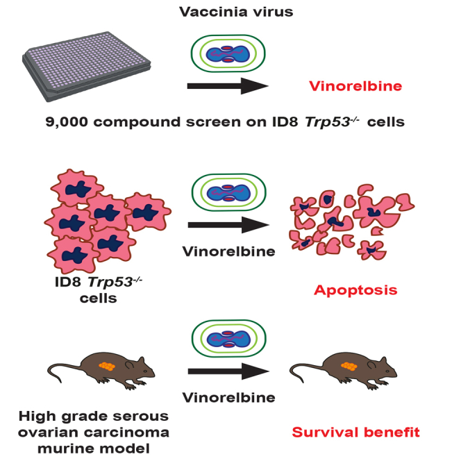 Figure 1
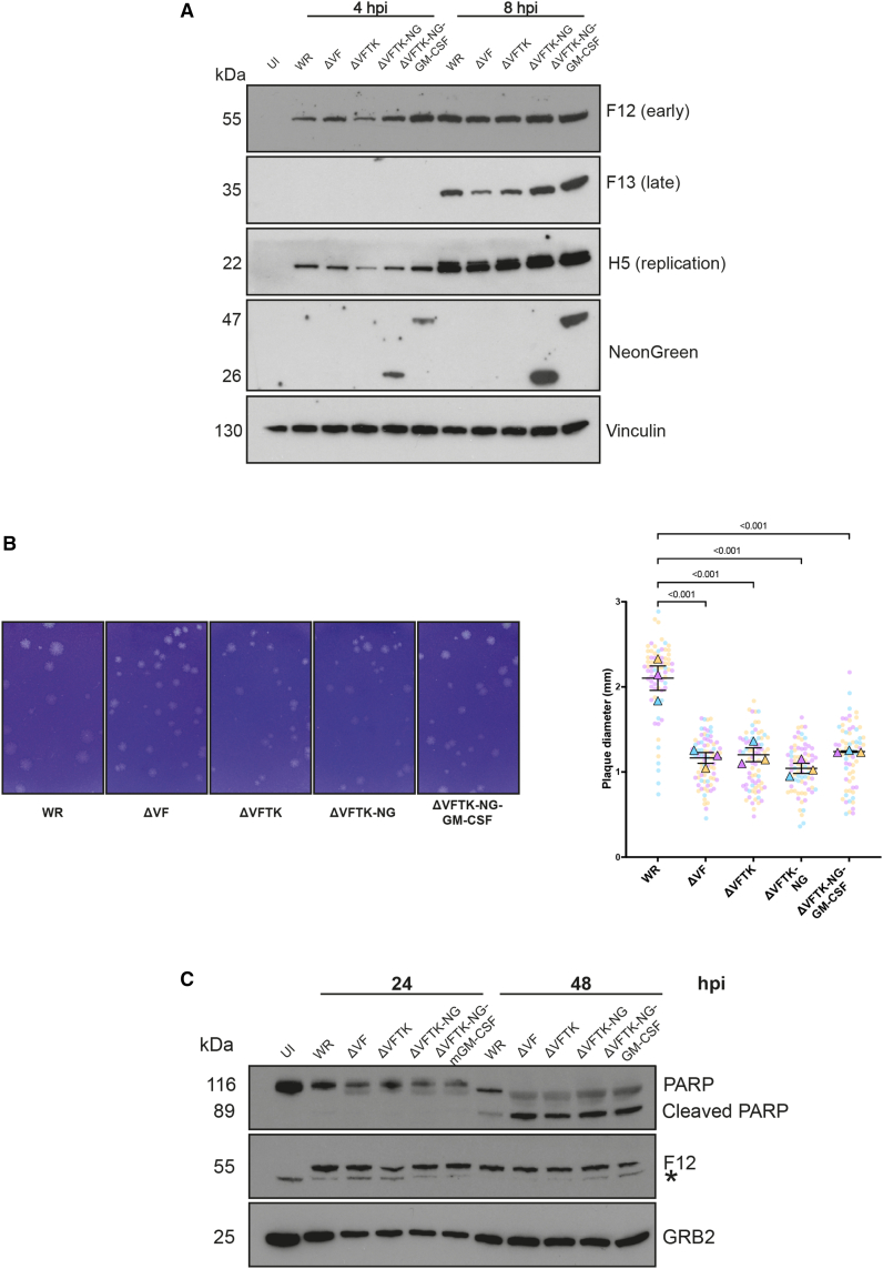 Figure 2
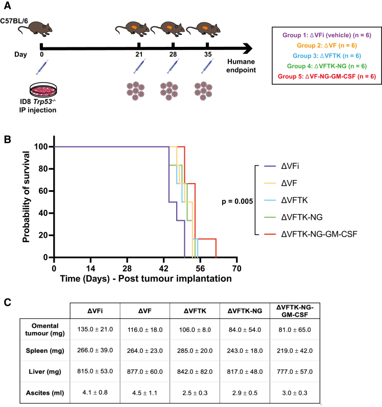 Figure 3
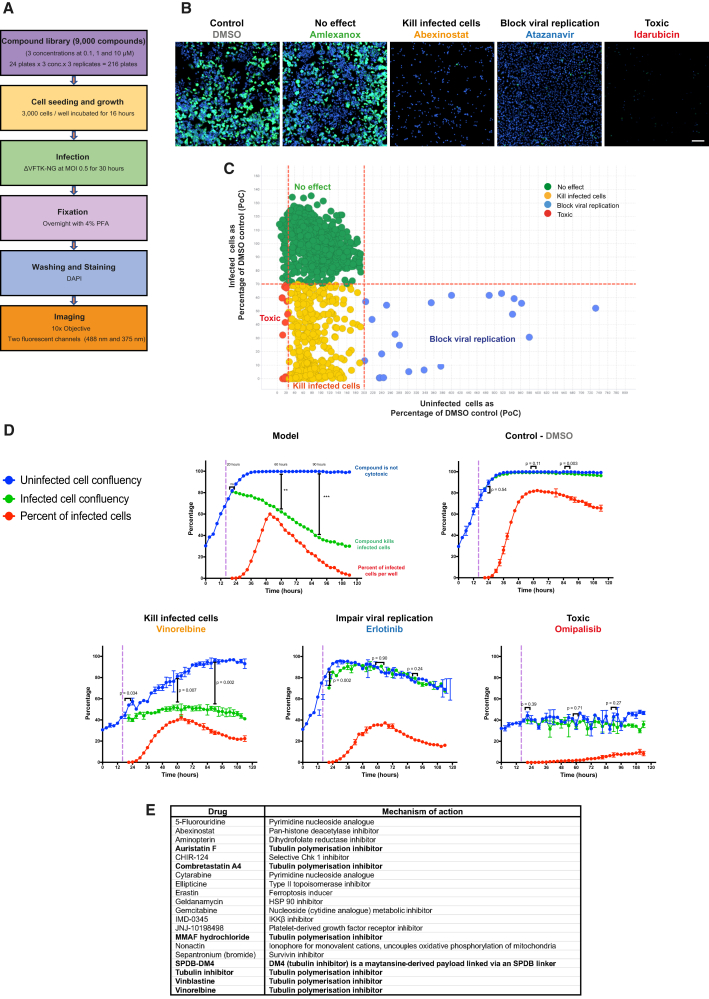 Figure 4
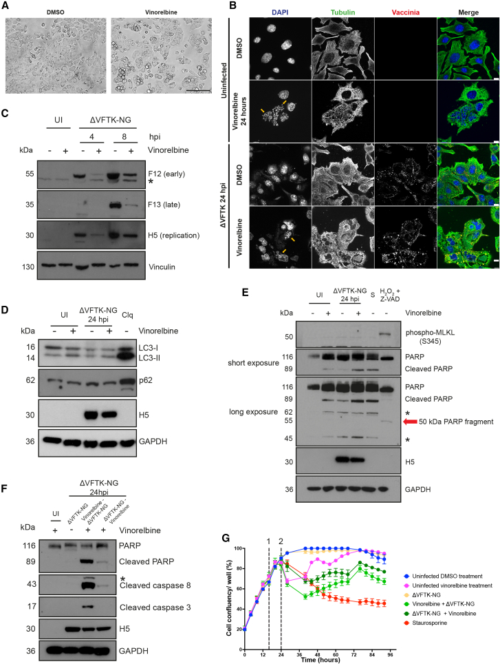 Figure 5
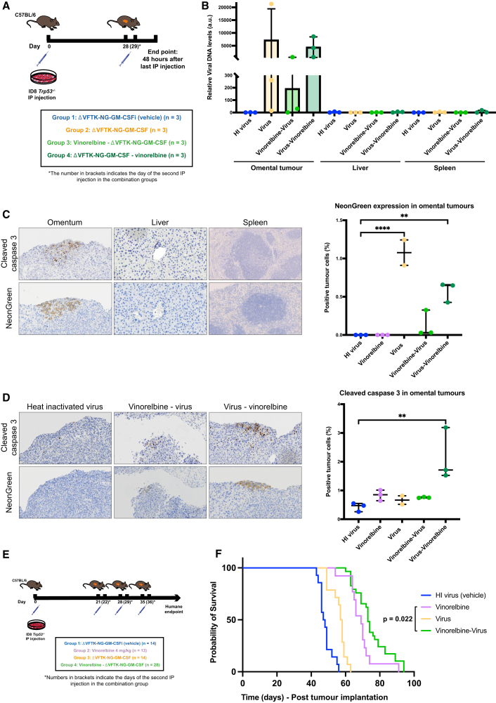 Figure 6
Peer Reviews
No public reviews on file for this paper yet. If you reviewed it on a platform where reviews are public (OpenReview, ICLR, NeurIPS, ICML), you can paste yours below so the community can read it here.
Videos
No videos yet. Explain this paper in a talk, walkthrough, or lecture? Add one.
Taxonomy
TopicsVirus-based gene therapy research · Immunotherapy and Immune Responses · Animal Virus Infections Studies
Introduction
Ovarian cancer (OC) is the sixth most common female malignancy in the United Kingdom and the leading cause of death from a gynecological cancer.1 It is a highly heterogeneous disease with histotypes varying in their genetic and molecular profiles, clinical phenotypes, and risk factors.2 Despite its heterogeneity, standard management has remained the same for decades and includes surgical debulking combined with platinum-taxane chemotherapy.3 Response rates are initially high, especially in high-grade serous carcinoma but 80% of patients with advanced disease recur, with all relapsed disease ultimately developing fatal therapy resistance.4 The introduction of bevacizumab (vascular endothelial growth factor [VEGF] inhibitor) and PARP inhibitors in treatment regimens based on tumor molecular biomarker status can extend progression-free survival.5^,^6 However, the failure of maintenance therapies to extend overall survival for the majority of patients highlights the need for new therapies that overcome chemoresistance.7^,^8
Vaccinia virus represents a promising oncolytic agent for OC due to its natural tropism for ovarian tissue.9 The virus is best known for its use as the vaccine for smallpox eradication in the 1980s, demonstrating its safety and tolerability in humans.10 It also preferentially infects, replicates, and kills primary and metastatic tumors over normal tissues.11^,^12 This tumor-specific cytotoxicity reduces the systemic side effects induced by the virus compared to existing chemotherapeutics.13 It also exclusively replicates in the cytoplasm, thus avoiding DNA insertions into the host genome.14 Moreover, the safety and tumor specificity of the virus has been further enhanced through genome manipulation, by deleting genes encoding for virulence factors and proteins regulating nucleotide metabolism.15 For example, loss of thymidine kinase (TK) and vaccinia growth factor (VGF) significantly reduces viral pathogenicity, reducing side effects in patients, while simultaneously increasing tumor specificity.16^,^17^,^18 Therefore, TK and VGF are deleted from most vaccinia strains used in clinical trials.19 Vaccinia can tolerate 25–40 kb foreign DNA insertions, and transgenes have been expressed in non-essential loci to improve its oncolytic properties and immunogenicity.15^,^20 Vaccinia kills cells by direct oncolysis and induces immunogenic cell death, an attractive feature that can be exploited and enhanced when used as an oncolytic virus against OC tumors.21^,^22
Olvi-Vec, derived from the Lister vaccinia strain, is the only oncolytic that has progressed to a phase III clinical trial for the treatment of platinum-resistant OC in combination with chemotherapy and bevacizumab.23^,^24 This trial provided evidence that patients with platinum-resistant disease can respond to platinum chemotherapy. In contrast, a phase II clinical trial using the attenuated modified vaccinia Ankara virus expressing 5T4 tumor-associated antigen (MVA-5T4, TroVax) monotherapy in asymptomatic women with recurrent OC failed to show a significant improvement compared to placebo.25 This trial highlights the importance of combination therapies for OC with vaccinia.
The primary aim of our study was to identify a combination regimen that enhances the efficacy of vaccinia in treating ovarian carcinoma in vivo. We took advantage of a recombinant vaccinia virus lacking F1, a viral inhibitor of apoptosis,26^,^27 and the VGF.28^,^29 The recombinant ΔVF virus induces increased cell death during infection compared to the parental Western Reserve (WR).30 This is the first study in which the oncolytic efficacy of ΔVF was assessed in vivo in a syngeneic high-grade serous ovarian carcinoma mouse model. Additional recombinant viruses have been constructed to improve the tumor specificity and immunogenicity of ΔVF. A high-throughput compound screen was also conducted to identify combination partners that enhance the efficiency of ΔVF and its derivatives in killing OC cells and inhibiting tumor progression.
Results
Targeting and arming ΔAVF recombinant virus
To improve the potential tumor specificity of the ΔVF virus, we deleted the thymidine kinase (TK) gene to generate the ΔVFTK virus. To facilitate visualization of infected cells, we also introduced NeonGreen (NG) into the TK locus, generating the ΔVFTK-NG virus. This virus was additionally armed with granulocyte-macrophage colony-stimulating factor (GM-CSF) (ΔVFTK-NG-GM-CSF). The inclusion of GM-CSF in this construct was not intended for mechanistic investigation of its immunological effects. Rather, the overall aim of this study was to identify small molecule inhibitors that enhance the antitumor efficacy of the recombinant viruses, rather than to assess GM-CSF-mediated immune responses. Characterization of the new recombinant viruses demonstrated they have similar viral protein expression and spread profiles to the ΔVF virus (Figures 1A and 1B). Immunoblot analysis confirmed NeonGreen (26 kDa) and the NeonGreen-GM-CSF fusion protein (47 kDa) were expressed in cells infected with the ΔVFTK-NG and ΔVFTK-NG-GM-CSF viruses (Figure 1A). Moreover, all viruses retained the pro-apoptotic activity of the parental ΔVF virus (Figure 1C).Figure 1. Characterization of recombinant vaccinia viruses(A) Immunoblot analysis comparing the expression levels of the indicated viral proteins in HeLa cells infected with the specified viruses. Vinculin is the cell loading control. The experiment was repeated three times, and a representative example is shown. (B) Representative images of plaque formation by the indicated virus strains in BS-C-1 cells at 72 hpi. The graph represents quantitative analysis of plaque diameter measured in millimeters for the indicated viral strains. The three independent plaque assays are represented by the three different colors in the SuperPlot. Each dot represents one plaque, and the triangles represent the medians of all the plaques measured in each experiment. Data are represented as mean ± SD. One-way ANOVA was used to determine significance between all groups with Tukey multiple comparisons post-hoc test. (C) Immunoblot analysis assessing PARP cleavage in ID8 Trp53^−/−^cells infected with the indicated viruses. F12 and GRB2 represent viral and cell loading controls, respectively. The asterisk (∗) indicates a non-specific band. Hpi, hours post-infection. The experiment was repeated three times, and a representative blot is shown.
The oncolytic efficacy of the new recombinant viruses was examined in vivo using a syngeneic mouse model of high-grade serous ovarian carcinoma (ID8 Trp53^−/−^) (Figure 2A). The heat-inactivated ΔVF (ΔVFi) virus was used as vehicle, and the median survival of mice in this group was 45.5 days. The median survival of mice inoculated with ΔVF, ΔVFTK, ΔVFTK-NG, and ΔVFTK-NG-GM-CSF viruses was 51.5, 51, 51, and 54 days, respectively. Log rank analysis demonstrated that only ΔVFTK-NG-GM-CSF (p = 0.005) significantly prolonged mouse survival (Figure 2B) compared to vehicle control. Viral inoculation of mice with the recombinant viruses had no effect on the size of their liver and spleen nor ascitic volumes (Figure 2C). NeonGreen expression did not affect mouse survival, as mice inoculated with ΔVFTK and ΔVFTK-NG had the same median survival of 51 days. An additional survival experiment in which viral treatment began 14 days post-intraperitoneal (IP) ID8 Trp53^−/−^ cell injection with the mice receiving four viral intraperitoneal (i.p.) doses instead of three was conducted. In this protocol, both ΔVFTK-NG and ΔVFTK-NG-GM-CSF viruses significantly prolonged mouse survival compared to the heat-inactivated ΔVF control (Figure S1). These data provide further support for the contribution of viral oncolysis to the observed therapeutic benefit and indicate that the ΔVFTK-NG-GM-CSF was the only recombinant virus that consistently improved survival. These experiments also illustrated the need to identify combination agents to improve the efficacy of vaccinia virus in the treatment of OC.Figure 2. Efficacy of recombinant vaccinia viruses in vivo(A) Schematic representation of the experimental design of the in vivo survival study. Five groups of mice were injected i.p. with ID8 Trp53^−/−^ cells on day 0 and subsequently inoculated with the indicated viruses on day 21, 28, and 35. ΔVFi is the control heat-inactivated virus. (B) Kaplan-Meier survival curve showing survival data for each virus analyzed by log rank test. (C) Quantification of omental tumor, spleen, and liver weights as well as ascitic volumes for each group. Data are represented as mean ± SD. One-way ANOVA was used to determine significance between all groups with Tukey multiple comparisons post hoc test.
Identifying a combination agent to improve the efficacy of oncolytic vaccinia
A high-throughput drug screen was conducted on ΔVFTK-NG-infected murine OC (ID8 Trp53^−/−^) cells (Figure 3A). The library consisted of 9,000 well-characterized compounds that are in clinical development or have been approved by Food and Drug Administration (FDA) and/or European Medicines Agency (EMA) and used in clinic. NeonGreen fluorescence was used to identify infected cells, and DAPI staining of nuclei was used to calculate the number of cells remaining at the end of the screen. After imaging of wells, compounds were allocated into one of four categories (“no effect,” “kill infected cells,” “block virus replication,” and “toxic”) based on the number of infected (green) and uninfected (non-green) cells relative to infected DMSO controls on the same plate (Figure 3B). The “no effect” are wells with similar numbers of infected and uninfected cells compared to the DMSO control, whereas “block virus replication” are those with reduced infected and increased uninfected cells due to cell proliferation. Toxic compounds resulted in a significant loss of infected and uninfected cells. The ideal compounds are the “kill infected cells” category, as they represent wells with fewer infected but similar numbers of uninfected cells compared to the DMSO control (Figure 3C). These compounds, in principle, enhance the efficacy of vaccinia in killing infected ID8 Trp53^−/−^ cells.Figure 3. Identifying targets for combination therapies with oncolytic vaccinia(A) Schematic summarizing the high-throughput drug screening strategy. (B) Representative images of ID8 Trp53^−/−^ cells infected with ΔVFTK-NG virus and treated with the indicated compounds belonging to the four categories. NeonGreen expression (green) indicates infected cells, and DAPI was used to stain nuclei (blue). Cells were imaged using Opera Phenix Plus with 10× air objective. Scale bars, 200 μm. (C) Graphical illustration of compound allocation in the four categories. (D) Representative graphs from the secondary validation screen of selected primary screen hits at 1 μM (omipalisib, erlotinib, and vinorelbine). The model represents an ideal compound, and DMSO is the negative control. The purple dashed line represents the time at which ΔVFTK-NG virus (MOI 0.5) was added (16 h after cell seeding). Statistical analysis was done by comparing uninfected against infected cell confluency at 20, 60, and 90 h after cell seeding using Student’s t test. Error bars represent standard deviation (SD). (E) List of hit compounds after the secondary validation screen.
Images from “kill infected cells” wells were reviewed, and a cutoff ratio of infected to uninfected cells of 0.3 was applied to generate the initial hit list of 60 compounds (Table 1). During the validation of these hits, an additional 60 compounds, absent from the initial library that belong to the same class of molecules and/or had a similar mechanism of action, were added to the hit list (Figure S2). A secondary live cell imaging screen was conducted to assess compound cytotoxicity as well as the impact on viral replication and spread. Based on this analysis, the compounds were categorized as “kill infected cells,” “impair viral replication,” and “toxic” (Figure 3D). Positive hits (“kill infected cells”) were identified based on the reduction in median percentage of infected cell confluency per well over time compared to uninfected cells. This analysis resulted in 20 compounds, seven of which are tubulin polymerization inhibitors (Figure 3E). We decided to focus on the seven compounds targeting the same process as this is likely to be indicative of true hits. The impact of these inhibitors was further confirmed in two additional human high-grade serous OC cell lines, OVCAR3 and OVCAR4 (Figures S3 and S4).Table 1. List of 60 primary screen hitsDrugMechanism of action2-Amino-8-[4-(2-hydroxyethoxy) cyclohexyl]-6-(6-methoxy-3-pyridinyl)-4-methylpyrido[2,3-d]pyrimidin-7(8H)-onePI3K/mTOR kinase inhibitor5-FluorouridinePyrimidine nucleoside analogueAbexinostatPan-histone deacetylase inhibitorActinomycinDNA repair inhibitorAlvespimycin (17-DMAG) hydrochlorideHSP 90 inhibitorAminopterinDihydrofolate reductase inhibitorAmsacrine hydrochlorideTopoisomerase II inhibitorAT13387HSP 90 inhibitorAuristatin FTubulin polymerization inhibitorBortezomibProteosome inhibitorCanertinibEGFR inhibitorCCT137690Aurora kinase inhibitorCephaelineEmetic alkaloidCH5138303HSP 90 inhibitorCHIR-124Selective Chk1 inhibitorChromomycin A3DNA replication and transcription inhibitorCombretastatin A4Tubulin polymerization inhibitorCytarabinePyrimidine nucleoside analogueCytochalasin EActin polymerization inhibitorD8-MMADTubulin polymerization inhibitorD8-MMAF hydrochlorideTubulin polymerization inhibitorDiacetoxyscirpenolRibosome inhibitorDiosgeninPrecursor of steroidal hormonesDp44mTIron chelatorEllipticineTopoisomerase II inhibitorEmetine hydrochloride hydrateRibosome inhibitorEnzastaurinProtein kinase C inhibitorErastinFerroptosis inducerErgosterolSterolFloxuridinePyrimidine nucleoside analogueFosbretabulin disodiumTubulin polymerization inhibitorGanetespibHSP90 inhibitorGeldanamycinHSK90 inhibitorGemcitabineNucleoside (cytidine analogue) metabolic inhibitorGSK3787PPARδ antagonistHSP990HSP90 inhibitorImidazole ketone erastinFerroptosis inducerMMADTubulin polymerization inhibitorMMAF hydrochlorideTubulin polymerization inhibitorMMAF-OMeADC cytotoxin and tubulin polymerization inhibitorNSC319726p53^R175^ mutant reactivatorNutlin-3P53-MDM2 interaction inhibitorNVP-TAE226Focal adhesion kinase inhibitorPazopanib hydrochlorideProtein tyrosine kinase inhibitorPDGF receptor tyrosine kinase inhibitor IVTyrosine kinase inhibitorPemetrexedFolate-dependent enzyme inhibitor. Inhibitor of nucleotide synthesisPodophyllotoxinTubulin polymerization inhibitorPralatrexateDihydrofolate reductase inhibitorPU-H71HSP90 inhibitorRaltitrexedThymidylate synthase inhibitorRosiglitazonePPAR agonistSilvestrolRNA helicase inhibitorTanespimycinHSP90 inhibitorThapsigarginSarco-endoplasmic reticulum Ca^2+^ ATPase inhibitorTRAM-34Potassium channel inhibitorTrifluridineNucleoside metabolic inhibitorTubulin inhibitor 1Tubulin polymerization inhibitorVinblastineTubulin polymerization inhibitorVincristine sulfateTubulin polymerization inhibitorVinorelbineTubulin polymerization inhibitor
Vinorelbine is a semi-synthetic second-generation vinca alkaloid with a broad spectrum antitumor activity that inhibits microtubule assembly.31^,^32 It has reduced side effects including neurotoxicity compared to other vinca alkaloids.33 An oral vinorelbine formulation is also available, making it an appealing combination agent.34 Vinorelbine is mainly used to treat non-small cell lung and breast cancers but also has some efficacy against OC.35^,^36^,^37^,^38 Based on this, we selected vinorelbine for mechanistic exploration and in vivo experiments.
Vaccinia and vinorelbine induce ID8 Trp53−/− cell death via apoptosis
Vinorelbine treatment of uninfected ID8 Trp53^−/−^ cells for 16 h results in the loss of their characteristic cobblestone appearance and cell-to-cell contacts as well as some cell rounding, indicative of possible progression to cell death (Figure 4A). Immunofluorescence analysis confirmed that vinorelbine results in loss of microtubules in ID8 Trp53^−/−^ cells, both with or without infection with the ΔVFTK virus (Figure 4B). Vinorelbine treatment also induced nuclear fragmentation and the formation of multinucleated cells suggestive of defects in cytokinesis or cell fusion.Figure 4. Vaccinia and vinorelbine induces apoptotic ID8 Trp53^−/−^ cell death(A) Representative phase contrast images of ID8 Trp53^−/−^ cells after 16 h treatment with DMSO or vinorelbine (1 μM). Scale bars, 150 μm. (B) Immunofluorescent images of microtubule cytoskeleton (tubulin, green) in ID8 Trp53^−/−^ cells treated with DMSO or vinorelbine with or without infection with ΔVFTK (vaccinia, red). DAPI (blue) was used to stain nuclei and cytoplasmic viral factories. Yellow arrows indicate fragmented nuclei. Scale bars, 10 μM. (C) Immunoblot of the indicated viral proteins following infection for 4 or 8 h with ΔVFTK-NG following 16 h pre-treatment with DMSO or vinorelbine. Vinculin is the cell loading control. (D) Immunoblot examining the levels of LC3 lipidation (LC3-II) and p62 in ID8 Tr53^−/−^ cells pretreated with vinorelbine or DMSO for 8 h and infected with ΔVFTK-NG for 24 h. Chloroquine (Clq) treatment for 3 h represents the positive control. (E) Immunoblot examining the levels pf MLKL phosphorylation and PARP cleavage in ID8 Tr53^−/−^ cells pretreated with vinorelbine or DMSO for 8 h and infected with ΔVFTK-NG for 24 h. Staurosporine (S) (8 h) and 0.1% hydrogen peroxide (H_2_O_2_) combined with Z-VAD (two hours) represent positive controls for apoptosis and necroptosis, respectively. (F) Immunoblot examining the levels of cleaved caspase-8, caspase-3, and PARP in ID8 Tr53^−/−^ cells treated with vinorelbine 8 h before or after infection with ΔVFTK-NG for 24 h. In (D, E, and F), H5 and GAPDH represent the viral and cell loading controls, respectively. For all experiments, uninfected DMSO-treated cells (UI) were the negative control. Asterisks (∗) indicate non-specific bands. Immunoblot experiments were repeated three times, and representative examples are shown. (G) Median percentage cell confluency over time for the indicated conditions. Cells were treated with the indicated compounds or ΔVFTK-NG at 16 h post-seeding (dashed line numbered 1). The dashed line numbered 2 (24 h after cell seeding) represents the time of addition of either vinorelbine or ΔVFTK-NG for the combination groups.
Vaccinia hijacks the microtubule cytoskeleton to facilitate its replication, intracellular transport, and spread.39^,^40^,^41^,^42 We previously found that depolymerization of microtubules with nocodazole reduces virus yield.43 Consistent with this, immunoblot analysis revealed that vinorelbine treatment reduced both early (F12) and late (F13) viral protein expression, as well as H5, which is required for viral replication (Figure 4C). Vinorelbine treatment prior to infection of ID8 Trp53^−/−^ cells clearly impairs viral gene expression but it is not immediately obvious why there is enhanced cell death or which programmed cell death pathway (autophagy, necroptosis, or apoptosis) is activated.
To assess whether vinorelbine induces autophagy in ID8 Trp53^−/−^ infected with the ΔVFTK-NG virus, we performed immunoblot analysis to examine the level of p62 expression and LC3 lipidation (LC3-II) (Figure 4D). In contrast to chloroquine-treated cells (positive control), there was no change in the level of p62 in infected or non-infected cells with or without vinorelbine. Likewise, the levels of LC3-II did not change, except in the positive control, indicating that autophagy is not responsible for increasing cell death of vinorelbine-treated ID8 Trp53^−/−^ cells.
To investigate the possible involvement of necroptosis, we performed immunoblot analysis to examine the level of MLKL phosphorylation (pMLKL) and look for the presence of 50 kDa PARP fragment generated by lysosomal proteases.44 We found pMLKL and the 50 kDa PARP fragment that are indicative of necroptosis are only present in the positive control (0.1% hydrogen peroxide and Z-VAD treatment) (Figure 4E). We did, however, see that vinorelbine induced cleavage of PARP to generate an 89 kDa fragment in infected cells, suggesting that apoptosis is responsible for their death.45 The presence of cleaved caspase-3 and -8 in immunoblots of infected cells treated with vinorelbine confirmed apoptosis was activated (Figure 4F). The combination of vinorelbine and infection is required to induce apoptosis, as cleavage of PARP, caspase-3, or caspase-8 is not observed in non-infected or infected cells with or without the drug, respectively. Pre-treating infected cells with vinorelbine also appears to be more effective at inducing PARP, caspase-3, or caspase-8 cleavage (Figure 4F). To examine if this translates into increased cell death, we performed live cell imaging of ID8 Trp53^−/−^ cells treated with vinorelbine before or after vaccinia infection using the same conditions used in the initial drug screen. We found that pre-treating ID8 Trp53^−/−^ cells with vinorelbine leads to increased initial cell death compared to adding vinorelbine after infection (Figure 4G). However, both conditions eventually result in similar levels of cell death.
Vinorelbine and ΔVFTK-NG-GM-CSF improve mouse survival
An in vivo distribution study was carried out to examine the tumor specificity of the ΔVFTK-NG-GM-CSF virus (Figure 5A). The ΔVFTK-NG-GM-CSF virus was selected for the combination studies with vinorelbine based on previous in vivo data showing that it was the only virus to consistently confer a survival benefit compared to the heat-inactivated virus. However, the role of GM-CSF transgene was not the focus of this investigation. Viral replication was detected in omental tumors but not in liver or spleen in mice inoculated intraperitoneally with virus with or without vinorelbine (Figures 5B and 5C). Pre-treatment with vinorelbine impacted viral replication in vivo consistent with our observations in ID8 Trp53^−/−^ cells in culture (Figures 5B and 5D). Vinorelbine also reduced the level of GM-CSF expression in omental tumors (Figure S5). To assess the efficacy of the combination treatment on the survival of mice, we treated mice with vinorelbine 24 h before injection of the ΔVFTK-NG-GM-CSF virus (Figure 5E). Log rank analysis demonstrated that this combination significantly prolonged mouse survival compared to vinorelbine (p = 0.022; median survival 73.5 vs. 69 days) (Figure 5F). Moreover, both conditions were also significantly better than virus alone (median 57 days). Our observations suggest that combining vinorelbine and ΔVFTK-NG-GM-CSF virus improves mouse survival against high-grade serous ovarian carcinomas.Figure 5. Assessing the efficacy of ΔVFTK-NG-GM-CSF in vivo(A) Schematic representation of the experimental design of the distribution study. Mice were injected i.p. with ID8 Trp53^−/−^ cells on day 0 and received their i.p. injection on day 28, and the combination groups received their second component (vinorelbine or virus) on day 29. Heat-inactivated (HI) virus (ΔVFTK-NG-GM-CSFi) is the negative control. (B) Quantification of viral DNA in omental tumor, liver, and spleen measured by qPCR and expressed relative to viral DNA levels of the liver sample in the HI virus group. Error bars represent mean ± SD. (C) Representative immunohistochemical images of the distribution of cleaved caspase-3 and NeonGreen in omental tumor, liver, and spleen harvested from a mouse in group 4. The graph shows the quantification of NeonGreen-positive cells in omental tumors. (D) Representative immunohistochemical images of the distribution of cleaved caspase-3 and NeonGreen in omental tumors from mice in groups 1, 3, and 4. The graph shows the quantification of cleaved caspase-3-positive cells in omental tumors. (E) Schematic representation of the experimental design of the vaccinia-vinorelbine combination study. Mice were injected with ID8 Trp53^−/−^ cells on day 0 and started receiving their i.p. treatment injections on day 21. Mice allocated to single-treatment groups received their inoculations on days 21, 28, and 35, and those allocated to the combination groups received vinorelbine and ΔVFTK-NG-GM-CSF 24 h apart. Heat-inactivated (HI) ΔVFTK-NG-GM-CSFi virus was the negative control. (F) Kaplan-Meier survival curve showing survival data for each group analyzed by log rank test. One mouse belonging to group 2 was excluded from the analysis, as after i.p. injection of ID8 Trp53^−/−^ cells omental tumor failed to form. The analysis is from the combination of two survival experiments following the same protocols.
Discussion
Vaccinia virus has the promise of being an ideal oncolytic agent, given its safety profile and ability to be genetically enhanced as well as combined with drug treatments.46^,^47 Clinical trials frequently use viruses deleted of genes encoding VGF (ΔVGF) and/or the thymidine kinase (ΔTK), as this increases their tumor specificity and improves safety.17^,^48 Deletion of F1, an inhibitor of apoptosis in combination with TK, also improves the oncolytic effectiveness of the virus, as it extends mouse survival in a human glioblastoma model and delays tumor growth in a syngeneic mouse colon model.26^,^27^,^49 Based on the available evidence, a virus lacking VGF, TK, and F1 is predicted to be safer and induce more tumor cell death.
We have now deleted VGF, TK, and F1 in the WR strain of vaccinia and also inserted GM-CSF to the genome to increase the immunogenicity of the virus. NeonGreen was also added, which like other reporters, can be used to monitor viral replication and distribution, detect metastatic disease, and assess toxicity clinically.50^,^51 Our in vivo survival studies demonstrated that our new recombinant viruses were well tolerated in mice bearing ID8 Trp53^−/−^ omental tumors but that only the ΔVFTK-NG-GM-CSF virus had consistently significant survival benefit (Figures 2B and S1). Moreover, when injected intraperitoneally, the ΔVFTK-NG-GM-CSF virus specifically replicates in omental tumors but not in the liver or spleen (Figures 5A–5C).
Multiple early phase clinical trials demonstrate that even with genomic modifications that enhance its tumor specificity, oncolytic activity, and immunogenicity, vaccinia clearly still needs a combination agent to achieve its full oncolytic potential.25^,^46 Given this, we performed a high-throughput drug screen on ID8 Trp53^−/−^ cells using 9,000 well-characterized compounds to identify those that increased the killing efficiency of our triple-deleted recombinant virus. By performing primary and secondary drug screens, we identified 20 hit compounds, seven of which were tubulin polymerization inhibitors (Figure 3E). Vinorelbine was our drug screen hit compound of choice, given it has some activity against OC.35^,^36^,^37
We found that the combination of vaccinia with vinorelbine induces ID8 Trp53^−/−^ cell death via apoptosis. This is not unexpected, as the ΔVFTK-NG virus lacks F1, the main inhibitor of intrinsic apoptosis during infection.26^,^27 Vinca alkaloids such as vinorelbine also induce apoptosis, which is often attributed to impaired spindle formation and subsequent prolonged mitotic arrest.52^,^53 However, these chemotherapeutics can additionally stimulate apoptosis independently of the cell cycle via alternative mechanisms including the activation of intrinsic apoptosis.54 Microtubule-targeting agents have been shown to upregulate pro-apoptotic Bcl-2 proteins (BAX, BAK, PUMA, Noxa, and Bad) as well as inactivate anti-apoptotic Bcl-2 proteins through phosphorylation (Bcl-2, Bcl-xL, Mcl-1, and Bcl-W).53^,^55^,^56 Vinorelbine and the ΔVFTK-NG virus both act on the intrinsic mitochondrial apoptotic pathway, and their effects complement each other, leading to enhanced apoptotic cell death compared to either agent alone. Therefore, vinorelbine pre-treatment will not only induce mitotic arrest but also activate BAK/BAX, leading to mitochondrial membrane pore formation, resulting in a pro-apoptotic “priming,” making cells more susceptible to vaccinia-induced apoptosis.57 Consistent with this notion, vinorelbine pre-treatment of cells resulted in increased PARP and caspase cleavage at 24 h post-infection compared to adding the drug after infection (Figure 4F). Live cell imaging also demonstrated that addition of vinorelbine before infection increased initial cell death compared to first adding the virus and then the drug (Figure 4G).
When tested in a mouse model of high-grade serous ovarian carcinoma, we found that the combination of vinorelbine followed by infection with the ΔVFTK-NG-GM-CSF virus produced a significant survival benefit compared to the control (median survival of 73.5 compared to 47.5 days). Our results are in line with previous studies demonstrating that a combination of vinorelbine and vesicular stomatitis virus (VSV) significantly prolonged the survival of aggressive subcutaneous 4T1 syngeneic mouse model of triple-negative breast cancer.58 Vinorelbine has good response rates in phase I and II clinical trials for recurrent OC, and the National Comprehensive Cancer Network recommended vinorelbine for recurrent epithelial, fallopian tube, or primary peritoneal cancers.59 It is evident from our in vivo experiments that vinorelbine at the dose used (4 mg/kg) has good efficacy against ID8 Trp53−/− tumors, suggesting that the drug represents a promising therapy for patients with high-grade serous carcinoma. Nevertheless, when vinorelbine is combined with the ΔVFTK-NG-GM-CSF virus, mouse survival is further increased.
Our findings suggest that the enhanced tumor cell killing observed with the combination of vinorelbine and ΔVFTK-NG-GM-CSF is primarily due to an additive or synthetic lethal interaction, rather than increased viral replication. Although vinorelbine initially suppressed cell proliferation in vitro, confluency recovered over time, indicating a predominantly cytostatic effect (Figure 4G). In contrast, the combination treatment led to a sustained reduction in confluency, consistent with induction of apoptosis as confirmed by cleaved caspase-3 and PARP immunoblotting (Figures 4F and 4G). Moreover, quantitative PCR (qPCR) analysis of viral gene expression demonstrated that vinorelbine pre-treatment reduced viral replication relative to infection, with virus followed by vinorelbine, demonstrating that vinorelbine impairs vaccinia replication when administered prior to viral infection. These data support the notion that the combination effect arises from enhanced cell death rather than increased viral propagation.
Further experiments are required to examine how reducing the number of doses and concentration of vinorelbine, together with higher viral titers, affects mouse survival. In this way, it should be possible to develop strategies with the ΔVFTK-NG-GM-CSF virus that are effective with lower vinorelbine doses to decrease systemic toxicity and the likelihood of patients becoming resistant to vinorelbine. In addition, adjusting the interval between viral and vinorelbine administration may allow greater viral replication and transgene expression, potentially improving therapeutic efficacy. A limitation of the present study is that the immunological effects of the GM-CSF-expressing oncolytic vaccinia virus were not investigated. While GM-CSF is known to modulate anti-tumor immunity, dissecting its contribution in our system would require extensive in vivo and immunological studies, which are beyond the scope of the current work. Our primary aim was to perform a drug screen to identify compounds that enhance the efficacy of the recombinant oncolytic vaccinia virus. GM-CSF expression was attenuated in the presence of vinorelbine, and although even low levels of GM-CSF might influence the tumor immune microenvironment, the combination efficacy observed here was not attributed to GM-CSF specifically. The immunomodulatory role of GM-CSF will be the subject of future studies directly comparing armed and unarmed viruses with vinorelbine. These studies should also include immune profiling of tumor-infiltrating dendritic cells, CD8^+^ T cells, and other relevant immune populations to clarify the role of GM-CSF in shaping therapeutic response. Future studies will also focus on validating these findings across additional syngeneic and human OC models to assess the robustness and generalizability of the therapeutic effect.
Materials and methods
Cells and culture
The ID8 Trp53^−/−^ cell line was provided by Professor Iain McNeish, Imperial College London (London, UK).60 HeLa and BS-C-1 cell lines were provided by the European Molecular Biology Laboratory (Heidelberg, Germany) and 143B TK^−/−^ from The Francis Crick Institute Cell Services (London, UK). All cell lines were mycoplasma tested and maintained in Dulbecco’s modified Eagle’s medium (DMEM) (Sigma-Aldrich, St. Louis, Missouri, #51435C) supplemented with 10% fetal bovine serum (FBS) (Thermo Fisher Scientific, Waltham, Massachusetts, #10270-106) and 1% penicillin/streptomycin (pen/strep) (Sigma-Aldrich, #P4333) at 37°C and 5% CO_2_.
Virus amplification and sucrose purification
HeLa cells were grown in 15 cm culture dishes (Corning Inc., Sigma Aldrich, #353025) at approximately 80% confluency and were infected with vaccinia at an MOI of 0.1 for 48–72 h. The cells were scraped in PBS and centrifuged at 500 × g at 4°C for 5 min. Cell pellets were resuspended in PBS and re-centrifuged. The resulting cell pellet was resuspended in 7 mL of Tris buffer (10 mM Tris HCl, 2 mM MgCl_2_, pH 9.0), disrupted by 20 strokes in a 7 mL Wheaton Dounce homogenizer (DWK Life Sciences, Millville, New Jersey, #357538) and centrifuged at 500 × g at 4°C for 5 min. The supernatant was collected and added on top of an 8 mL 35% sucrose cushion in a Beckman SW40 ultracentrifuge tube (Beckman Coulter, Brea, California #3117-0380). The solution was centrifuged at 192, 000 × g at 4°C for 30 min in a Beckman Optima l-100 XP ultracentrifuge, using an SW32Ti swing-out rotor. The virus pellet was resuspended in a desired amount of Tris buffer, titrated using plaque assays, aliquoted, and stored at −20°C (short term) or −80°C (long term).
Viral plaque assays and heat inactivation
Confluent monolayers of BS-C-1 cells were infected with vaccinia virus in serum-free media at the required dilutions for 1 h at 37°C, 5% CO_2_. The media was then replaced with 2 mL of semi-solid media (1:1 ratio of 3% carboxy-methyl cellulose sodium salt dissolved in water and 2× modified Eagle’s medium (MEM) supplemented with 10% FBS and 1% pen/strep). After 72 h, the cells were fixed by adding 1 mL of 8% formaldehyde to the media for 30 min at room temperature. The media and formaldehyde were removed, and cells were stained with 1 mL crystal violet (1:5 dilution in PBS) for 30 min. Subsequently, the plates were rinsed with cold water and left to air dry. To determine the plaque size, plaque diameter was measured in millimeters using Fiji line tool, available at imagej.net. For heat inactivation, aliquots of viruses were incubated for 3 h at 60°C and then cooled on ice for 1 h. Heat inactivation was confirmed by the absence of plaque-forming units using plaque assays.
Generation of recombinant viruses
Recombinant viruses were constructed in the ΔVF virus background, which is derived from the WR strain.30 The transient dominant selection (TDS) method was used to delete TK from the ΔVF genome.61^,^62 To create the targeting vector for TDS, a DNA fragment containing the left (387bp) and right (321bp) TK recombination arms was cloned into the pSSGB vector containing the selectable markers GFP-bsd under the control of a synthetic vaccinia promoter (pSS).62 The fluorescent ΔVFTK virus expressing NG (ΔVFTK-NG) and the ΔVFTK virus expressing NG and GM-CSF (ΔVFTK-NG-GM-CSF) under the control of the synthetic early/late vaccinia promoter (pEL) at the TK locus were created via homologous recombination between ΔVF virus and the pBSΔTK-NG- and pBSΔTK-NG-GM-CSF-targeting vectors, respectively. These targeting vectors were created using BlueScript (PBS(+)) as the backbone vector and DNA fragments containing sequences for pEL-NG and pEL-NG-GM-CSF as the inserts. For all new recombinant viruses, the targeting vectors contained TK recombination arms flanking the DNA sequence to be inserted in the vaccinia genome. A small portion of the 5′ open reading frame of J2R (gene encoding for TK) was retained within the left TK recombination arm to ensure the correct transcription of J1R. These DNA fragments were synthesized using the IDT gBlocks gene synthesis service (Coralville, Iowa). Subsequently, HeLa cells, infected with ΔVF at MOI of 0.05, were transfected with the relevant targeting vectors using Lipofectamine 2000 (Thermo Fisher Scientific, #52887) and incubated for 48 h at 37°C and 5% CO_2_. The viruses were harvested by scraping the cells and subjecting them to three freeze-thaw cycles. The harvested viruses were used to infect confluent monolayers of 143B TK^−/−^ cells with BrdU selection or BS-C-1 cells with or without blasticidin selection. Recombinant viruses, which are shown schematically in Figure S6, were isolated by identifying and picking fluorescent green plaques over at least three rounds of purification. For all three recombinant viruses, TK deletion and/or insertions of NG or NG-GM-CSF were confirmed by PCR analysis after each round of plaque purification and sequencing. The recombinant viruses were amplified and purified using a 35% sucrose gradient.
Quantification of viral DNA and GM-CSF mRNA
Total DNA was isolated from mouse tissues using DNeasy Blood and Tissue Kit (QIAGEN, Hilden, Germany), as per manufacturer’s protocol and quantified using a NanoDrop (Thermo Fisher Scientific). Real-time qPCR was performed in triplicates in 384-well reaction plates (Applied Biosystems, Waltham, Massachusetts). Each PCR reaction (total volume 10 μL) contained 20 ng DNA, 5 μL Power SYBR-Green PCR Master Mix (Applied Biosystems), and 0.5 μM of forward and reverse primers. Viral DNA was amplified using primers specific to a region for H5 vaccinia gene (H5-F: 5′-GTAAGAAGTAAATGCGTGC-3′, H5-R: 5′-CCACGTTTGTTCATATACTAC-3′), and APP1 (APP1-F: 5′-CGGAAACGACGCTCTCATG-3′ and APP1-R: 5′-CCAGGCTGAATTCCCCAT-3) was amplified as a nuclear gene standard reference. Changes in viral DNA amount were calculated using the 2^−ΔΔCt^ method and represented as fold changes relative to the indicated control.63 For quantification of GM-CSF mRNA, 20 mg of omental tumor tissue was homogenized in TRIzol reagent using a Precellys Evolution tissue homogenizer (Bertin Technologies, Montigny-le-Bretonneux, France). Following chloroform addition and centrifugation, the aqueous phase containing RNA was combined with an equal volume of 70% ethanol and purified using RNeasy Mini Kit columns, according to the manufacturer’s instructions (QIAGEN). One-step real-time qPCR was then performed on 50 μg of RNA template using the QuantiNova SYBR Green RT-PCR Kit (QIAGEN) with 0.5 μM of gene-specific primers: GM-CSF (Csf2-F: CTACTACCAGACATACTGCC; Csf2-R: GCATTCAAAGGGGATATCAG) and GAPDH as the housekeeping control (GAPDH-F: TCTTGTGCAGTGCCAGCCT; GAPDH-R: CAATATGGCCAAATCCGTTCA).
Immunoblotting
Cells were collected and lysed in 21 μL of PBS supplemented with protease/phosphatase inhibitors (Cell Signaling, #5871) and 5U benzonase nuclease (Millipore, #E1014). SDS was added at a final concentration of 1% followed by 22 μL of 2× SDS loading buffer (Thermo Fisher Scientific, #LC2676). The samples were heated at 95°C for 3 min, loaded onto Bolt 4-12% or 10% Bis-Tris Plus pre-cast gels (Thermo Fisher Scientific) and run in MOPS SDS running buffer (Thermo Fisher Scientific, #NP0001) for 55 min at 150 V. SeeBluePlus2 protein standard (Thermo Fisher Scientific, #LC5925) was used as reference for protein molecular weight. The proteins were transferred to nitrocellulose membranes (Thermo Fisher Scientific, #IB23001), blocked in 5% milk in PBS with 0.1% Tween 20 (PBS-T) (Sigma-Aldrich, #P9416) for 1 h at room temperature and incubated overnight with primary antibodies. Primary antibodies used were F12 (1:4,000,64), F13 (1:6,000,39), H5 (1:10,000,65), GRB2 (1:1,000, Santa Cruz, Dallas, Texas, #sc-255), Vinculin (1:10,000, Sigma-Aldrich, #V9264), GAPDH (1:1,000, Santa Cruz, #sc-32233), PARP (1:1,000, Cell Signaling, Danvers, Massachusetts, #9542), cleaved caspase-8 (1:1,000, Cell Signaling, #8592), cleaved caspase-3 (1:1,000, Cell Signaling, #9664), LC3-B (1:1,000, Abcam [#ab48394], Cambridge, UK), p62/SQSTM1 (1:1,000, Novus Biologicals, Centennial, Colorado, #NBP1-42822), and NeonGreen (1:1,000, Cell Signaling, #41236). Goat anti-rabbit (#111035003) and anti-mouse (#115005003) secondary HRP antibodies, obtained from Jackson ImmunoResearch (West Grove, Pennsylvania), were used at 1:10,000 in 5% milk with PBS-T. For the examination of phosphorylated proteins, 1 mM orthovanadate (New England Biolabs, Ipswich, Massachusetts, #P0758) was added during blocking primary and secondary antibody incubation. The membranes were incubated with SuperSignal West Pico PLUS Chemiluminescence reagent (Thermo Fisher Scientific, #34580) for 1 min at room temperature and exposed to UltraCruz Autordiography Film (Santa Cruz, #sc-201697).
Drug screen
Chemical library
A small molecule drug library of 9,000 well-characterized compounds, assembled from commercial libraries by the high-throughput screening (HTS) science technology platform (STP) of the Francis Crick Institute was used for the primary screen. This chemical library can be accessed (https://hts.crick.ac.uk/db/view/libraryView.php) under the database name “Full Chemical Collection V6”. A customized library of 120 compounds was purchased form MedChemExpress (Monmouth Junction, New Jersey) for the secondary screen.
Primary screen
The small molecule library, resuspended in dimethyl sulfoxide (DMSO), was transferred into intermediate low dead volume (LDV; Labcyte, #LPS-0200) 384-well plates at a concentration of 10 and 1 mM. An acoustic liquid handler (Echo 550 Beckman-Coulter) was then used to dispense compounds into Greiner microclear 384-well plates (Greiner, #781091) so they reach a final concentration of 0.1, 1, and 10 μM. Each assay plate was prepared in triplicate. Prior to cell seeding, the compounds were diluted in 10 μL of complete DMEM per well. A volume of 40 μL of cells, corresponding to 3,000 cells, was dispensed into the 384-well assay plates, which were incubated for 16 h at 5% CO_2_ and 37°C. The ΔVFTK-NG virus was dispensed in each well at an MOI 0.5, and the plates were incubated at 5% CO_2_ and 37°C for 30 h. Cells were fixed using 4% paraformaldehyde (PFA), permeabilized, and stained by adding 0.01% Triton X-100 and DAPI. The cells were imaged on Opera Phenix Plus (Revvity, Waltham, Massachusetts) using 10× air NA 0.3 objective. The microscope was equipped with a set of lasers and filters for the excitation and emission wavelengths specific for DAPI (excitation: 375 nm and emission: 435–480 nm) and NeonGreen (excitation: 488 nm and emission: 500–550 nm).
Primary drug screen analysis
Five fields per well were imaged and analyzed using Harmony (v.5.0). Screen data were analyzed with the cellHTS2 R package.66^,^67 Raw measurements were scaled relative to the mean of the within-plate DMSO controls to report a percentage-of-control (PoC) for each feature, and replicates were summarized as a median value. The replication between technical replicates was assessed using the Spearman correlation. Tibco Spotfire (v.14.0) software was used to process the large datasets. Hit determination was based on how the compounds compared against PoC in terms of infected and uninfected cells.
Animal experiments
All animal experiments were carried out in accordance with the UK Home Office regulations under the project license PA780D61A and PP1321516 (from May 2024) and were approved by the Imperial College Animal Welfare and Ethical Review Body. Female, 6- to 7-week-old C57BL/6 mice were purchased from Charles River Laboratories (Harlow, UK) and acclimatized for a week prior to cell injection. They had standard laboratory diet and free access to water. Mice were inoculated intraperitoneally (IP) with ID8 Trp53^−/−^ cells (5 × 10^6^) in 200 μL sterile PBS on day 0. The tumors were allowed to grow for 14, 21, or 28 days depending on the protocol of each study. Details of the experimental designs of these studies are provided in Figures 2, 5, and S1. Day 21 was used as the standard tumor establishment time point used in most in vivo survival studies, as it reflects late-stage disease. In the survival study presented in Figure S1, viral treatment began on day 14, and mice received four intraperitoneal (IP) viral injections instead of three. This modified protocol was designed to assess whether initiating treatment earlier, when tumor burden is lower, and increasing the number of viral doses could enhance therapeutic efficacy. Day 28 was used exclusively for the viral distribution study. This later time point was chosen to allow for greater tumor progression, providing larger tissue samples suitable for assessing viral localization within the tumor. Sample sizes were established with the help of the experimental design assistant of NC3Rs to achieve statistical power while minimizing animal use. All viruses were injected i.p. at a titer of 1 × 10^7^ pfu/mL in 200 μL sterile PBS. Vinorelbine tartrate was obtained from Hammersmith hospital pharmacy, Imperial Collage Healthcare NHS Trust (London, UK) and was injected i.p. at 4 mg/kg in 200 μL sterile PBS. The mice were weighed daily, monitored regularly for adverse side effects, and were killed when reached humane endpoints. These included any of the following: swelling restricting movement, piloerection, hunched posture, reduced activity, facial grimace, dehydration lasting >24 h, 15% weight loss, altered respiration, and self-mutilation. All decisions on animal welfare were made by staff blinded to treatment allocation. The schedule 1 method followed was cervical dislocation and exsanguination by decapitation. Omental tumors, liver, and spleen were dissected and placed in 10% neutral buffered formalin for 48 h and then transferred to 70% ethanol.
Histopathology
Harvested tissues, embedded in paraffin to create blocks, were sectioned at 3 μm thickness and attached on glass microscope slides for examination. Histochemical hematoxylin and eosin (H&E) staining was carried out using Tissue-Tek Prisma Plus Automated Slide Stainer (Leica Biosystems, Deer Park, Illinois). Blinded histopathological analysis was performed by Professor Priestnall and Dr. Suarez-Bonnet, board-certified veterinary pathologists associated with the Experimental Histopathology STP of The Francis Crick Institute. For immunohistochemistry (IHC), samples were stained for NeonGreen (1:200, Cell Signaling, #41236) and cleaved caspase 3 (1:250, Cell signaling, #9579S) antibodies, respectively. IHC staining was performed on the Leica Bond Rx autostainer platform (Leica Biosystems, #3498240), and a DAB kit, which included a secondary antibody, DAB, and hematoxylin counterstain, was used (BOND Polymer Refine Detection, Leica Biosystems, #DS9800). The slides were scanned with a Zeiss Axio Scan Z1 Slide Scanner operated by ZEN Lite Software.
Quantitative analysis of immunohistochemistry slides was performed using QuPath (v.0.3.0). Slides were scanned and imported from a secure server. Cell detection was carried out using default DAB channel parameters, matched to the chromogen used. Stain separation was optimized by estimating stain vectors. Tumor and non-tumor regions were annotated to train a random forest classifier for compartment classification. Cleaved caspase-3 and vaccinia staining were analyzed using intensity thresholds of 0.15 and 0.2, respectively (Cell: DAB OD Mean). A script was developed to calculate the percentage of marker expression in tumor versus non-tumor areas, with results reported as intra-tumoral expression percentages.
Immunofluorescence
For fixed cell imaging, cells were seeded on 0.1% fibronectin-coated (Sigma-Aldrich, #F0895) coverslips. Cells were infected with vaccinia virus, and at experimental endpoint, cells were fixed with 1mL of iced cold methanol for 20 min at −20°C and then washed with PBS. Cells were incubated in blocking buffer (1% bovine serum albumin and 2% fetal calf serum) for 30 min followed by the primary antibody for 1 hour. Primary antibodies used were as follows: B5 (1:1,00068) and ɑ-Tubulin (1:500, Sigma-Aldrich, #T6074). Cells were incubated in Alexa-Fluor-488- and Alexa-Fluor-568-conjugated secondary antibodies (1:1,000 dilution in blocking buffer) for a further 40 min and stained with DAPI (300 nM in PBS) for 5 min. Coverslips were mounted on glass microscope slides using 5 μL Mowiol. Mounted coverslips were imaged on a Zeiss Axio Observer spinning-disk microscope equipped with a Plan-Apochromat 100×/1.46 oil lens, an Evolve 512 cameral, and a Yokagawa CSUX spinning disk. The microscope was controlled by Slidebook software (3i Intelligent Imaging Innovations). Images were analyzed using Fiji Imaging Analysis Software.
Live cell imaging
ID8 Trp53^−/−^ cells were seeded at the desired confluency in 384-well plates and were transferred to the Incucyte S3 live cell imaging system (Sartorius) to capture phase-contrast and green-fluorescent images every 3 h using a 4× objective. Cells were removed from the Incucyte S3 at the required time points for drug treatment and/or infection and quickly replaced in the Incucyte to continue imaging. Whole wells were imaged and analyzed using the integrated software module “Basic Analyser.” The median percentage confluency of infected and uninfected cells and the ratio of green fluorescence per well area were quantified. Raw data were exported for further statistical analysis.
Live cells to assess morphological changes following vinorelbine treatment were imaged using Invitrogen Evos M5000 microscope (Invitrogen) equipped with advanced LED illumination and a range of objective lenses to achieve various magnifications. Images were captured using the integrated software.
Statistical analysis
Statistical analysis was performed using Prism 10 (GraphPad Software). In all graphs, data are represented by the mean and standard deviation (SD) or median and interquartile range (IQR) from three independent experiments, unless stated otherwise. Data were tested for normality of distribution using normality and lognormality tests in Prism 10. For parametric data, Student’s t test was used to compare two datasets and one-way ANOVA for multiple datasets followed by either a Tukey’s (comparing samples to each other) or Dunnett’s (comparing samples to control) post hoc correction. For non-parametric data, Kruskal Wallis test was used for multiple datasets followed by Dunn’s post hoc test. p values <0.05 were considered significant. For survival data, Kaplan Meier-survival analysis was used, and log rank Mantel-Cox test was applied to compare two or more survival curves. Pairwise comparisons of individual survival curves were performed manually, and a Bonferroni correction was applied to determine the new threshold of significance.
Data availability
All data were stored on the internet server of The Francis Crick Institute and can be requested from the corresponding author.
Acknowledgments
This project was supported by 10.13039/501100000289Cancer Research UK (C422/A29942). M.W. is also supported by the 10.13039/100010438Francis Crick Institute, which receives its core funding from Cancer Research UK (CC2096), the 10.13039/501100000265UK Medical Research Council (CC2096), and the 10.13039/100010269Wellcome Trust (CC2096). I.A.M. also acknowledges support from 10.13039/501100000299Ovarian Cancer Action (grant number PSN418). For the purpose of open access, the authors have applied a CC BY public copyright license to any author-accepted manuscript version arising from this submission.
Author contributions
S.D., conceptualization, data curation, formal analysis, investigation, and writing—original draft. C.J.Q., data curation, formal analysis, investigation, methodology, and writing—review & editing. K.E.T., in vivo investigation, methodology, and validation. L.A.S., conceptualization and writing—review & editing. A.P., conceptualization and writing—review & editing. I.D.R., supervision, formal analysis, methodology, and writing—review & editing. D.P.E., formal analysis. M.H., supervision, resources, and methodology. I.A.M., conceptualization, funding acquisition, resources, supervision, and writing—review & editing. M.W., conceptualization, funding acquisition, resources, supervision, and writing—review & editing.
Declaration of interests
The authors declare no competing interests.
The reference list from the paper itself. Each links out to its DOI / PubMed record.
- 1CRUK Cancer Research Uk, Ovarian cancer survival statisticshttps://www.cancerresearchuk.org/health-professional/cancer-statistics/statistics-by-cancer-type/ovarian-cancer/survival#ref 2019
- 2De Leo A.Santini D.Ceccarelli C.Santandrea G.Palicelli A.Acquaviva G.Chiarucci F.Rosini F.Ravegnini G.Pession A.What Is New on Ovarian Carcinoma: Integrated Morphologic and Molecular Analysis Following the New 2020 World Health Organization Classification of Female Genital Tumors Diagnostics 1120216973391974110.3390/diagnostics 11040697 PMC 8070731 · doi ↗ · pubmed ↗
- 3González-Martín A.Harter P.Leary A.Lorusso D.Miller R.E.Pothuri B.Ray-Coquard I.Tan D.S.P.Bellet E.Oaknin A.Newly diagnosed and relapsed epithelial ovarian cancer: ESMO Clinical Practice Guideline for diagnosis, treatment and follow-up Ann. Oncol.3420238338483759758010.1016/j.annonc.2023.07.011 · doi ↗ · pubmed ↗
- 4Pokhriyal R.Hariprasad R.Kumar L.Hariprasad G.Chemotherapy Resistance in Advanced Ovarian Cancer Patients Biomark. Cancer 1120191179299 x 1986081510.1177/1179299 X 19860815 PMC 661306231308780 · doi ↗ · pubmed ↗
- 5Haunschild C.E.Tewari K.S.Bevacizumab use in the frontline, maintenance and recurrent settings for ovarian cancer Future Oncol.1620202252463174622410.2217/fon-2019-0042 PMC 7036749 · doi ↗ · pubmed ↗
- 6Konstantinopoulos P.A.Norquist B.Lacchetti C.Armstrong D.Grisham R.N.Goodfellow P.J.Kohn E.C.Levine D.A.Liu J.F.Lu K.H.Germline and Somatic Tumor Testing in Epithelial Ovarian Cancer: ASCO Guideline J. Clin. Oncol.382020122212453198606410.1200/JCO.19.02960 PMC 8842911 · doi ↗ · pubmed ↗
- 7Vasan N.Baselga J.Hyman D.M.A view on drug resistance in cancer Nature 57520192993093172328610.1038/s 41586-019-1730-1PMC 8008476 · doi ↗ · pubmed ↗
- 8Wang L.Wang X.Zhu X.Zhong L.Jiang Q.Wang Y.Tang Q.Li Q.Zhang C.Wang H.Zou D.Drug resistance in ovarian cancer: from mechanism to clinical trial Mol. Cancer 232024663853916110.1186/s 12943-024-01967-3PMC 10976737 · doi ↗ · pubmed ↗