Oxidative and Nitrosative Stress Biomarkers in Personnel of Radiodiagnostic Laboratory Services Exposed to Ionizing Radiation
Unal Öztürk, Mujde Aksimsek, Ergul Belge Kurutas

TL;DR
This study found that workers exposed to medical radiation show signs of increased stress in their bodies, suggesting a need for better protection and monitoring.
Contribution
The study provides empirical evidence linking occupational ionizing radiation exposure to oxidative and nitrosative stress in radiodiagnostic personnel.
Findings
Exposed workers had significantly lower catalase activity and higher superoxide dismutase activity.
Malondialdehyde, nitric oxide, and 3-nitrotyrosine levels were elevated in the exposed group.
These biomarker changes indicate increased oxidative and nitrosative stress in radiodiagnostic personnel.
Abstract
Background: Ionizing radiation (IR) is widely used in medical imaging and interventional procedures. However, chronic occupational exposure may result in oxidative and nitrosative stress, contributing to long-term health risks. Objective: This study aimed to evaluate oxidative and nitrosative stress biomarkers in radiodiagnostic laboratory personnel occupationally exposed to IR compared to non-exposed healthy controls. Methods: A cross-sectional study was conducted, including 100 radiodiagnostic laboratory personnel (exposed group) and 100 age- and sex-matched healthy controls. Venous blood samples were analyzed for catalase (CAT), superoxide dismutase (SOD), malondialdehyde (MDA), nitric oxide (NO), and 3-nitrotyrosine (3-NTx). Standard spectrophotometric and enzyme-linked immunosorbent assay (ELISA) methods were used. Group comparisons were performed using Student’s t-test and…
Genes, proteins, chemicals, diseases, species, mutations and cell lines named across the full text — each resolved to its canonical identifier and authoritative record.
Click any figure to enlarge with its caption.
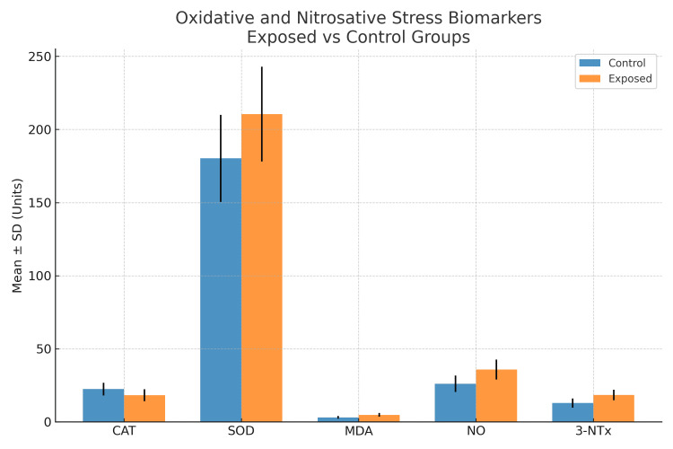 Figure 1
| Variable | Exposed group (n=100) | Control group (n=100) | p-value |
| Age (years) | 35.9 ± 7.1 | 36.2 ± 6.8 | 0.742 |
| Sex (Male %) | 70 (70%) | 68 (68%) | 0.815 |
| Work duration (years) | 10.9 ± 7.1 | – | – |
| Lead apron use (%) | 69 (69%) | – | – |
| Thyroid shield use (%) | 59 (59%) | – | – |
| Dosimeter use (%) | 80 (80%) | – | – |
| Biomarker | Exposed group (n=100) | Control group (n=100) | t-value | p-value |
| CAT (U/mg Hb) | 18.25 ± 4.07 | 22.46 ± 4.34 | 5.12 | 0.001 |
| SOD (U/mg Hb) | 210.52 ± 32.38 | 180.21 ± 29.71 | 6.45 | 0.004 |
| MDA (µmol/L) | 4.84 ± 1.21 | 3.12 ± 0.92 | 5.98 | 0.002 |
| NO (µmol/L) | 35.74 ± 6.81 | 26.05 ± 5.67 | 6.01 | 0.003 |
| 3-NTx (ng/mL) | 18.41 ± 3.59 | 12.89 ± 3.14 | 6.75 | 0.001 |
Peer Reviews
No public reviews on file for this paper yet. If you reviewed it on a platform where reviews are public (OpenReview, ICLR, NeurIPS, ICML), you can paste yours below so the community can read it here.
Videos
No videos yet. Explain this paper in a talk, walkthrough, or lecture? Add one.
Taxonomy
TopicsRadiation Dose and Imaging · Effects of Radiation Exposure · Chemical Safety and Risk Management
Introduction
Ionizing radiation (IR) has become an indispensable tool in modern medicine, particularly in diagnostic and interventional radiology. With the increasing use of radiological modalities such as X-ray, computed tomography (CT), and angiographic imaging, healthcare professionals working in radiodiagnostic laboratories are continuously exposed to low doses of radiation. Although these exposures are typically within recommended safety limits, cumulative effects over time may lead to significant biological consequences [1,2].
The primary mechanism of IR-induced biological damage involves the generation of reactive oxygen species (ROS) and reactive nitrogen species (RNS) through radiolysis of water and interaction with biomolecules. Excessive production of these free radicals overwhelms the antioxidant defense systems of the cell, resulting in oxidative and nitrosative stress. This imbalance may lead to structural and functional alterations in lipids, proteins, and DNA, thereby contributing to mutagenesis, apoptosis, and carcinogenesis [3,4].
Oxidative stress is commonly assessed by measuring lipid peroxidation products such as malondialdehyde (MDA) and enzymatic antioxidants, including catalase (CAT) and superoxide dismutase (SOD). Nitrosative stress, on the other hand, can be evaluated by nitric oxide (NO) and 3-nitrotyrosine (3-NTx) levels, which reflect RNS-mediated protein modifications [5]. Several studies have suggested that chronic low-dose radiation exposure may disrupt oxidant/antioxidant homeostasis in healthcare workers, but data remain limited, particularly in regions with varying radiation safety practices [6,7].
The Eastern Mediterranean region, encompassing provinces such as Adana, Hatay, and Kahramanmaraş in Türkiye, hosts a considerable number of radiodiagnostic laboratories where healthcare workers are routinely exposed to IR. Despite the widespread use of radiological equipment in these centers, there is a paucity of data on occupational oxidative and nitrosative stress biomarkers in this population.
Therefore, the present study aimed to determine the levels of oxidative and nitrosative stress biomarkers in radiodiagnostic laboratory personnel in the Eastern Mediterranean region of Türkiye. By comparing radiation-exposed healthcare workers with unexposed controls, this study sought to provide evidence on the potential health risks associated with occupational IR exposure and to emphasize the importance of radiation protection practices.
Materials and methods
Study design and population
This cross-sectional study was conducted between August 2020 and June 2021 in the Eastern Mediterranean region of Türkiye, covering the provinces of Adana, Hatay, and Kahramanmaraş (Appendices A-C). A total of 200 participants were recruited, including 100 radiodiagnostic laboratory personnel occupationally exposed to ionizing radiation (IR) and 100 age- and sex-matched healthy controls with no known radiation exposure. The exposed group consisted of radiology technicians, interventional radiology staff, and angiography laboratory personnel.
The sample size was calculated using G*Power software (Ver. 3.1, Heinrich-Heine-Universität Düsseldorf, Düsseldorf, Germany) based on an effect size of 0.5, a power of 90%, and α=0.05. This estimation was supported by previous studies investigating oxidative stress biomarkers in radiation-exposed healthcare workers [8,9]. Inclusion criteria for the exposed group were: (i) active employment in radiodiagnostic laboratories for at least one year, and (ii) willingness to participate with informed consent. Exclusion criteria included: (i) history of chronic systemic disease (e.g., diabetes mellitus, chronic kidney disease, or autoimmune disorders), (ii) use of antioxidant supplements or medications within the last three months, and (iii) prior history of therapeutic radiation exposure. The same exclusion criteria were applied to the control group. All participants completed a structured questionnaire (Appendix D) prepared by the investigators, including sociodemographic information (age, sex, education, income, marital status), lifestyle habits (smoking, alcohol use), occupational history (work duration, radiation exposure site), and protective measures (use of lead apron, thyroid shield, and dosimeter). Health-related complaints such as fatigue, headache, irritability, and reproductive outcomes were also recorded. Following overnight fasting (at least eight hours), 5 mL of venous blood was collected from each participant into ethylenediaminetetraacetic acid (EDTA) tubes. Plasma and erythrocyte fractions were separated by centrifugation at 3000 rpm for 10 minutes at 4 °C.
Oxidative Stress Biomarkers
CAT activity was determined spectrophotometrically based on the decomposition of hydrogen peroxide at 230 nm, whereas SOD activity was measured using the inhibition of nitroblue tetrazolium reduction at 505 nm [10,11]. MDA, an indicator of lipid peroxidation, was quantified using the thiobarbituric acid-reactive substances (TBARS) method [12].
Nitrosative Stress Biomarkers
NO levels were measured by the Griess reaction, while 3-NTx concentrations were determined spectrophotometrically as a marker of protein nitration [13,14].
All measurements were performed in duplicate in the Research Laboratory of the Department of Medical Biochemistry, Kahramanmaraş Sütçü İmam University Faculty of Medicine, using validated protocols.
CAT activity was measured using a commercially available spectrophotometric assay kit (Cayman Chemical, Ann Arbor, MI, USA), with an analytical sensitivity of 0.1 U/mL. The intra- and inter-assay coefficients of variation (CVs) were <5% and <8%, respectively. SOD activity was determined using a colorimetric assay kit based on the inhibition of nitroblue tetrazolium reduction (Cayman Chemical, USA), with a detection limit of 0.05 U/mL and intra- and inter-assay CVs of <6% and <9%, respectively. MDA levels were quantified using the thiobarbituric acid-reactive substances (TBARS) assay (Sigma-Aldrich, St. Louis, MO, USA), with a sensitivity of 0.1 µmol/L and intra- and inter-assay CVs of <7% and <10%, respectively. NO concentrations were measured using a colorimetric Griess reaction-based assay kit (Cayman Chemical, USA), with a detection limit of 1.0 µmol/L and CVs below 8%. Serum 3-nitrotyrosine (3-NTx) levels were measured using a commercially available enzyme-linked immunosorbent assay (ELISA) kit (Abcam, Cambridge, UK), with a minimum detectable concentration of 0.1 ng/mL. The intra- and inter-assay CVs were <6% and <9%, respectively. All assays were performed in duplicate according to the manufacturer’s protocols, and calibration curves were generated using certified standards provided with each kit.
Occupational exposure was defined as active employment in radiodiagnostic laboratories with routine contact with ionizing radiation sources. Work duration was recorded as a descriptive indicator of occupational history and potential cumulative exposure, rather than as a precise quantitative measure of individual radiation dose.
The study protocol was approved by the Non-interventional Clinical Research Ethics Committee of Kahramanmaraş Sütçü İmam University (Date: February 1, 2021, Decision No: 88). Written informed consent was obtained from all participants prior to enrollment, and the study was conducted in accordance with the Declaration of Helsinki.
Statistical analysis
Data were analyzed using IBM SPSS Statistics for Windows, version 25 (IBM Corp., Armonk, NY, USA). Continuous variables were expressed as mean ± standard deviation (SD) or median (interquartile range) as appropriate. Categorical variables were presented as frequencies and percentages. Intergroup comparisons were performed using the independent samples t-test or Mann-Whitney U test for continuous variables and chi-square test for categorical variables. Pearson’s correlation analysis was applied to evaluate the relationship between duration of occupational exposure and oxidative/nitrosative stress biomarkers. A p-value <0.05 was considered statistically significant.
Results
A total of 200 participants were included in the study, comprising 100 radiodiagnostic laboratory personnel (exposed group) and 100 healthy individuals without occupational radiation exposure (control group). The mean age of the exposed group was 35.9 ± 7.1 years, while the control group had a mean age of 36.2 ± 6.8 years. The proportion of male participants was similar in both groups (70% vs. 68%). There were no statistically significant differences between the groups in terms of age or sex distribution (p=0.742 and p=0.815, respectively). The average duration of employment in radiodiagnostic laboratories among the exposed group was 10.9 ± 7.1 years, reflecting occupational history rather than measured cumulative radiation dose. Regarding radiation protection practices, 69% of the exposed group reported regular use of lead aprons, 59% reported using thyroid shields, and 80% reported using personal dosimeters (Table 1).
Oxidative and nitrosative stress biomarkers
Comparisons of oxidative and nitrosative stress biomarkers between groups are presented in Table 2. CAT activity was significantly lower in the exposed group compared to the controls (18.25 ± 4.07 vs. 22.46 ± 4.34 U/mg Hb, p=0.001). SOD activity was significantly higher in the exposed group (210.52 ± 32.38 vs. 180.21 ± 29.71 U/mg Hb, p=0.004). MDA levels, a marker of lipid peroxidation, were significantly elevated in exposed workers (4.84 ± 1.21 vs. 3.12 ± 0.92 µmol/L, p=0.002). NO concentrations were significantly higher in the exposed group compared to controls (35.74 ± 6.81 vs. 26.05 ± 5.67 µmol/L, p=0.003). Levels of 3-NTx were also significantly elevated in the exposed group (18.41 ± 3.59 vs. 12.89 ± 3.14 ng/mL, p=0.001). See Appendix E.
These findings indicate that occupational exposure to ionizing radiation was associated with increased oxidative and nitrosative stress, reflected by elevated MDA, NO, and 3-NTx levels, increased SOD activity, and decreased CAT activity (Figure 1).
Comparison of oxidative and nitrosative stress biomarkers between exposed and control groupsBar graph showing mean ± SD values of oxidative and nitrosative stress biomarkers in radiodiagnostic laboratory personnel (exposed) and healthy controls. Catalase (CAT) activity was significantly lower in the exposed group, whereas superoxide dismutase (SOD) activity, malondialdehyde (MDA), nitric oxide (NO), and 3-nitrotyrosine (3-NTx) levels were significantly higher compared to controls. Error bars represent standard deviation.
Discussion
In this study, we investigated oxidative and nitrosative stress biomarkers in radiodiagnostic laboratory personnel exposed to ionizing radiation (IR) in the Eastern Mediterranean region of Türkiye. Our findings demonstrate that exposure to IR is associated with decreased CAT activity, increased SOD activity, and elevated levels of MDA, NO, and 3-NTx. These results suggest that occupational IR exposure disrupts the oxidant-antioxidant balance, leading to oxidative and nitrosative stress. The observed reduction in CAT activity in exposed workers may reflect enzyme consumption as a consequence of increased hydrogen peroxide formation during radiation-induced oxidative stress. Similar decreases in CAT activity have been reported among medical radiation workers, supporting the hypothesis that chronic low-dose IR may impair antioxidant defenses [15]. Conversely, we observed an increase in SOD activity, which may represent a compensatory upregulation in response to excess superoxide anion production. Previous studies have also shown that SOD activity is often elevated in individuals with chronic oxidative stress as a cellular adaptation mechanism [16].
MDA levels were significantly higher in the exposed group, indicating enhanced lipid peroxidation. This finding aligns with earlier research demonstrating that IR exposure increases lipid peroxidation products, which serve as reliable markers of oxidative damage [17,18]. Lipid peroxidation can compromise cell membrane integrity, promote mutagenesis, and ultimately contribute to carcinogenesis, underscoring its clinical relevance in occupational radiation safety. We also found significant elevations in NO and 3-NTx levels in radiation-exposed workers. NO is a reactive nitrogen species with dual physiological and pathological roles. While NO at physiological concentrations regulates vasodilation and neurotransmission, its overproduction leads to peroxynitrite formation, which in turn nitrates tyrosine residues to form 3-NTx. Elevated 3-NTx levels observed in our study indicate protein nitration and nitrosative stress. Prior investigations have reported similar increases in nitrotyrosine levels among individuals exposed to IR, further supporting our findings [19,20].
Taken together, our results suggest that chronic occupational IR exposure induces a state of persistent oxidative and nitrosative stress. This imbalance may have long-term health implications. Oxidative stress has been implicated in the pathogenesis of cancer, cardiovascular disease, and neurodegenerative disorders, conditions that may occur with higher prevalence among healthcare workers exposed to radiation [21]. Nitrosative stress, particularly peroxynitrite-mediated protein modifications, has been associated with autoimmune phenomena and tissue injury, raising concerns about possible systemic effects in chronically exposed populations [22]. Our findings are consistent with several international reports. Ionizing radiation is known to trigger cellular damage through mechanisms such as oxidative stress and radiation-induced bystander effects [23]. Mitochondria function as key regulators of cellular stress responses and apoptosis, underscoring their critical role in pathways of cellular injury [24]. Collectively, these studies strengthen the evidence that even low-dose occupational radiation exposure, when accumulated over years, may have measurable biological effects.
Another important implication of our study is the observed gap in radiation safety practices. Despite high reported rates of dosimeter and protective equipment usage, biomarker alterations were still evident. This highlights the need for regular training programs, stricter monitoring of cumulative doses, and potential dietary or pharmacological interventions aimed at reinforcing antioxidant defenses. Antioxidant supplementation, such as vitamins C and E, has been suggested as a potential protective strategy against radiation-induced oxidative stress, although more randomized controlled trials are needed to validate their efficacy [25]. Nevertheless, this study has certain limitations. First, its cross-sectional design precludes establishing causality between radiation exposure and oxidative/nitrosative stress. Second, we did not assess genetic polymorphisms of antioxidant enzymes, which may influence inter-individual variability in response to IR. Third, the study population was limited to three provinces, which may not fully represent all radiology workers in Türkiye. Future longitudinal studies with larger cohorts and mechanistic investigations are warranted to confirm these findings.
Another important consideration is the external validity of the present findings. Radiation safety practices, including the routine use of personal protective equipment, availability of dosimeters, institutional safety culture, and adherence to radiation protection guidelines, may vary substantially between healthcare institutions and countries. In addition, differences in equipment age, maintenance standards, and procedural workload (particularly in high-volume interventional radiology units) may influence cumulative occupational exposure levels. As the present study was conducted in selected centers within the Eastern Mediterranean region of Türkiye, these regional and institutional characteristics should be taken into account when extrapolating the results to other settings. Therefore, caution is warranted when generalizing our findings to radiodiagnostic personnel working under different safety protocols or technological conditions.
Importantly, the present study did not evaluate DNA damage, clinical outcomes, or symptom-biomarker associations. Therefore, the findings should not be interpreted as evidence of increased disease risk but rather as biochemical alterations associated with occupational exposure to radiation-related work environments.
An additional limitation of this study is the heterogeneity of the radiation-exposed group. Radiodiagnostic personnel encompass diverse professional roles, including interventional radiology staff, computed tomography technicians, and conventional X-ray operators, who are known to differ substantially in terms of radiation dose intensity and exposure patterns. Due to the limited sample size within each occupational category and the lack of comprehensive individual cumulative dose data, subgroup analyses according to professional role were not feasible in the present study. This heterogeneity may have contributed to variability in biomarker levels and should be considered when interpreting the results. Future studies with larger sample sizes, standardized dosimetry records, and stratified analyses are needed to elucidate role-specific oxidative and nitrosative stress responses.
Several limitations of this study should be acknowledged. First, the cross-sectional design limits the ability to establish temporal or causal relationships between occupational ionizing radiation exposure and alterations in oxidative and nitrosative stress biomarkers. Consequently, it cannot be determined whether the observed biomarker elevations preceded radiation exposure or developed as a result of cumulative exposure over time. In addition, oxidative and nitrosative stress biomarkers are known to exhibit biological variability and may fluctuate over time. Longitudinal studies incorporating repeated biomarker measurements and cumulative dose assessment are, therefore, warranted to provide more definitive evidence regarding temporal dynamics and causality.
Another important limitation relates to exposure assessment. In this study, occupational exposure was categorized based on employment in radiodiagnostic laboratories, and work duration was used as a surrogate indicator of occupational history rather than a direct measure of radiation dose. Therefore, the observed biomarker alterations cannot be attributed exclusively to ionizing radiation exposure, as other occupational or environmental factors (such as workload, shift patterns, psychosocial stress, or non-radiation-related chemical exposures) may also contribute. Consequently, the findings should be interpreted as associations with radiation-related occupational environments rather than definitive radiation-induced effects.
In summary, our study provides evidence that occupational exposure to ionizing radiation is associated with increased oxidative and nitrosative stress, as reflected by altered biomarker profiles. These findings underscore the importance of reinforcing radiation protection strategies and considering antioxidant interventions to safeguard the health of healthcare workers.
Conclusions
This study showed that radiodiagnostic laboratory personnel chronically exposed to ionizing radiation had significantly altered oxidative and nitrosative stress biomarker profiles. Lower catalase activity, higher superoxide dismutase activity, and elevated levels of malondialdehyde, nitric oxide, and 3-nitrotyrosine suggest a disrupted oxidant-antioxidant balance.
These findings highlight the potential long-term health risks of occupational radiation exposure and emphasize the importance of strict radiation protection measures. Routine biomarker monitoring and antioxidant-based preventive strategies may help mitigate adverse effects, but further longitudinal studies are needed to confirm these observations.
The reference list from the paper itself. Each links out to its DOI / PubMed record.
- 1Reduction in primary operator radiation dose exposure during coronary angioplasty using radiation absorbing drape Cureus Sharma AB Agrawal R 015202310.7759/cureus.46619 PMC 1062656437937037 · doi ↗ · pubmed ↗
- 2Ionising radiation and cancer risks: what have we learned from epidemiology?Int J Radiat Biol Gilbert ES 4674828520091940190610.1080/09553000902883836 PMC 2859619 · doi ↗ · pubmed ↗
- 3Free radicals, metals and antioxidants in oxidative stress-induced cancer Chem Biol Interact Valko M Rhodes CJ Moncol J Izakovic M Mazur M 14016020061643087910.1016/j.cbi.2005.12.009 · doi ↗ · pubmed ↗
- 4Ionizing radiation-induced metabolic oxidative stress and prolonged cell injury Cancer Lett Azzam EI Jay-Gerin JP Pain D 486032720122218245310.1016/j.canlet.2011.12.012PMC 3980444 · doi ↗ · pubmed ↗
- 5healthcare workers exposure to ionizing radiation: oxidative stress and antioxidant response Indian J Occup Environ Med Bolbol SA Zaitoun MF Abou El-Magd SA Mohammed NA 72772520213442124110.4103/ijoem.IJOEM_198_20PMC 8341408 · doi ↗ · pubmed ↗
- 6Blood oxidative stress parameters in hospital workers occupationally exposed to low doses of ionizing radiation: A systematic review and meta-analysis Heliyon Mansouri B Moradi A Saba F 010202410.1016/j.heliyon.2024.e 39989 PMC 1169390239748987 · doi ↗ · pubmed ↗
- 7Chronic low dose exposure of hospital workers to ionizing radiation leads to increased micronuclei frequency and reduced antioxidants in their peripheral blood lymphocytes Int J Radiat Biol Siama Z Zosang-Zuali M Vanlalruati A Jagetia GC Pau KS Kumar NS 6977099520193066821310.1080/09553002.2019.1571255 · doi ↗ · pubmed ↗
- 8Radiation protection dosimetry for diagnostic radiology patients Radiat Prot Dosimetry Wall BF 40941910920041527335910.1093/rpd/nch 317 · doi ↗ · pubmed ↗