A theoretical and experimental model of flow characteristics in subretinal injections
Reza Ladha, Benjamin Merveille, Roland Wyns, Benoit Scheid, François Willermain, Marc D. de Smet

TL;DR
This study models subretinal injection flow to optimize parameters for safer gene therapy delivery in retinal diseases.
Contribution
A combined theoretical and experimental model to quantify flow dynamics and residual flow in subretinal injections.
Findings
Jet speed increases with the square root of injection pressure, matching theoretical predictions.
Residual flow persists for 28–47 seconds and increases logarithmically with injection pressure.
The 'lock-and-load' priming method reduces jet speed and increases residual flow compared to 'load-and-lock'.
Abstract
Subretinal injections (SI) are used to deliver gene therapies for inherited retinal diseases, yet the optimal injection parameters remain undefined. This study used theoretical and experimental models to quantify the relationship between injection pressure, flow dynamics, and residual flow. A theoretical model (TM) was developed based on the Hagen-Poiseuille law and the theory of a jet immersed in the same liquid. An experimental model (EM) was constructed to allow for measuring flow and residual flow across injection pressures ranging from 0 to 20 psi. We assessed the effects of ambient pressure, injection system tubing length, and syringe priming technique. A minimum pressure of 6 psi was required to generate a detectable flow in the EM. Jet speed increased with the square root of injection pressure, aligning with theoretical predictions. Residual flow persisted for 28–47 seconds…
Genes, proteins, chemicals, diseases, species, mutations and cell lines named across the full text — each resolved to its canonical identifier and authoritative record.
Click any figure to enlarge with its caption.
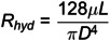 Figure 1
 Figure 2
 Figure 3
 Figure 4
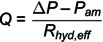 Figure 5
 Figure 6
 Figure 7
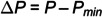 Figure 8
 Figure 9
 Figure 10
 Figure 11
 Figure 12
 Figure 13
 Figure 14
 Figure 15
 Figure 16
 Figure 17
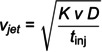 Figure 18
 Figure 19
 Figure 20
 Figure 21
 Figure 22
 Figure 23
 Figure 24
 Figure 25
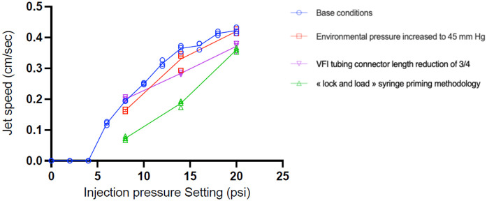 Figure 26
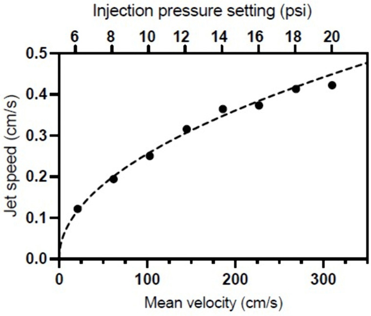 Figure 27
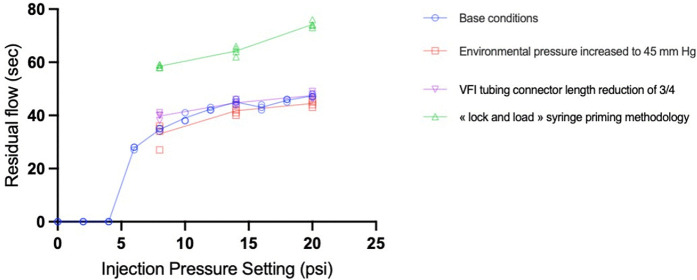 Figure 28
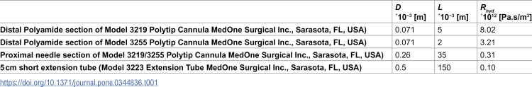 Figure 29
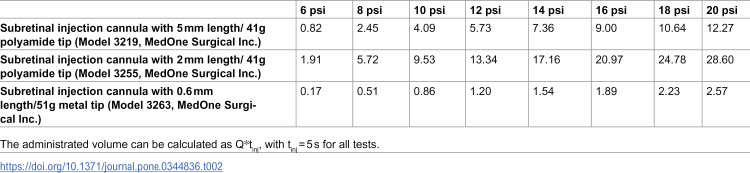 Figure 30
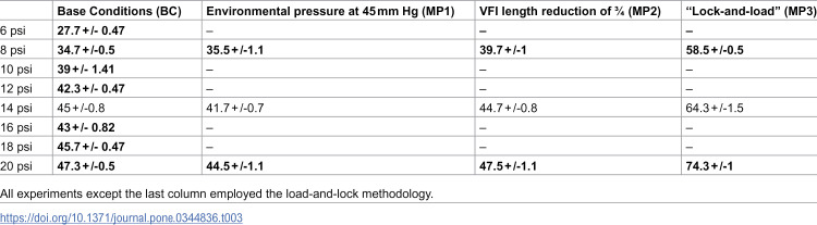 Figure 31
Peer Reviews
No public reviews on file for this paper yet. If you reviewed it on a platform where reviews are public (OpenReview, ICLR, NeurIPS, ICML), you can paste yours below so the community can read it here.
Videos
No videos yet. Explain this paper in a talk, walkthrough, or lecture? Add one.
Taxonomy
TopicsNeuroscience and Neural Engineering · Retinal and Macular Surgery · Retinal Development and Disorders