TEM-based Study of the Phenotype of Astrocytes Differentiated from Induced Pluripotent Stem Cells from a Healthy Donor and a Patient with Parkinson’s Disease
K.A. Kutukova, M.V. Ivanov, E.V. Novosadova, A.V. Brydun, E.L. Arsenyeva, L.V. Novosadova, I.V. Kokorev, I.A. Grivennikov, V.S. Sukhorukov, S.N. Illarioshkin

TL;DR
This study uses electron microscopy to compare astrocytes from healthy and Parkinson’s disease patients, revealing mitochondrial and structural differences linked to the disease.
Contribution
The study demonstrates the utility of TEM in identifying ultrastructural changes in astrocytes derived from iPSCs with a PD-associated LRRK2 mutation.
Findings
Astrocytes from PD patients showed mitochondrial damage, including swelling and cristae destruction.
Cells with LRRK2 mutations exhibited vacuole accumulation and cytoskeletal changes.
Despite ultrastructural differences, no significant differences in cell area or nuclear-cytoplasmic ratios were observed.
Abstract
The aim of this study was to study the role of transmission electron microscopy (TEM) in assessment of the phenotype of astrocytes obtained with the directed differentiation technique from induced pluripotent stem cells (iPSCs) from a healthy donor and from a patient with a hereditary form of Parkinson’s disease (PD). Monolayer astrocyte cultures differentiated from iPSCs from a healthy donor and a PD patient having the G2019S mutation in the LRRK2 gene were used in the study. The obtained glial cultures were characterized using real-time PCR and immunocytochemical staining for glia-specific genes and proteins. TEM was used to examine astrocyte ultrastructure. PCR analysis and immunocytochemical staining demonstrated that cell lines received from a healthy donor and a PD patient expressed the required pattern of glia-specific genes and synthesized astrocyte-specific proteins. However,…
Genes, proteins, chemicals, diseases, species, mutations and cell lines named across the full text — each resolved to its canonical identifier and authoritative record.
Click any figure to enlarge with its caption.
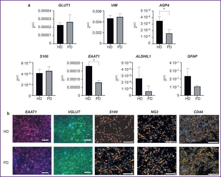 Figure 1
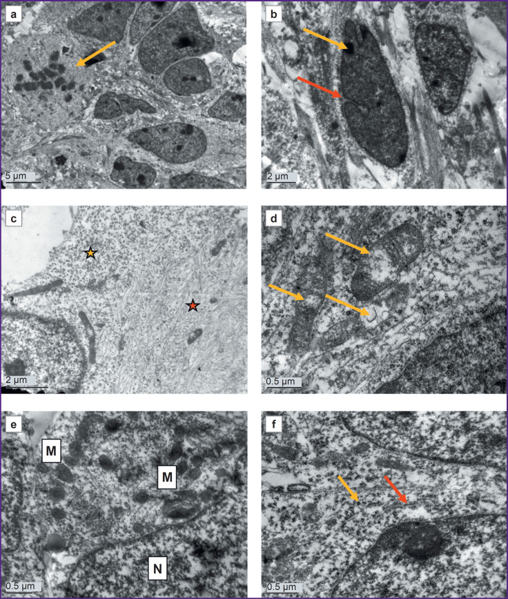 Figure 2
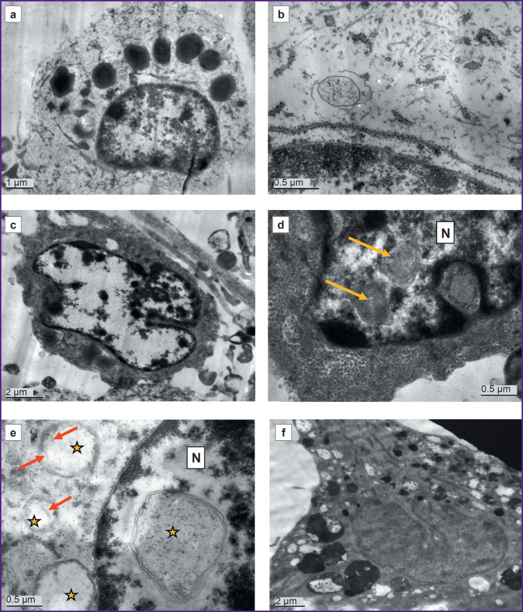 Figure 3
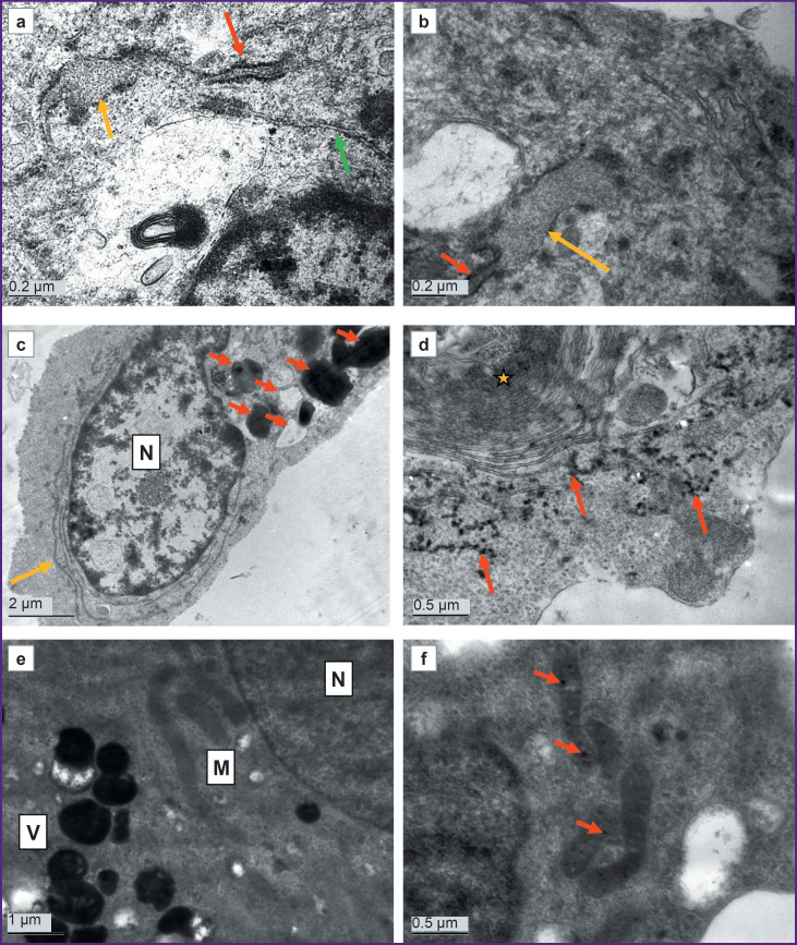 Figure 4
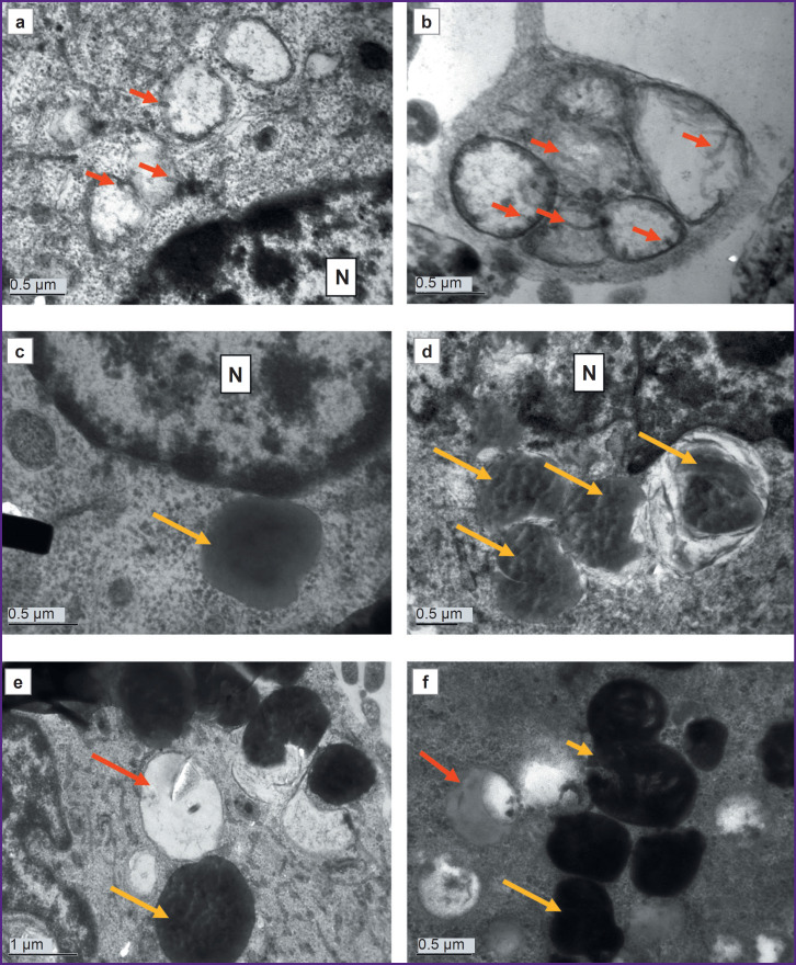 Figure 5
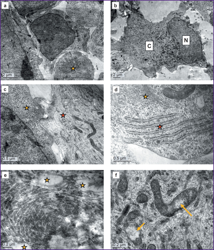 Figure 6
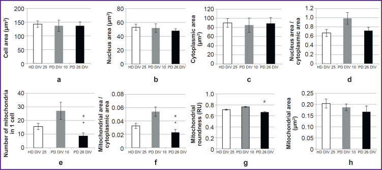 Figure 7
Peer Reviews
No public reviews on file for this paper yet. If you reviewed it on a platform where reviews are public (OpenReview, ICLR, NeurIPS, ICML), you can paste yours below so the community can read it here.
Videos
No videos yet. Explain this paper in a talk, walkthrough, or lecture? Add one.
Taxonomy
TopicsPluripotent Stem Cells Research · Neurogenesis and neuroplasticity mechanisms · Mesenchymal stem cell research