Quantitative assessment of ophthalmic viscosurgical device retention during phacoemulsification and aspiration: an ex vivo analysis
Ippei Watanabe, Hirotaka Hoshi, Kanna Cho, Hirokazu Mukuno

TL;DR
This study measures how much of a surgical eye gel stays in the eye during cataract surgery, showing how injection volume affects retention.
Contribution
The paper introduces a quantitative method to assess residual ophthalmic viscosurgical device (OVD) in ex vivo eyes during phacoemulsification and aspiration.
Findings
Using 0.1 mL of dispersive OVD with the soft shell technique, about 60 μL remained after 2 minutes of PEA.
Injecting 0.15 mL of dispersive OVD left approximately 60 μL after 5 minutes of PEA.
Injecting 0.05 mL of dispersive OVD resulted in less than 30 μL remaining after 0.5 minutes, offering insufficient corneal protection.
Abstract
To quantify the amount of residual ophthalmic viscosurgicaldevice (OVD) during phacoemulsification and aspiration (PEA). A fluorescein-stained dispersive OVD consisting of 3% hyaluronic acid (HA) and 4% chondroitin sulfate (CS) was injected into porcine eyes in volumes of 0.05, 0.1, and 0.15 mL. Subsequently, a cohesive OVD containing 1% HA was injected, and the soft shell technique (SST) was used. Porcine eyes filled with 0.4 mL of dispersive OVD alone were also evaluated. PEA was performed for 0.5, 1, 2, 3, 4, and 5 min, and the amount of dispersive OVD remaining in the eye at each time point was quantified by measuring sulfate ions contained in the CS molecules. Using the SST with 0.1 mL of dispersive OVD, the corneal endothelium was covered for up to 2 min of PEA, and approximately 60 μL of dispersive OVD remained. With the SST using 0.15 mL of dispersive OVD, approximately 60 μL…
Genes, proteins, chemicals, diseases, species, mutations and cell lines named across the full text — each resolved to its canonical identifier and authoritative record.
Click any figure to enlarge with its caption.
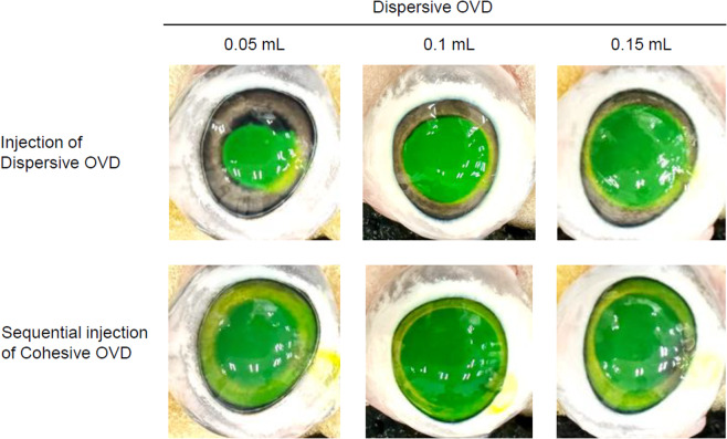 Figure 1
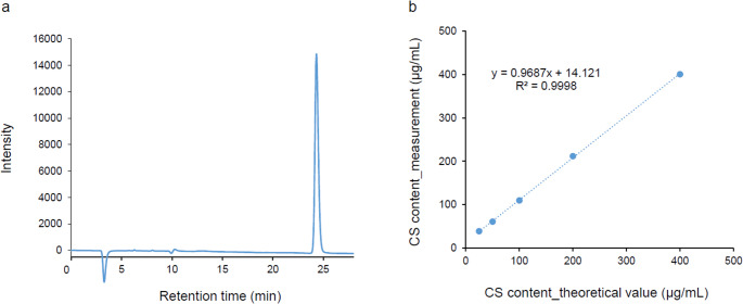 Figure 2
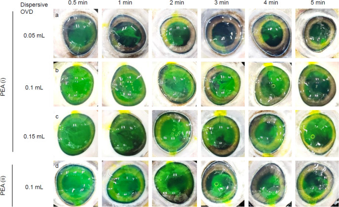 Figure 3
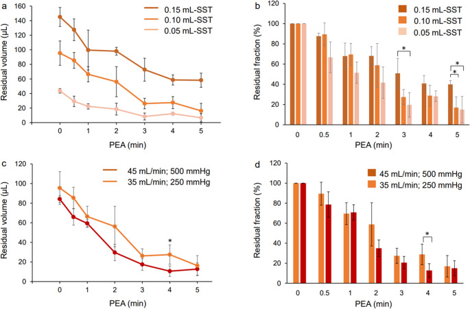 Figure 4
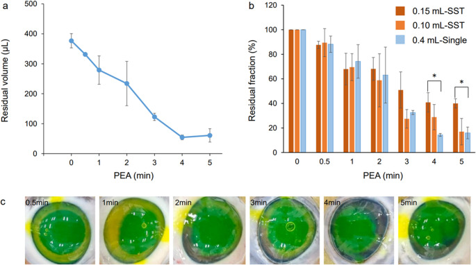 Figure 5
Peer Reviews
No public reviews on file for this paper yet. If you reviewed it on a platform where reviews are public (OpenReview, ICLR, NeurIPS, ICML), you can paste yours below so the community can read it here.
Videos
No videos yet. Explain this paper in a talk, walkthrough, or lecture? Add one.
Taxonomy
TopicsRetinal and Macular Surgery · Intraocular Surgery and Lenses · Ocular Infections and Treatments