An exploratory study of altered regional homogeneity in Parkinson’s disease with depression
Shihua Liu, Xudong Zhu, Yan Chen, Chao Zhang, Xiaowei Zhu, Rumeng Zhang, Lei Chen, Bin Li, Ping Zhong

TL;DR
This study uses brain scans to find patterns of brain activity linked to depression in Parkinson’s disease patients, identifying potential biomarkers.
Contribution
The study identifies specific brain regions with altered regional homogeneity in Parkinson’s disease with depression, suggesting potential neuroimaging biomarkers.
Findings
DPD showed increased ReHo in the left inferior temporal gyrus and decreased ReHo in the right middle frontal gyrus, left insula, and left hippocampus.
ReHo in the left insula and hippocampus showed strong correlations with depression severity and could distinguish DPD with high accuracy.
Altered ReHo in specific brain regions may serve as potential biomarkers for depression in Parkinson’s disease.
Abstract
Depression is a prevalent non-motor symptom in Parkinson’s disease (PD), yet its pathogenesis is unclear and biomarkers are lacking. This rs-fMRI study used Regional Homogeneity (ReHo) to explore neural correlates in PD with depression (DPD). We included 23 DPD, 24 non-depressed PD (NDPD), and 20 healthy controls (HC). ReHo analysis was applied to identify regional brain activity differences. Correlations between ReHo values and depression severity (HAMD scores) were examined. ROC analysis assessed the diagnostic utility of ReHo changes. Compared to NDPD, DPD showed increased ReHo in the left inferior temporal gyrus (ITG) and decreased ReHo in the right middle frontal gyrus (MFG), left insula, and left hippocampus. ReHo in left ITG positively correlated with HAMD scores (r = 0.4347, P = 0.0023), while right MFG (r = -0.5262, P = 0.0001), left insula, and left hippocampus (r = -0.4049,…
Genes, proteins, chemicals, diseases, species, mutations and cell lines named across the full text — each resolved to its canonical identifier and authoritative record.
Click any figure to enlarge with its caption.
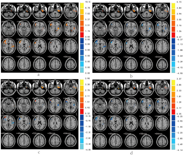 Figure 1
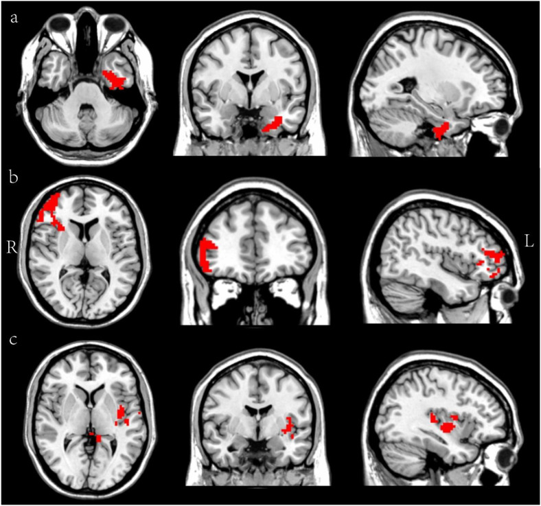 Figure 2
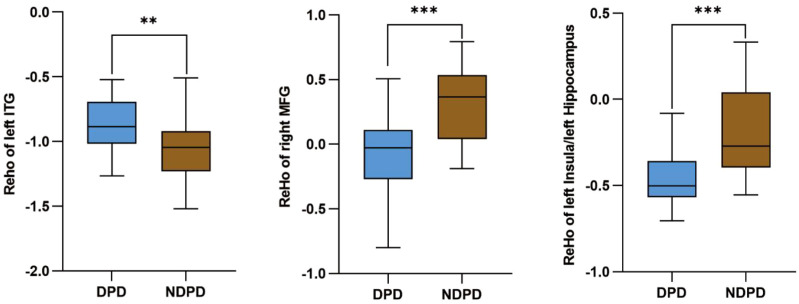 Figure 3
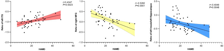 Figure 4
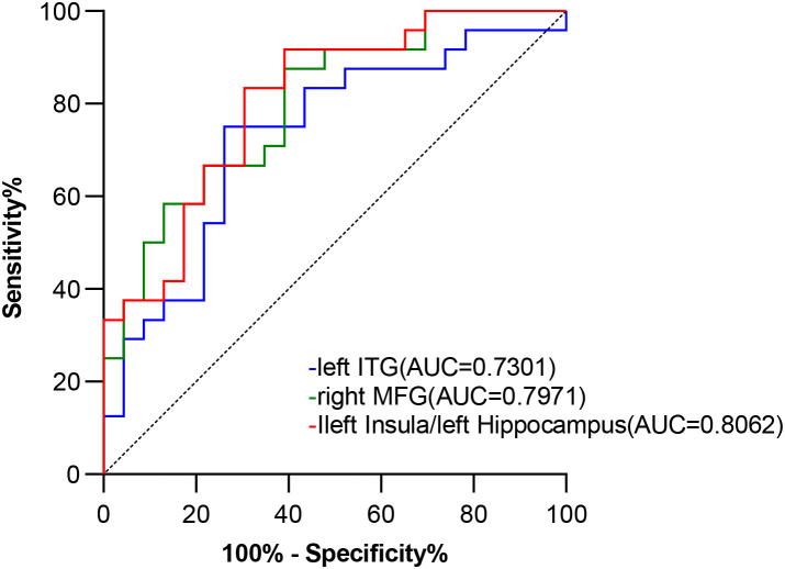 Figure 5
Peer Reviews
No public reviews on file for this paper yet. If you reviewed it on a platform where reviews are public (OpenReview, ICLR, NeurIPS, ICML), you can paste yours below so the community can read it here.
Videos
No videos yet. Explain this paper in a talk, walkthrough, or lecture? Add one.
Taxonomy
TopicsParkinson's Disease Mechanisms and Treatments · Neurological disorders and treatments · Functional Brain Connectivity Studies