Case Report: A case of craniopharyngioma in the cerebellopontine angle
Mulin Zhang, Chengyue Zhu, Yuping Diao, Yin Mo

TL;DR
A rare case of craniopharyngioma was found in the cerebellopontine angle of a 36-year-old man and successfully treated with surgery.
Contribution
This case report highlights the unusual location of craniopharyngioma in the cerebellopontine angle, expanding clinical understanding of this tumor's presentation.
Findings
The tumor was identified using CT and MRI scans, showing specific imaging characteristics.
Surgical resection confirmed the diagnosis of craniopharyngioma.
The case emphasizes the importance of considering rare tumor locations in differential diagnoses.
Abstract
We report a rare case of craniopharyngioma located in the cerebellopontine angle. A 36-year-old male patient presented with relevant clinical symptoms. Computed tomography (CT) revealed a slightly hypodense mass in the left cerebellopontine angle extending to the ambient cistern. Magnetic resonance imaging (MRI) demonstrated an irregular lesion in the same region, which was slightly hyperintense to isointense on T1-weighted images and heterogeneously hyperintense on T2-weighted images. Diffusion-weighted imaging (DWI) showed mixed signals. The lesion was surgically resected, and the final pathological diagnosis confirmed it as a craniopharyngioma.
Genes, proteins, chemicals, diseases, species, mutations and cell lines named across the full text — each resolved to its canonical identifier and authoritative record.
Click any figure to enlarge with its caption.
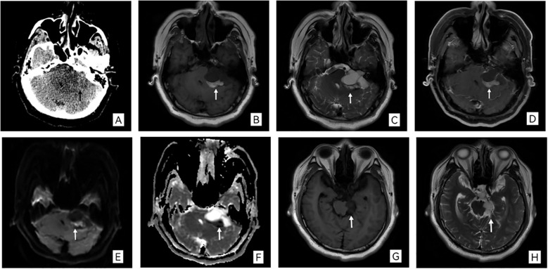 Figure 1
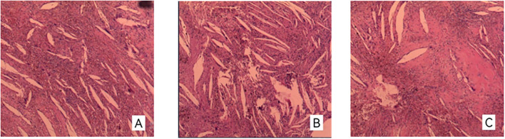 Figure 2
Peer Reviews
No public reviews on file for this paper yet. If you reviewed it on a platform where reviews are public (OpenReview, ICLR, NeurIPS, ICML), you can paste yours below so the community can read it here.
Videos
No videos yet. Explain this paper in a talk, walkthrough, or lecture? Add one.
Taxonomy
TopicsPituitary Gland Disorders and Treatments · Meningioma and schwannoma management · Teratomas and Epidermoid Cysts