What can we learn from the features and presentation of retinal pigment epithelium hypertrophy?
Gustavo Rosa Gameiro, Maura Abraham-Marin, Zelia Maria Correa

Abstract
Genes, proteins, chemicals, diseases, species, mutations and cell lines named across the full text — each resolved to its canonical identifier and authoritative record.
Click any figure to enlarge with its caption.
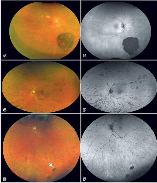 Figure 1
Peer Reviews
No public reviews on file for this paper yet. If you reviewed it on a platform where reviews are public (OpenReview, ICLR, NeurIPS, ICML), you can paste yours below so the community can read it here.
Videos
No videos yet. Explain this paper in a talk, walkthrough, or lecture? Add one.
Taxonomy
TopicsRetinal Development and Disorders · Retinal and Optic Conditions · Retinal Imaging and Analysis
Dear Editor,
We read with great interest the article by Carvalho et al., describing the use of congenital hypertrophy of the retinal pigment epithelium (CHRPE) as a phenoty-pic marker for familial adenomatous polyposis (FAP)^(1)^. The study highlights the importance of CHRPE as a risk factor for FAP, particularly in cases with bilateral fish-tail lesions^(2^,^3)^. This is an informative and important finding that motivated us to add further insights into the clinical spectrum of retinal pigment epithelium (RPE) hypertrophy through a review of the three distinct presentations. Knowledge of these is critical for accurate diagnosis and differentiation.
Figure 1. Clinical spectrum of retinal pigment epithelium (RPE) hypertrophy. Solitary RPE hypertrophy (A and B). A. A wide-field color fundus photograph showing a large flat dark gray to black lesion with scalloped margins in the inferotemporal periphery associated with lacunae skin pigmentation; B. Autofluorescence (AF) showing sharp lesion margins and blockage of the autofluorescence despite the areas of lost pigment; Bear-track lesions (C and D). C. A wide-field color fundus photograph showing diffuse small light-gray patches of retinal pigment epithelium hyperplasia in a clumping pattern consistent with the “bear-track” configuration; D. AF image in which lesions completely block the fundus fluorescence. Fish-tail lesions (E and F). E. A wide-field color fundus photograph showing two small gray-black flat lesions in the inferior periphery with associated whitish fish-tail edges (indicated by the white arrow); F. AF image in which the lesions block some of the AF showing. The hyper-AF associated with the fish-tail effect can be seen at the lesion margins.
We believe a more detailed discussion of these forms of RPE hypertrophy would add to the utility of Carvalho et al.’s findings and assist your readers in distinguishing between FAP-associated lesions and other RPE abnormalities.^(2)^
We also feel that the use of the term “congenital” in this context is problematic as there is no evidence to indicate, which if any of these variations are truly congenital. The lesions are known to grow in more than 50% of cases. In clinics, patients with RPE hypertrophy merit an annual fundus evaluation to monitor for lesion enlargement and the other aforementioned risks.
We commend the authors for their significant contribution and for raising awareness of CHRPE as an important screening finding. We hope the points we have made will broaden the discussion of this condition and further emphasize the importance of early diagnosis and multidisciplinary approaches to hereditary syndromes like FAP.
The reference list from the paper itself. Each links out to its DOI / PubMed record.
- 1Carvalho AA Crespo TS Násser LS Maia CM Fonseca CA Silveira CM Use of congenital hypertrophy of the retinal pigment epithelium as a clinical sign of familial adenomatous polyposis Arq Bras Oftalmol 2024883 S 0004-27492025000300300.10.5935/0004-2749.2023-0115 PMC 1299759339607153 · doi ↗ · pubmed ↗
- 2Traboulsi EI Maumenee IH Krush AJ Giardiello FM Levin LS Hamilton SR Pigmented ocular fundus lesions in the inherited gastrointestinal polyposis syndromes and in hereditary nonpolyposis colorectal cancer Ophthalmology 1988957964969284532210.1016/s 0161-6420(88)33093-9 · doi ↗ · pubmed ↗
- 3Bonnet LA Conway RM Lim LA Congenital Hypertrophy of the Retinal Pigment Epithelium (CHRPE) as a screening marker for Familial Adenomatous Polyposis (FAP): systematic literature review and screening recommendations Clin Ophthalmol 2022167657743532104210.2147/OPTH.S 354761 PMC 8934868 · doi ↗ · pubmed ↗
- 4Salmon B Schalenbourg A “De novo” appearance of a choroi-dal melnoma during 5 years’ follow-up for CHRPE Klin Monbl Augenheilkd 202223945905923547281210.1055/a-1785-5349 · doi ↗ · pubmed ↗
- 5Trichopoulos N Augsburger JJ Schneider S Adenocarcinoma arising from congenital hypertrophy of the retinal pigment epithelium Graefes Arch Clin Exp Ophthalmol 200624411251281598381810.1007/s 00417-005-0011-x · doi ↗ · pubmed ↗