Full-thickness macular hole associated with drusenoid pigment epithelial detachment in age-related macular degeneration
Kemal Tekin, Cemile Ucgul Atilgan

Abstract
Genes, proteins, chemicals, diseases, species, mutations and cell lines named across the full text — each resolved to its canonical identifier and authoritative record.
Click any figure to enlarge with its caption.
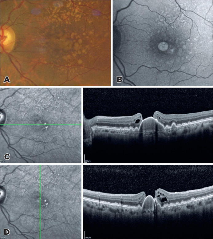 Figure 1
Peer Reviews
No public reviews on file for this paper yet. If you reviewed it on a platform where reviews are public (OpenReview, ICLR, NeurIPS, ICML), you can paste yours below so the community can read it here.
Videos
No videos yet. Explain this paper in a talk, walkthrough, or lecture? Add one.
Taxonomy
TopicsRetinal Diseases and Treatments · Retinal Imaging and Analysis · Ocular Diseases and Behçet’s Syndrome
A fundus image of a 70-year-old woman who has been followed up for age-related macular degeneration showed widespread soft drusen, aggregation of confluent drusen, and central macular hole with pigment epithelial detachment (A). The macular hole and drusen were hyperautofluorescence in fundus autofluorescence (B). Horizontal and vertical optical coherence tomography scans passing through the fovea showed subretinal pigment epithelium deposits, a large drusenoid pigment epithelial detachment combined with full-thickness macular hole^(1)^ with a detached posterior hyaloid (C,D).
The reference list from the paper itself. Each links out to its DOI / PubMed record.