Atypical presentation of sector retinitis pigmentosa
Mariana Calheira Gontijo, Mariana Gouveia Bastos Meirelles, Ricardo Luz Leitão Guerra

Abstract
Genes, proteins, chemicals, diseases, species, mutations and cell lines named across the full text — each resolved to its canonical identifier and authoritative record.
Click any figure to enlarge with its caption.
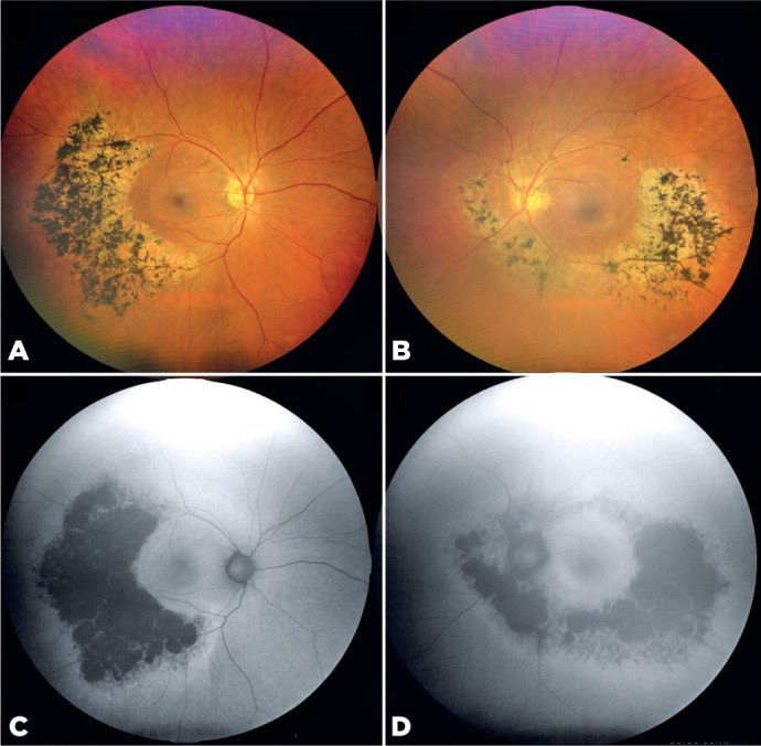 Figure 1
Peer Reviews
No public reviews on file for this paper yet. If you reviewed it on a platform where reviews are public (OpenReview, ICLR, NeurIPS, ICML), you can paste yours below so the community can read it here.
Videos
No videos yet. Explain this paper in a talk, walkthrough, or lecture? Add one.
Taxonomy
TopicsRetinal Development and Disorders · Retinopathy of Prematurity Studies · Ocular Disorders and Treatments
A 58-year-old woman presented with bilateral, asymmetric sector retinitis pigmentosa (RP) (Figure A, B). Fundus autofluorescence imaging revealed hyperautofluorescent rings surrounding hypoautofluorescent regions corresponding to bone spicule pigmentation and retinal degeneration (Figure C, D). The left eye also exhibited nuclear cataract–induced media opacities. Despite the asymmetry, macular integrity was relatively well-preserved in both eyes. Asymmetric and sector RP is a rare variant of the disease characterized by localized retinal involvement, typically affecting specific quadrants and displaying less symmetry compared with the classic diffuse form of RP^(1^,^2)^.
The reference list from the paper itself. Each links out to its DOI / PubMed record.
- 1Lal T Yu ZX Guan B Bender C Chan CC Cukras CA Clinical and histopathologic correlates of asymmetric retinitis pigmentosa JAMA Ophthalmol.202113991029323435138110.1001/jamaophthalmol.2021.2688 PMC 8343521 · doi ↗ · pubmed ↗
- 2Coussa RG Basali D Maeda A De Benedictis M Traboulsi EI Sector retinitis pigmentosa: Report of ten cases and a review of the literature Mol Vis.2019258698931908405 PMC 6937219 · pubmed ↗