Deficiencies in the Fanconi anemia or homologous recombination pathway enhance the antitumor effects of the hypoxia-activated prodrug CP-506
Lesley Schuitmaker, Alexander M.A. van der Wiel, Natasja G. Lieuwes, Rianne Biemans, Nikki A.M. Mutsters, Jennifer Jung, Victoria Claudino Bastos, Èlia Prades Sagarra, Sheng Kuang, Jeremy Setton, Sabine A.S. Langie, Kim R. Kampen, Jan Theys, Ala Yaromina, Ludwig J. Dubois

TL;DR
This study shows that tumors with defects in specific DNA repair pathways respond better to a new cancer drug called CP-506, especially in low-oxygen environments.
Contribution
The study identifies Fanconi anemia and homologous recombination pathway deficiencies as biomarkers for CP-506 efficacy in hypoxic tumors.
Findings
FA- or HR-deficient cells and xenografts showed increased sensitivity to CP-506 compared to parental controls.
CP-506 increased γH2AX expression in FA- and HR-deficient models but not in NHEJ-deficient models.
Deficiencies in FA or HR, but not NHEJ or NER, determine CP-506 sensitivity through a synthetic-lethal interaction.
Abstract
The novel hypoxia-activated prodrug CP-506 selectively targets the hypoxic, treatment-resistant tumor microenvironment. Given the alkylating effector metabolites of CP-506, we hypothesized that defects in interstrand crosslink (ICL) and double-strand break repair influence treatment efficacy. In vitro and in vivo isogenic cancer models proficient or deficient in the Fanconi anemia (FA), homologous recombination (HR), or non-homologous end joining (NHEJ) pathway were used to assess CP-506-induced cytotoxicity and DNA damage. Viability and clonogenic assays demonstrated enhanced sensitivity to CP-506 in FA- or HR-deficient cells compared to parental cells, which was confirmed by spheroid growth inhibition studies. In vivo, CP-506 caused greater enhancement ratios in FA- and HR-deficient xenografts versus parental controls (p < 0.0001) but not in NHEJ-deficient xenografts (p = 0.18).…
Genes, proteins, chemicals, diseases, species, mutations and cell lines named across the full text — each resolved to its canonical identifier and authoritative record.
Click any figure to enlarge with its caption.
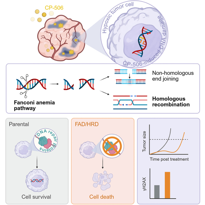 Figure 1
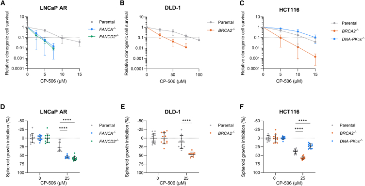 Figure 2
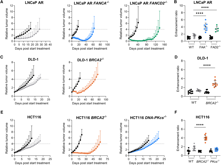 Figure 3
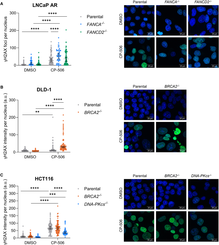 Figure 4
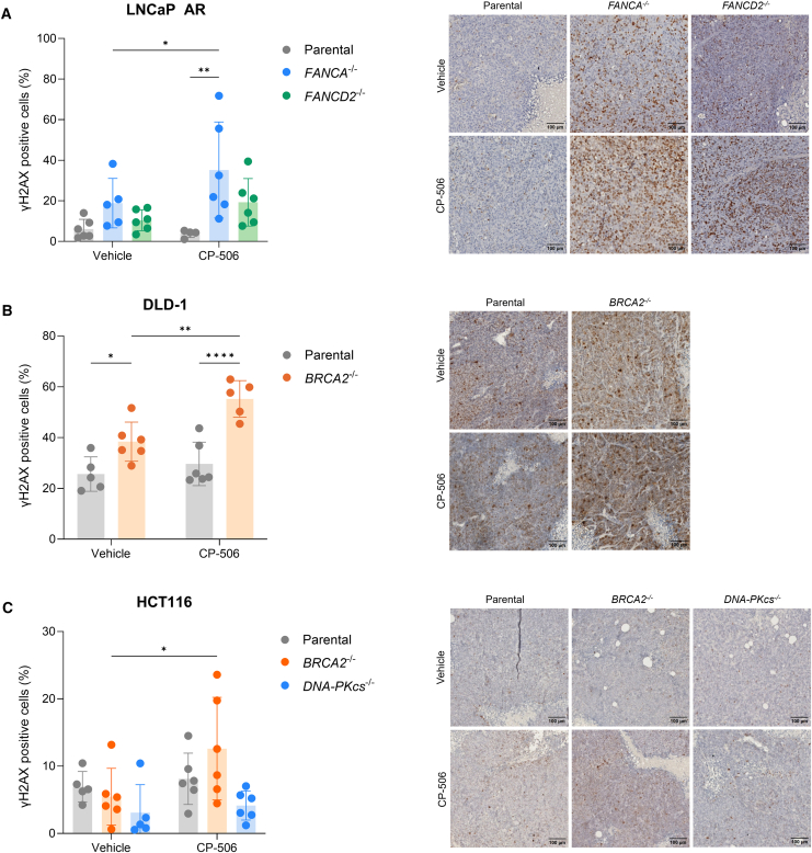 Figure 5
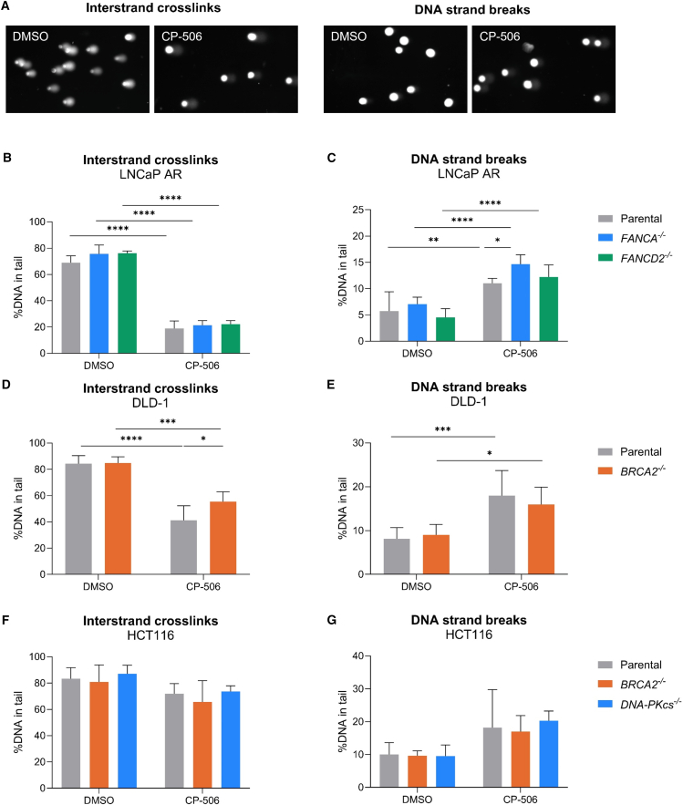 Figure 6
Peer Reviews
No public reviews on file for this paper yet. If you reviewed it on a platform where reviews are public (OpenReview, ICLR, NeurIPS, ICML), you can paste yours below so the community can read it here.
Videos
No videos yet. Explain this paper in a talk, walkthrough, or lecture? Add one.
Taxonomy
TopicsCancer, Hypoxia, and Metabolism · DNA Repair Mechanisms · Cell death mechanisms and regulation
Introduction
Hypoxia-activated prodrugs (HAPs) are a class of cytotoxic agents that selectively target and eliminate hypoxic tumor cells, which are associated with disease progression1 and resistance to conventional anti-cancer therapies.2 Several HAPs have been evaluated in both preclinical and clinical settings.3^,^4 Despite highly encouraging preclinical results, implementation of HAPs in the clinic has not been successful to date, which can be, at least in part, attributed due to a lack of patient stratification in the design of these clinical trials.5 The identification of key factors influencing the tumoral response to HAPs and respective biomarkers of response is therefore essential for successful clinical implementation.
At least three factors are proposed to influence the antitumor effects of a HAP5: first, the degree and severity of tumor hypoxia; second, the levels and activity of endogenous oxidoreductases effecting an initial activation step to yield an oxygen-sensing intermediate; and third, the intrinsic sensitivity of the tumor cell to the effector molecules of the HAP.6
CP-506 is a second-generation HAP with more favorable properties compared to its predecessor PR-104.7 Firstly, CP-506 is resistant to AKR1C3 metabolism, an off-target effect that limited the clinical development of PR-104, causing dose-limiting myelotoxicity in phase 1 clinical trials.8 Secondly, CP-506 has favorable pharmacological properties compared to its predecessor, such as resistance to glucuronidation, a major clearance pathway of PR-104A in humans,9 bioavailability of CP-506 mesylate, and the bystander potential of the effector metabolites.10 Finally, CP-506 is solely activated under severe hypoxic conditions (<0.1% O_2_).11
Recently, we have validated the hypoxia selectivity of CP-506, demonstrating potent antitumor effects in vivo in a broad range of hypoxic tumor xenograft models. Furthermore, a causal relationship between tumor oxygenation and its therapeutic efficacy was established. Moreover, we identified the one-electron reductases cytochrome P450 oxidoreductase (POR), methionine synthase reductase (MTRR), novel diflavin oxidoreductase 1 (NDOR1), and inducible nitric oxide synthase 2A (NOS2A) as likely candidates for the required initial activation step of CP-506.11
As bifunctional alkylators, the hydroxylamine and amine effector metabolites of CP-506 induce various forms of DNA damage, including interstrand crosslinks (ICLs) and monoadducts, as evidenced by the hypoxia-selective formation of DNA adducts.11^,^12 ICLs are extremely toxic DNA lesions because the covalent linkage between the two DNA strands prevents DNA strand separation and thereby interferes with DNA replication and transcription. Repair of ICLs involves a complex and highly coordinated response of components of the Fanconi anemia (FA) pathway or nucleotide excision repair (NER).13 ICL repair proceeds via the formation of double-strand DNA break (DSB) intermediates with subsequent error-free repair by homologous recombination (HR) in the S-phase or by error-prone repair by non-homologous end joining (NHEJ) in all phases of the cell cycle.14^,^15^,^16
The intrinsic sensitivity of tumor cells to the effector molecules of CP-506 is therefore likely determined by the integrity of the DNA damage response (DDR), able to recognize and repair the induced DNA damage.17 Supportive of this, we recently discovered that the MDA-MB-468 cell line—most responsive to CP-506 treatment in a panel of 15 different in vivo xenograft models11—is defective in the FA pathway, harboring a truncating mutation (Q869∗) in the FA complementation group A (FANCA) gene. In addition, several studies have demonstrated that cells and tumors deficient in HR are more sensitive to DSB- and crosslink-inducing chemotherapies,18^,^19^,^20 poly (ADP-ribose) polymerase inhibitors (PARPi),21^,^22 and PR-104 and TH-302,23^,^24^,^25 HAPs with a similar mechanism of action as CP-506.
The present study aimed to evaluate the role of different DNA repair pathways in determining the antitumor efficacy of CP-506. We hypothesized that cancer cells deficient in DNA repair pathways involved in the repair of ICLs and DSBs may exhibit increased sensitivity to CP-506, consistent with the concept of synthetic lethality. In vitro, we first determined the sensitivity of isogenic cancer cell lines, proficient or deficient in the FA, HR, or NHEJ pathway, to CP-506 using cell viability assays and clonogenic cell survival assays, after which we validated our findings in 3D spheroid models. In vivo, we further characterized the role of these DNA repair pathways in the antitumor effects of CP-506 in isogenic xenograft models. Lastly, DNA damage and repair were assessed by γH2AX—a marker of DSB—expression26 and by alkaline comet assays.27^,^28
Results
Cells deficient in the FA or HR pathway are more sensitive to CP-506
To investigate the role of DNA repair pathways on the efficacy of CP-506, we first assessed cell viability of the different isogenic cancer cell lines under normoxic and anoxic conditions upon increasing concentrations of CP-506. For all isogenic cell lines tested, normoxic IC_50_ values were consistently higher than anoxic IC_50_ values resulting in hypoxia-cytotoxicity ratios (HCRs) ranging from 3.2 to 20.3, supportive of the hypoxia-dependent metabolism and cytotoxicity of CP-506. Deficiencies in FA resulted in higher sensitivity to CP-506 compared to parental cells (Table 1). Similarly, isogenic cancer cells deficient in HR were more sensitive to CP-506 under anoxic conditions compared to their respective parental controls. The isogenic cancer cells deficient in NHEJ (DNA-PKcs^−/−^) were less sensitive to CP-506 under anoxic conditions, as indicated by higher anoxic IC_50_ values compared to their respective parental control (Table 1). Complete dose-response curves under normoxic and anoxic conditions are shown in Figure S1.Table 1IC_50_ values of in vitro monolayer cultures of isogenic cancer cell lines proficient or deficient in DNA repair pathwaysCell lineCancer typeDNA repair pathwayNIC_50_ (μM)AIC_50_ (μM)HCRp valueLNCaP ARprostateparental471.573.26.4–LNCaP AR FANCA^−/−^prostateFA>500.036.4>13.7nsLNCaP AR FANCD2^−/−^prostateFA302.025.711.8nsDLD-1colorectalparental>500.0158.6>3.2–DLD-1 BRCA2^−/−^colorectalHR>500.072.5>6.9<0.0001HCT116colorectalparental>500.065.2>7.7–HCT116 BRCA2^−/−^colorectalHR387.819.120.3<0.001HCT116 DNA-PKcs^−/−^colorectalNHEJ>500.0103.9>4.8nsCell viability of isogenic cancer cells exposed to increasing concentrations of CP-506 under normoxic (21% O_2_) or anoxic (≤0.02% O_2_) conditions.NIC_50_, normoxic IC_50_ value; AIC_50_, anoxic IC_50_ value; HCR, hypoxia-cytotoxicity ratio. p values are determined by comparison of curve fit parameters between the isogenic cancer cell lines and their respective parental cell lines.
These findings were validated using clonogenic cell survival assays. Under normoxic conditions, CP-506 exposure only marginally affected clonogenic cell survival in any of the isogenic cancer cell lines tested (Figure S2). Under anoxic conditions, clonogenic survival decreased with increasing CP-506 concentrations (Figures 1A–1C). Compared to their respective parental controls, clonogenic cell survival was significantly decreased in LNCaP AR cancer cells deficient in FANCA (p < 0.05) and FANCD2 (p < 0.001; Figure 1A). Similarly, deficiencies in BRCA2, but not in DNA-PKcs, sensitized HCT116 (p < 0.01) and DLD-1 cancer cells (p < 0.0001) to CP-506 treatment (Figures 1B and 1C).Figure 1. Deficiencies in the Fanconi anemia or the homologous recombination pathway sensitize cancer cells and spheroids to CP-506Clonogenic cell survival upon CP-506 treatment of LNCaP AR (A), DLD-1 (B), and HCT116 (C) isogenic cell lines proficient or deficient in FA, HR, or NHEJ under anoxic conditions. Spheroid growth inhibition 7 days post start of CP-506 treatment of isogenic spheroids proficient or deficient in FA, HR, or NHEJ (D–F). Per condition, 10–12 spheroids were used. Data represent mean ± SD of ≥3 independent experiments. ∗∗∗∗p < 0.0001.
To test the involvement of the NER pathway in repairing CP-506-induced DNA damage, we assessed clonogenic cell survival upon CP-506 treatment in AA8 cells proficient or deficient in XPD, involved in the NER pathway, or XRCC3, involved in the HR pathway.29 CP-506 treatment under normoxic conditions did not decrease clonogenic cell survival in AA8 parental and AA8 UV5 cells and only marginally in AA8 IRS1sf cells at the highest CP-506 concentration tested (Figure S3). Clonogenic cell survival upon anoxic CP-506 treatment was not significantly different between AA8 parental and XPD-deficient AA8 UV5 cells (p = 0.85). XRCC3-deficient AA8 IRS1sf cells, however, showed enhanced sensitivity to CP-506 treatment under anoxic conditions as compared to parental (p < 0.001) and AA8 UV5 cells (p < 0.001; Figure S3).
Hypoxic 3D spheroid cultures were used to further confirm the role of DNA repair pathways in the cytotoxicity of CP-506. In all spheroid cultures, CP-506 induced a spheroid growth inhibition (SGI) at 7 days post start of treatment. FA-deficient LNCaP AR spheroids were significantly more sensitive to CP-506 as indicated by a more pronounced SGI in FANCA- (54.5% ± 3.8%; p < 0.0001) and in FANCD2-deficient spheroids (58.4% ± 6.2%; p < 0.0001) when compared to parental LNCaP AR spheroids (26.0% ± 13.7%) (Figure 1D). In DLD-1 spheroids, CP-506 exposure induced a stronger SGI in spheroids deficient in BRCA2 (46.9% ± 6.6%) compared to parental spheroids (11.2% ± 18.8%; p < 0.0001) (Figure 1E). In HCT116 spheroids, BRCA2 deficiency also resulted in a stronger SGI (57.4% ± 4.3%) compared to parental spheroids (38.1% ± 6.4%; p < 0.0001). In line with the 2D in vitro results, spheroids deficient in DNA-PKcs were significantly less sensitive (SGI: 22.8% ± 6.5%; p < 0.0001) to CP-506 treatment as compared to parental HCT116 spheroids (Figure 1F). These data further confirm that cells and spheroids deficient in FA and HR, but not in NHEJ or NER, exhibit increased sensitivity to CP-506.
Primary glioblastoma (GBM) spheroids were assessed for their DNA repair capacity (supplemental methods, Figure S4; Table S1). U3056MG and U3013MG displayed the highest expression of FA and HR repair-related genes, whereas U3021MG and U3085MG showed the most aberrations in these genes (Table S1). A dose-dependent decrease in cell viability was observed in all GBM cell lines treated with CP-506; however, the largest reductions in cell viability at 7 days post treatment were observed in U3021MG and U3085MG spheroids (p < 0.01; Figure S4B). Spheroid survival analyses (Kaplan-Meier), as well as spheroid growth delay (SGD) and SGI calculations, demonstrated dose-dependent delays in reaching the treatment endpoint for all GBM cell lines (Figure S4C). While SGD analyses identified U3085MG as the most sensitive model, SGI analyses revealed growth suppression across multiple spheroid models, including those with higher FA and HR repair-related gene expression (Table S1).
The antitumor effects of CP-506 are enhanced in xenografts deficient in the FA or the HR pathway
To further evaluate the role of DNA repair pathways in the single-agent antitumor activity of CP-506 in vivo, mice bearing subcutaneous isogenic xenografts were treated with CP-506. As a functional validation of the DNA repair-deficient models, mice were treated with the non-hypoxia-activated alkylating agent chlorambucil. In all models tested, CP-506 and chlorambucil were well tolerated with only transient body weight loss during the treatment period (Figure S5). Tumor hypoxia—an essential factor for CP-506 activation—was confirmed in all models tested as determined by pimonidazole positivity (Figure S6).
Treatment with CP-506 resulted in tumor growth inhibition (TGI) for LNCaP AR parental (52.3% ± 56.4%; p < 0.05), FANCA^−/−^ (98.6% ± 1.5%; p < 0.0001), and FANCD2^−/−^ tumors (97.7% ± 3.2%; p < 0.0001; Figure 2A). CP-506 increased the time to reach four times the starting volume (T4×SV) compared to vehicle-treated controls in LNCaP AR parental (p = 0.29), LNCaP AR FANCA^−/−^ (p < 0.0001), and LNCaP AR FANCD2^−/−^ (p < 0.0001). The resulting enhancement ratios (ERs) were significantly higher for LNCaP AR FANCA^−/−^ (ER: 4.0 ± 1.1; p < 0.0001) and LNCaP AR FANCD2^−/−^ (3.4 ± 0.8; p < 0.0001) compared to LNCaP AR parental (ER: 1.5 ± 0.5) xenografts (Figure 2B; Table S2). FA-deficient LNCaP AR xenografts were also significantly more sensitive to chlorambucil (Figure S7).Figure 2. The antitumor effects of CP-506 are enhanced in tumors deficient in the Fanconi anemia or homologous recombination pathwayMice bearing isogenic tumor xenografts, proficient or deficient in FA (FANCA^−/−^ and FANCD2^−/−^), HR (BRCA2^−/−^), or NHEJ (DNA-PKcs^−/−^), were treated with vehicle (black circles) or CP-506 (colored circles) after which tumor growth was monitored (A, C, and E), and time to reach four times starting volume (T4×SV) and corresponding enhancement ratio (ER) were determined (B, D, and F). Data represent mean ± SD (n = 5–10 animals per group). ∗∗∗∗: p < 0.0001.
CP-506 effectively inhibited tumor growth when compared to vehicle treatment in DLD-1 parental (37.9% ± 10.3%; p < 0.01) and DLD-1 BRCA2^−/−^ xenografts (77.8% ± 11.0%; p < 0.0001; Figure 2C). For DLD-1 parental xenografts, T4×SV was not significantly different between vehicle and CP-506-treated animals (p = 0.49), whereas CP-506 treatment significantly prolonged T4×SV in BRCA2-deficient xenografts (p < 0.0001; Table S2). Treatment of DLD-1 BRCA2^−/−^ xenografts with CP-506 resulted in a significantly higher ER (2.9 ± 0.7; p < 0.0001) when compared to parental DLD-1 xenografts (ER: 1.3 ± 0.2; Figure 2D). Similar responses were observed in HCT116 xenografts. BRCA2-deficient xenografts showed significantly enlarged ERs (4.0 ± 0.6; p < 0.0001) compared to HCT116 parental xenografts (1.7 ± 0.6; Figures 2E and 2F). In HCT116 DNA-PKcs^−/−^ xenografts, however, CP-506 induced a TGI of 33.2% ± 17.3% (p = 0.15) and significantly increased T4×SV (17.5 ± 3.6) compared to vehicle-treated controls (12.6 ± 2.7; p < 0.05; Figure 2E; Table S2). However, the ER of HCT116 DNA-PKcs^−/−^ xenografts (1.4 ± 0.3; p = 0.18) was not significantly different from HCT116 parental xenografts (Figure 2F; Table S2). In line with these findings, the antitumor effects of chlorambucil were also more pronounced in HCT116 and DLD-1 xenografts deficient in HR (Figure S7). Taken together, these data demonstrate that in vivo tumors deficient in FA or HR, but not NHEJ, are more sensitive to CP-506.
CP-506 induces phosphorylation of histone H2AX
To explore the extent of residual DNA damage after CP-506 exposure in more detail, γH2AX expression was assessed using immunofluorescence in in vitro isogenic cancer cells and immunohistochemistry in ex vivo isogenic xenografts. Upon anoxic exposure to CP-506, γH2AX expression presented as distinct nuclear foci in isogenic LNCaP AR cells (Figure 3A). 48 h after CP-506 exposure, all isogenic LNCaP AR cells showed elevated foci counts compared to vehicle treatment (p < 0.0001). FANCA- (55.0 [interquartile range (IQR) = 28.0–84.0]; p < 0.0001) and FANCD2-deficient (37.0 [IQR = 9.5–76.8]; p = 0.06) LNCaP AR cells expressed a higher number of nuclear foci compared to LNCaP AR parental cells (28.0 [IQR = 11.5–53.0]). The number of foci per nucleus remained higher in FANCD2-deficient cells 72 h post treatment (50.0 [IQR = 32.0–89.0]; p < 0.0001), whereas γH2AX foci in FANCA-deficient cells (34.5 [IQR = 16.0–59.8]; p = 0.99) reverted to parental level (36.0 [IQR = 10.0–52.0]; Figure S8A).Figure 3CP-506 induced persistent DNA damage in Fanconi anemia- or homologous recombination-deficient isogenic cancer cellsγH2AX foci count per nucleus for LNCaP AR isogenic cells (A) and quantification of γH2AX immunofluorescence intensity per nucleus for DLD-1 (B) and HCT116 (C) isogenic cancer cells with representative images 48 h post start of treatment under anoxic conditions. Scale bar: 20 μm. Blue: Hoechst; green: γH2AX. n ≥ 42 cells per condition. Data represent median (IQR). ∗∗p < 0.01, ∗∗∗p < 0.001, ∗∗∗∗p < 0.0001.
Interestingly, in isogenic DLD-1 and HCT116 cells, γH2AX expression presented as pan-nuclear staining after anoxic exposure to CP-506 (Figures 3B and 3C). Therefore, instead of counting distinct foci, γH2AX fluorescence intensity per nucleus was quantified. Exposure to CP-506 resulted in an elevated (p < 0.0001) expression of γH2AX in DLD-1 BRCA2-deficient cells when compared to DLD-1 parental cells (Figure 3B). The differences in γH2AX expression levels between parental and BRCA2-deficient DLD-1 cells remained at 72 h after treatment (p < 0.01; Figure S8B). In HCT116 cells, BRCA2 deficiency did not result in elevated γH2AX expression levels neither after 48 h (p = 0.53; Figure 3C) nor after 72 h post treatment (p = 0.61; Figure S8C). HCT116 DNA-PKcs^−/−^ cells exhibited a significant decrease in H2AX phosphorylation at 48 h (p < 0.001; Figure 3C) and 72 h (p < 0.0001; Figure S8C) post treatment compared to their respective parental cancer cells.
Next, γH2AX positivity was assessed in isogenic tumors excised 48 h post treatment. For LNCaP AR parental and FANCD2-deficient tumors (Figure 4A), the percentage of γH2AX-positive cells after CP-506 treatment (3.8% ± 1.9% and 19.3% ± 11.7%, respectively) was similar compared to vehicle-treated tumors (6.1% ± 4.8%; p = 0.78 and 10.4% ± 5.2%; p = 0.24). In contrast, in FANCA-deficient tumors, the percentage of γH2AX-positive cells was significantly increased after exposure to CP-506 (35.2% ± 23.7%) compared to vehicle exposure (18.8% ± 12.2%; p < 0.05). Furthermore, LNCaP AR FANCA^−/−^ tumors (p < 0.01), but not LNCaP AR FANCD2^−/−^ tumors (p = 0.16), exhibited significantly higher percentages of γH2AX-positive cells when compared to LNCaP AR parental tumors in line with in vitro γH2AX results.Figure 4CP-506 induced persistent DSB damage in isogenic tumor xenografts deficient in Fanconi anemia or homologous recombination pathwayPercent of γH2AX-positive cells in LNCaP AR (A), DLD-1 (B), and HCT116 (C) isogenic tumors with representative images of immunohistochemistry staining of γH2AX 48 h post treatment. Scale bar: 100 μm. n = 4–6 animals per group. Data represent mean ± SD. ∗p < 0.05, ∗∗p < 0.01, ∗∗∗∗p < 0.0001.
For DLD-1 tumors (Figure 4B), the baseline level of γH2AX-positive cells was higher in BRCA2-deficient tumors (38.4% ± 7.7%) compared to parental tumors (25.6% ± 6.8%; p < 0.05). For parental tumors, no differences were observed in the amount of γH2AX-positive cells after CP-506 exposure (29.7% ± 8.5%; p = 0.40) compared to vehicle-treated tumors. In contrast, the percentage of γH2AX-positive cells in CP-506-treated DLD-1 BRCA2^−/−^ tumors (55.2% ± 7.2%; p < 0.01) was significantly increased compared to vehicle exposure. Similar results were obtained for HCT116 tumors (Figure 4C). For HCT116 parental tumors, no significant difference was found between the vehicle- (7.0% ± 2.3%) and CP-506-treated (8.1% ± 3.8%; p = 0.67) tumors. For HCT116 BRCA2^−/−^ tumors, there was a significant increase in the percentage of γH2AX-positive cells post CP-506 treatment (12.6% ± 7.6%) compared to the vehicle-treated HCT116 BRCA2^−/−^ tumors (5.5% ± 4.2%; p < 0.05), indicating residual DNA damage within these HR-deficient tumors. In line with the in vitro data (Figure S8), HCT116 DNA-PKcs^−/−^ tumors showed the lowest levels of γH2AX-positive cells upon vehicle (3.1% ± 4.2%) and CP-506 (4.2% ± 2.2%) treatment, with a slight but not significant (p = 0.70) treatment-induced increase (Figure 4C). These results demonstrate that cancer cells and tumors deficient in FA or HR show a higher level of residual DNA damage 48 h after CP-506 treatment as compared to their respective parental counterparts.
The alkaline comet assay confirms the presence of ICLs and DNA breaks upon CP-506 treatment
To gain more insights into the type of CP-506-induced DNA damage and the potential underlying DNA repair mechanisms, the standard alkaline comet assay to detect DNA strand breaks and the modified alkaline comet assay to detect ICLs were performed. In the standard alkaline comet assay, the extent of DNA migration into the tail (%DNA in tail) is proportional to the number of DNA strand breaks, whereas in the modified alkaline comet assay, a decrease in %DNA in tail is indicative of an increase in ICL since these lesions inhibit DNA migration (Figure 5A).27Figure 5CP-506 induced ICLs and DNA strand breaks in isogenic cancer cellsComet assay analysis of isogenic LNCaP AR, DLD-1, and HCT116 cells 48 h post treatment under anoxic conditions. Representative comets of cells exposed to CP-506 or DMSO (A). Cells were either assessed for interstrand crosslinks (ICLs; B, D, and F) or DNA strand breaks (SSB and DSB; C, E, and G). Medians from two biological repeats with one or more technical repeats were averaged ±SD. ∗p < 0.05, ∗∗p < 0.01, ∗∗∗p < 0.001, ∗∗∗∗p < 0.0001.
As expected, CP-506 induced ICLs, as shown by the reduction of the %DNA in tail compared to vehicle treatment for LNCaP AR (p < 0.0001), HCT116 (p = 0.27), and DLD-1 (p < 0.05) cancer cells at 48 h (Figures 5B, 5D, and 5F) and 72 h (Figures S9A, S9C, and S9E) post start of treatment. At 48 and 72 h post CP-506 treatment, DNA breaks were evident in all isogenic models (Figures 5C, 5E, 5G, S9B, S9D, and S9F).
Upon CP-506 exposure of LNCaP AR cells, no differences in the reduction of the %DNA in tail, and thus the presence of ICLs, were observed between FANCA- (21.3% ± 3.6%; p = 0.73) or FANCD2-deficient (22.2% ± 2.7%; p = 0.57) and parental cells (18.9% ± 5.7%; Figures 5B and S9A). DNA strand breaks were increased in FANCA-deficient cells (14.7% ± 1.8%; p < 0.05) compared to parental cells (11.0 ± 0.9 %DNA in tail; Figure 5C). This difference remained, although non-significant at 72 h post treatment (Figure S9B). In LNCaP AR FANCD2^−/−^ cells, the amount of DNA strand breaks was not increased compared to parental cells upon CP-506 exposure (12.2% ± 2.3%; p = 0.69).
The presence of ICLs was significantly increased in DLD-1 parental cells as evidenced by the 14.2% (p < 0.05) and 16% (p < 0.01) reduction in %DNA in tail compared to BRCA2-deficient cells at 48 and 72 h post CP-506 exposure, respectively (Figures 5D and S9C). DLD-1 BRCA2^−/−^ cells displayed lower levels of DNA strand breaks as compared to DLD-1 parental cells at 48 h (16% ± 4% vs. 18% ± 5.7%; p = 0.38; Figure 5E) and 72 h (20.4% ± 2.6% vs. 25.3% ± 2.7%; p < 0.05; Figure S9D). In HCT116 isogenic cancer cells, although showing a slight increase, the presence of ICLs or DNA strand breaks upon exposure to CP-506 was not significantly different compared to vehicle-treated cells (Figures 5F and 5G). There were no differences in the amount of CP-506-induced ICLs present in HCT116 parental (71.9% ± 7.6%) compared to BRCA2- (65.6% ± 16.2%; p = 0.56) or DNA-PKcs-deficient (73.6% ± 65.6%; p = 0.96) cells (Figures 5F and S9E). There was no difference in the amount of DNA strand breaks in DNA-PKcs-deficient cells compared to parental cells at 48 h (20.3% ± 2.9% vs. 18.2% ± 11.5%; p = 0.87; Figure 5G) or 72 h (20.5% ± 6% vs. 12.5% ± 3.2%; p = 0.17; Figure S9F) after exposure to CP-506. These findings confirm the formation of ICL and DNA damage upon CP-506 exposure; however, they were unable to explain the differences in antitumor efficacy of CP-506 between isogenic xenograft models with deficiencies in various DNA repair pathways.
Discussion
In the present study, we evaluated the role of DNA repair pathways in determining the cytotoxicity and antitumor effects of the novel hypoxia-activated DNA crosslinking agent CP-506. The FA and HR pathways are crucial for high-fidelity repair of ICLs and DSBs.13^,^16 Here, we provided evidence that deficiencies in FA or HR, but not in NHEJ or NER, repair pathways resulted in enhanced sensitivity to CP-506.
We first showed in vitro that FA- or HR-deficient cell lines were 2.0- to 3.4-fold more sensitive to CP-506 under anoxic conditions. Interestingly, HCT116 DNA-PKcs^−/−^ cells deficient in NHEJ, a pathway also involved in repair of DSBs and associated with resistance to chemotherapy and radiotherapy,30^,^31 were 1.6-fold more resistant to CP-506. Lastly, we confirmed—in 3D in vitro hypoxic spheroid cultures, which more closely represents the tumor environment32—that a deficiency in FA or HR, but not NHEJ, significantly increased the cytotoxicity of CP-506. This was confirmed in primary patient-derived GBM 3D spheroid models, with models characterized by aberrations in FA- or HR pathway-related genes presenting enhanced sensitivity to CP-506 treatment. Furthermore, we showed in vivo that CP-506 demonstrated monotherapeutic antitumor activity in all isogenic tumor xenograft models tested. In accordance with our in vitro data, CP-506 was more effective in delaying tumor growth in FA- and HR-deficient tumors compared to their parental tumors. Our findings are in line with previous studies demonstrating that cells or tumors deficient in FA or HR were more sensitive to HAPs such as PR-10423^,^24 and TH-30224^,^25 with similar mechanisms of action compared to CP-506. Furthermore, previous studies showed that the FA-deficient MDA-MB-468 triple-negative breast cancer xenograft model was highly sensitive to CP-506 with a 90% curative response.11 Lastly, FA- and HR-deficient tumors were also significantly more sensitive to chlorambucil,18^,^33 a non-hypoxia-activated DNA crosslinking agent with a similar mechanism of action as CP-506, further evidencing ICL induction to be the mechanism of action of CP-506. Altogether, these data further support the involvement of FA and HR in determining the cytotoxicity and antitumor effects of CP-506.
Mechanistically, we have demonstrated that CP-506-induced phosphorylation of H2AX was markedly increased in FA- or HR-deficient cancer cells and tumors compared to their respective parental counterparts. This is in agreement with other studies showing that, in DNA repair-proficient cells, the levels of γH2AX foci restored to baseline level 48 h after exposure to cisplatin or nitrogen mustard HN2, both non-hypoxia-activated ICL-inducing agents.34 Similarly, γH2AX foci remained persistent 48 h post treatment in cancer cells deficient in HR (XRCC3^−/−^) or NER (ERCC1^−/−^).34
Previous studies have observed that models with a high response to CP-506, including FaDu xenografts, showed persistent expression of γH2AX levels even 72 h after exposure, while γH2AX levels reverted to baseline in models with a more resistant phenotype, such as the UT-SCC-5 xenografts.35 Our results therefore suggest that parental cancer cells can repair the CP-506-induced DNA damage, contrary to FA- or HR-deficient cancer cells. Unlike radiation, the γH2AX response after exposure to ICL-inducing agents occurs with delayed kinetics, with a peak induction at 12–24 h and a gradual decrease until 48 h.34^,^36^,^37^,^38 These delayed kinetics could be explained by need of cells to progress into the S-phase before γH2AX induction15 or by other causes, i.e., not related to DNA repair.
Phosphorylation of H2AX on Ser139 is widely used as a marker for DSBs; however, its functionality extends beyond its role in DNA repair. While H2AX is phosphorylated at sites of DSBs, its presence does not necessarily indicate the occurrence of DSBs.39 Increased or persistent γH2AX expression has been found in situations of replication stress due to replication fork stalling or damage,40^,^41^,^42 apoptosis,43^,^44 or cell senescence.45 To unravel the underlying cause of the persistent γH2AX expression in FA- and HR-deficient cells upon CP-506 treatment, we performed alkaline comet assays to assess the presence of ICLs and DNA strand breaks. The modified alkaline comet assay confirmed the presence of CP-506-induced ICLs and DNA strand breaks in all isogenic cell lines, in line with our previously published in vitro and in vivo work showing that CP-506 caused induction of ICLs and DSBs specifically under severe hypoxic conditions.11^,^12 Despite the essential role of the FA pathway in sensing and coordinating the unhooking of ICLs,46^,^47 we observed similar levels of ICLs in FA-deficient and parental LNCaP AR cells, which may reflect the slow and replication-dependent nature of ICL repair.46 Since the presence of ICLs in CP-506-treated cells did not revert to the levels of the vehicle control within 72 h after treatment, the repair of CP-506-induced ICLs may require more than 72 h to resolve, possibly explaining the lack in observed differences in ICL repair between parental and FA- or HR-deficient models. This is supported by previous studies, which provided evidence for distinct unhooking and repair mechanisms of ICLs induced by different crosslinking agents.48
Despite similar ICL levels, FA-deficient cells displayed persistent γH2AX expression both in vitro and ex vivo. One explanation for the enhanced H2AX phosphorylation could be the accumulation of stalled replication forks resulting from ICL-induced replication stress.42 The presence of γH2AX at stalled replication forks is required for the recruitment and accumulation of FANCD2, which plays a crucial role in stabilizing stalled replication forks and promoting restart.49^,^50 In the absence of a functional FA pathway, unresolved stalled replication forks from ICL-induced replication stress may collapse, leading to the formation of DSBs,51^,^52 thereby resulting in persistent γH2AX levels and increased DNA strand breaks observed in FA-deficient compared to parental cells upon CP-506 treatment. Despite the vast evidence of the involvement of the FA pathway in the recognition and unhooking of ICLs, our findings could also be explained by previous studies suggesting that the FA pathway plays an important role in the more downstream ICL repair, including replication fork stabilization and repair of DSBs from ICL processing.53^,^54
Unexpectedly, DLD-1 BRCA2-deficient cells contained fewer detectable ICLs than parental cells, whereas no such difference was observed in HCT116 parental versus BRCA2-deficient cells. At present, we do not have a clear reason explaining why parental DLD-1 cells display higher levels of detectable ICLs than their BRCA2-deficient counterparts. Future experiments, such as the enzyme-modified comet assay,55 could address this by directly comparing the capacity of parental and BRCA2-deficient cells to process and repair ICL-containing DNA by means of isolating DNA repair proteins from these cell lines and incubating them with substrate DNA containing ICL or other DNA lesions. These experiments could provide insights into the DNA repair capacity of the proteins present within parental and BRCA2-deficient cells and give more information on specific DNA lesions.55
Moreover, it has been suggested that the NER pathway is responsible for the detection and incision of ICLs formed upon exposure to crosslinking agents,14^,^54^,^56^,^57 indicating that the removal of ICL is not solely dependent on the FA pathway. However, our results show no increased sensitivity to CP-506 in AA8 cells deficient in XPD, an important DNA helicase in the NER pathway, compared to parental AA8 cells, therefore suggesting that the NER pathway is not involved in the repair of CP-506-induced DNA damage.
We hypothesized that HR-deficient cells would show delayed repair of ICL-induced DSB intermediates upon CP-506 exposure compared to their respective parental cells. In addition, BRCA2 is known to stabilize and protect stalled replication forks, and its deficiency can lead to replication fork degradation, collapse, and DNA strand breaks.58^,^59^,^60^,^61 Contrary to our initial hypothesis, no differences in DNA strand breaks were observed between HCT116 parental and BRCA2-deficient cells, whereas DLD-1 BRCA2^−/**−^ cells showed lower levels of DNA strand breaks compared to their parental cells. The bifunctional effector metabolites of CP-506 induce complex DNA damage, including monoadducts and intrastrand crosslinks in addition to ICLs and ICL-repair-associated DSB intermediates,11^,^12 which cannot be discriminated by the alkaline comet assay. Furthermore, the high pH of the alkaline comet assay unwinds the double-stranded DNA and causes hydrolysis of alkali-labile sites into single-strand DNA breaks (SSBs), such that the DNA strand break readout includes SSBs, DSBs, and SSBs derived from alkali-labile sites.62 As a result, interpretation of DNA strand break levels in CP-506-treated cells may be complicated by the combined contribution of different lesion types.
DNA-PKcs-deficient cells displayed elevated DNA strand breaks following CP-506 treatment, likely reflecting slower repair due to the absence of the fast NHEJ pathway. However, this increase did not translate into enhanced sensitivity, as DNA-PKcs cells and tumors were generally more resistant to CP-506. Additionally, ATM- and CHEK2-deficient cells (L.S., R.B, and L.J.D., unpublished data) were also not hypersensitive to CP-506 treatment. ATM and CHEK2 are upstream DNA damage signaling kinases.63 In line with these findings, research by Kuligina et al. found that mutations in ATM and CHEK2, among others, are unlikely to cause severe homologous recombination deficiency (HRD) in prostate tumors in the clinic.64 Together, these findings suggest that CP-506 cytotoxicity is not driven by defects in canonical DSB repair pathways or loss of upstream DNA damage signaling proteins but rather by failure to repair CP-506-induced ICLs and subsequent replication stress resulting from deficiencies in FA (FANCA and FANCD2) or HR (BRCA2 and XRCC3, a RAD51 paralog) proteins directly required for ICL processing and repair of DSB intermediates.
A limitation of our study is that the alkaline comet assay is unable to differentiate between SSBs or DSBs.65 Since the effector metabolites of CP-506 are bifunctional alkylating agents, this might complicate the interpretation of our results. Furthermore, high concentrations of CP-506 could lead to cytotoxic effects, which may have introduced an underestimation in the detection of ICLs and DNA damage using the alkaline comet assay, although lower concentrations did not produce detectable levels of ICLs. Another potential limitation of our study is the difference in analysis of γH2AX staining between cell lines, driven by the occurrence of γH2AX foci in LNCaP AR cells, whereas in HCT116 and DLD-1 cells, a pan-nuclear γH2AX staining was observed. Overall, our data support γH2AX expression as a more sensitive marker of treatment response to cytotoxic concentrations of CP-506, as opposed to the alkaline comet assays, which showed variability and were unable to provide a more mechanistic understanding on the differences in therapeutic sensitivity between isogenic cell lines. Future studies should, therefore, include additional markers of apoptosis, necrosis, and cell cycle to discriminate between persistent γH2AX due to the inability of cells to repair DSB or collapsed replication forks or whether the persistent phosphorylation of H2AX is associated with different cellular processes. Additionally, use of other markers of DNA damage such as RAD51 and 53BP1, which are directly related to HR,66^,^67^,^68^,^69 and incorporating DNA adductomics analyses70 to directly measure DNA adducts could further increase our understanding of kinetics of DNA damage induction and repair after CP-506 exposure.
As previously shown by published studies,11 tumor hypoxia is an important, but not sole, determinant of CP-506 antitumor efficacy. While tumor hypoxia was confirmed in the isogenic models in the present study, those exhibiting the greatest antitumor response to CP-506 did not display the largest hypoxic fraction (HF), suggesting that tumor hypoxia alone does not fully account for the differential responses to CP-506 treatment. These findings are consistent with the hypothesis that the intrinsic sensitivity—i.e., the HRD status—of tumor cells plays a major role in determining the ultimate efficacy of CP-506, given that the tumors are hypoxic (5). In line with the enhanced sensitivity of HRD cells to ICL-inducing chemotherapies,18^,^19^,^20 PARPi,21^,^22 PR-104,23^,^24 and TH-302,24^,^25 our results suggest that CP-506 similarly leverages synthetic lethality as a therapeutic strategy. This highlights the importance of HRD status as a clinical biomarker for patient stratification. Pan-cancer analyses have shown that HRD is prevalent not only in ovarian and breast cancers but also in other malignancies such as for example prostate, pancreatic, and endometrial cancer. However, reported frequencies varied widely (6%–20%),71^,^72^,^73^,^74 which can be attributed to methodology—such as whether HRD was defined by mutational status of HR-related genes or genomic scars as reviewed in van der Wiel et al.75—and biological variability including inter-patient differences within the same cancer type74 or the presence of reversion mutations within individual patients.76 This underlines the importance of further exploring genetic HRD testing to improve patient stratification and guide treatment decisions in future clinical applications.
Despite the promising results of CP-506 as monotherapy, its therapeutic efficacy is expected to be the greatest when combined with complementary treatment modalities targeting well-oxygenated tumor cells,77 similarly as has been proposed for other HAPs, including TH-302,24^,^78^,^79^,^80^,^81 PR-104,24^,^82 and tirapazamine.83^,^84^,^85^,^86 Moreover, CP-506 has been proven to enhance the therapeutic efficacy of radiation, particularly using hypofractionation schedules that do not allow for reoxygenation.35 However, these published studies showed variability in the therapeutic response of combining radiation with CP-506 between different tumor models. This is likely attributable to differences in the intrinsic sensitivity of the models, as suggested by our data and evidenced by the differential response to CP-506 and mitomycin c in cell viability assays.35 Given the role of hypoxia in immunotherapy resistance,87^,^88^,^89 targeting tumor hypoxia with CP-506 may help to overcome this challenge, enhancing immunotherapy treatment efficacy, as previously has been shown for TH-302.88^,^90^,^91 Moreover, exploring this combination in HRD tumors would be of interest since these tumors show high genomic instability and mutational burden,92^,^93^,^94 resulting in an increased neoantigen load.95 Clinical studies have shown that HRD tumors may respond better to PD-1/PD-L1 and/or CTLA-4 immune checkpoint inhibitors.96^,^97 Further studies are warranted to investigate the potential of CP-506 in combination with immunotherapy in these contexts.
In conclusion, CP-506 is a novel hypoxia-activated DNA crosslinking agent that is selectively activated only under severe hypoxic conditions. Several HAPs have previously been evaluated in both preclinical and clinical settings, but despite promising preclinical findings, implementation of HAPs into the clinic has yet to be successful, with a lack of patient stratification at least in part accountable for this failure. Identification of key factors influencing the tumoral response to HAPs is therefore essential for their successful clinical application. Here, we demonstrated in several in vitro and in vivo models that isogenic cells and tumors deficient in FA or HR, but not in NHEJ, are markedly more sensitive to CP-506. Based on these findings, we propose that CP-506 is expected to be most effective in tumors that are both hypoxic and FA and/or HR deficient. CP-506 is currently being evaluated in an ongoing phase 1/2a clinical trial (NCT04954599), in which tumor hypoxia and DNA repair status will be assessed.
Materials and methods
Compounds
CP-506 (2-[(2-bromoethyl)-5-[(4-ethyl-1-piperazinyl)carbonyl]-2-(methylsulfonyl)-4-nitroanilino]ethyl methanesulfonate) was manufactured by Mercachem employing synthetic routes developed at the University of Auckland.7 Chlorambucil (4-[bis(2-chloroethyl)amino]benzenebutyric acid, 4-(4-[bis(2-chloroethyl)amino]phenyl)butyric acid) was purchased from Sigma-Aldrich. For in vitro experiments, compounds were prepared in dimethyl sulfoxide (DMSO) and stored at −20°C. For in vivo experiments, CP-506 was dissolved in water for injection (WFI) and chlorambucil in 48% PEG-400 in WFI (v/v).
Cell culture
Cells were cultured at 37°C in a humidified 5% CO_2_ air atmosphere and were short tandem repeat authenticated and confirmed to be mycoplasma-free by using the MycoAlert Mycoplasma Detection Kit (Lonza). Tissue of origin, genetic mutation and corresponding DNA repair pathway affected, provider, and culture medium are reported in Table S3. Culture medium for all 2D assays was pre-incubated 24 h before use in normoxic or anoxic conditions in a cell culture incubator (HERAcell 150 CO_2_ incubator; 21% O_2_, 5% CO_2_) or an anoxic workstation (A35 Don Whitley, Don Whitley Scientific; <1 ppm O_2,_ 10% H_2_, 5% CO_2_, residual N_2_). Upon overnight attachment, cells were transferred to normoxic or anoxic conditions and received pre-incubated culture medium. After 24 h, cells were exposed to CP-506-containing pre-incubated medium for 4 h.
Cell viability assays
Cells were seeded in 96-well plates in optimized cell densities. After CP-506 treatment, plates were transferred to normoxic conditions, washed with PBS, and received fresh culture medium. Cell viability was assessed 72 h after the start of treatment using the alamarBlue reagent according to the manufacturer’s instructions. Treatment response was quantified as IC_50_, i.e., the concentration of CP-506 that resulted in a 50% reduction in cell viability.
Clonogenic cell survival assays
Cells were seeded in 60 mm glass dishes in optimized cell densities. After CP-506 incubation, cells were transferred to normoxic conditions, washed, harvested, and seeded as single cells to assess clonogenic cell survival after ∼12 days. Colonies (>50 cells) were manually counted to determine plating efficiency, after which survival fractions were calculated.
Spheroid culture
Spheroids were grown as described previously.98 Spheroid growth was monitored using an IX81 inverted microscope (Olympus), equipped with an ORCA-Fusion C14440 20-UP camera (Hamamatsu), using the μManager open-source microscopy software.99 Spheroid volume was determined using the MATLAB-based open-source SpheroidSizer software.100 Once spheroids were hypoxic, i.e., reaching a spheroid volume of ca. 0.20 mm^3^ as determined by pimonidazole positivity previously,11 spheroids were treated with CP-506 for 24 h. Afterward, spheroids were washed and received fresh culture medium. Treatment response was quantified as SGI 7 days post start of treatment, calculated as
Animals
All animal experiments were conducted in accordance with institutional guidelines of Maastricht University for animal welfare and with appropriate ethical approval by the Central Committee for Animal Experiments (AVD1070020198905). Mouse strains are specified in Table S2.
Therapeutic response study
Tumor models were developed as previously described.11 A total of 1.5 × 10^6^ isogenic cancer cells (Table S2) were resuspended in 50 μL Matrigel (BD Biosciences) and injected subcutaneously into the right flank of 8–10 weeks old female (HCT116) or male (DLD-1 and LNCaP AR) mice. Body weight and tumor volume were monitored at least 3 times per week. When tumors reached the treatment starting volume (SV; LNCaP AR: 290.3 ± 92.5 mm^3^, HCT116: 220.3 ± 34.7 mm^3^, and DLD-1: 213.2 ± 33.1 mm^3^), mice were randomly assigned to the following treatment groups: vehicle (WFI), chlorambucil (3 mg/kg), or CP-506 (600 mg/kg) for 5 consecutive days (QD5, intraperitoneal [i.p.]). Tumor growth was assessed by measuring the tumor in three dimensions using a Vernier caliper and using
where a, b, and c are orthogonal diameters of the tumor and 0.5 mm is a correction for the thickness of the skin. Tumor response was quantified as (1) TGI, defined as
at the day respective control animals reached four times SV (4×SV) and (2) as the ER defined as the ratio of T4×SV of CP-506-treated animals to T4×SV of vehicle-treated animals.
Histology study
Isogenic xenograft-bearing mice were randomized to assess DNA damage at 48 h post start of treatment. Upon reaching a tumor volume of 223.9 ± 30.1 mm^3^, mice were treated (QD1, i.p.) with vehicle (WFI) or CP-506 (600 mg/kg). Pimonidazole (60 mg/kg dissolved in 0.9% saline, i.p.) was injected 1 h before tumor excision. Half of a tumor was fixed in 4% (v/v) formalin and embedded in paraffin for the detection of γH2AX, and the other half was snap-frozen in liquid nitrogen and stored at −80°C for the detection of pimonidazole to assess the HF as described previously35 (supplemental methods).
Assessment of DNA damage
In vitro γH2AX immunofluorescence
Cells were seeded in 35-mm glass dishes in optimized cell densities. Isogenic cancer cell lines were treated with anoxic IC_50_ values of respective parental cell lines (LNCaP AR: 73.2 μM, DLD-1: 158.6 μM, and HCT116: 65.2 μM). After CP-506 treatment under anoxic conditions, cells were transferred to normoxic conditions, washed, and received fresh culture medium. For immunofluorescence detection of γH2AX, cells were fixed 48 and 72 h post start of treatment using methanol (−20°C) for 15 min. Thereafter, cells were washed and permeabilized in 0.2% (v/v) Triton X-100 (Thermo Fisher Scientific) in PBS for 10 min at room temperature (RT). Next, non-specific binding was blocked using 5% (v/v) normal goat serum (Thermo Fisher Scientific) in 0.02% (v/v) Triton X-100 in PBS for 20 min at RT. Cells were incubated with primary anti-phospho-H2A.X (Ser139) antibody (clone JBW301, Merck, 1:500) for 2 h, after which cells were washed and incubated with Alexa Fluor 488-conjugated goat anti-rabbit IgG antibody (1:500; Invitrogen) for 1 h all in a humidified box at 37°C. Cells were washed and nuclei were stained using Hoechst (1:5,000; Thermo Fisher Scientific) for 10 min at RT. Slides were imaged using a Leica DMI 4000 confocal microscope (Leica) using a 60× oil immersion objective.
Image fluorescence was quantified using ImageJ software version 1.54f (National Institutes of Health)101 in a semi-automated manner. First, maximum intensity projections of z stacks were made to construct 2D images. For each constructed image, single-channel Hoechst images were used to determine nuclei as regions of interest. Next, to differentiate between the foci and background, manual thresholds were set by two independent researchers (N.A.M.M. and L.S.). Finally, γH2AX foci per nucleus were counted for LNCaP AR images, and γH2AX fluorescence intensity per nucleus was quantified for HCT116 and DLD-1 images.
Ex vivo γH2AX immunohistochemistry
Paraffin-embedded tumor material from isogenic xenografts was sectioned to assess γH2AX expression 48 h post start of treatment, as previously described.35 Tumor sections (7 μm) were deparaffinized (xylene, 20 min) and rehydrated using a graded ethanol series. For antigen retrieval, slides were microwaved in sodium citrate buffer solution for 20 min and cooled on ice. Endogenous peroxidase blocking was performed using 3% peroxidase solution (5 min), after which slides were washed in tris-buffered saline (TBS) with 0.2% (v/v) Tween 20 (Merck, TBS-Tw). To prevent unspecific antibody binding, slides were incubated for 30 min with 3% (w/v) bovine serum albumin (Carl Roth) in TBS-Tw. Slides were incubated with a mouse monoclonal anti-phospho-Histone H2A.X (Ser139) antibody (clone JBW301, biotin conjugate, Merck, 1:250) overnight at 4°C, followed by incubation with the VECTASTAIN Elite ABC Kit reagents (Vector Laboratories) according to the manufacturer’s instructions. As chromogen, 3′3′-diaminobenzidin (Sigma-Aldrich) was used, and counterstaining was performed using hematoxylin (Klinipath). Finally, the slides were mounted with coverslips using DPX mounting medium (Brunschwig Chemie).
Tumor sections were imaged using a Precipoint M8 microscope and scanner equipped with a 20× objective. For the quantification of γH2AX staining, ImageJ version 1.54f and QuPath version 0.4.3 (31) were used. First, vital tumor regions, excluding necrotic areas, connective tissue, and processing or staining artifacts, were segmented in an automated manner using a deep learning DynUNet model (supplemental methods). If necessary, manual corrections of the automated vital masks were performed in ImageJ, after which the vital masks were overlaid with the original image. Overlaid images, solely containing the vital tumor regions, and an original image per isogenic tumor model were imported in QuPath (Figure S10). The staining vectors were set on the original image, after which the tissue boundaries of the overlaid images were detected by means of the simple tissue detection function, followed by the detection of individual cells and nuclei using the positive cell detection function with optimized parameters per isogenic tumor model (Table S4). To define γH2AX-positive nuclei, the thresholds were set manually per isogenic tumor model according to signal intensity (DAB OD intensity in positive cell detections) and background staining (DAB OD intensity in negative cell detections) by one investigator (L.S.) blinded to subject coding. After setting the thresholds, the overlap of positive cell detections in QuPath was confirmed based on DAB staining in the original DAB image. The percentage of γH2AX-positive nuclei was calculated as the number of nuclei with γH2AX intensity above the set threshold divided by the total amount of nuclei detected in the vital region of the tumor section. Additionally, an object classifier was trained on representative images of the DLD-1 isogenic model with dedicated annotations to differentiate between tumor regions, necrotic regions, and connective tissue that were not detected by the machine learning model and too demanding to exclude manually.
Alkaline comet assay for detection of ICLs and DNA strand breaks
To assess SSB and DSB DNA damage, the standard alkaline comet assay was employed.27 In parallel, detection of ICL was performed using a modified alkaline comet assay, which can detect ICLs by challenging the cells in the gels with H_2_O_2_ as described previously.28 Isogenic cancer cells were treated under anoxic conditions with CP-506 (50 μM for HCT116; 100 μM for DLD-1 and LNCaP AR). Concentrations were selected based on preliminary dose-response experiments, corresponding to those at which ICLs and DNA damage were detectable using the (modified) alkaline comet assay. CP-506 and vehicle-treated cells were harvested 48 and 72 h post start of treatment and slowly frozen to −80°C in freezing medium (50% FBS, 45% culture medium, and 5% DMSO) until the (modified) alkaline comet assay was performed.
Single-cell suspensions (in cold PBS) were mixed with low melting point agarose at 37°C to a final concentration of 0.7% with 5 × 10^4^ cells/mL. Then, droplets (7 μL) of the cell-suspension-agarose mixtures were carefully placed on normal melting point agarose pre-coated microscope slides in duplicate, with a total of 12 mini-gels per slide. Once the mini-gels were set (2 min on cold plate), half of the slides were exposed to 100 mM hydrogen peroxide in ice-cold PBS for 5 min for the modified alkaline comet assay. After a cold PBS wash, all slides were exposed to lysis solution (2.5 M NaCl, 0.1 M Na_2_EDTA, 10 mM Trizma base, pH 10, and 1% [v/v] Triton X-100) for 1 h at 4°C. Slides were transferred to the electrophoresis tank and immersed in electrophoresis solution (0.3 M NaOH and 1 mM EDTA-Na_2_) for 40 min at 4°C for DNA unwinding. Electrophoresis was performed at 0.94 V/cm for 20 min at 4°C. Afterward, slides were washed in PBS and MilliQ for neutralization. The mini-gels were then dehydrated with 70% and 100% ethanol for 5 min each. After air-drying, the mini-gels were stained with 100 μL of 3× GelRed stain (Millipore) and covered using a coverslip for visualization. The fluorescence microscope Cytation III (BioTek, Agilent) equipped with a 10× objective was used to acquire images. The comets were analyzed using the semi-automated image analysis software Comet Assay IV (Instem Perceptive Instruments). All analyses were performed by one investigator (R.B.) blinded to experimental labeling. Tail intensity was quantified as a percentage of DNA in tail (%DNA in tail), i.e., the pixel intensity of the tail respective to the total DNA content. 50 random nuclei per mini-gel, 100 per experimental condition, were scored.
Statistics
Statistical analyses were performed using GraphPad Prism 10.1.2 software (GraphPad Software, Inc.). Cell viability and clonogenic cell survival curves were fitted to an inhibitory dose-response curve and linear quadratic model as a function of CP-506 concentration, respectively, after which the parameters of the curves were compared between the isogenic cancer cell lines with their respective parental control. Two-sided t test or one-way ANOVA with Dunnett’s multiple comparison test was performed to assess statistical significance between curve fit parameters. A two-way ANOVA was used to evaluate statistical significance in SGI, TGI, ER, T4×SV, percentage of γH2AX-positive cells, and %DNA in tail, the means of the parental and respective isogenic xenografts or cells for the different treatment arms followed by the Dunnet’s or Tukey’s multiple comparisons test. A non-parametric Kruskal Wallis test with Dunn’s multiple comparison was performed to test differences in γH2AX immunofluorescence staining. Data are reported as mean ± SD or median (IQR). Results were considered significant if the p value was <0.05 (∗), <0.01 (∗∗), <0.001 (∗∗∗), or <0.0001 (∗∗∗∗).
Data and code availability
The datasets generated and analyzed by the authors are available from the corresponding author on reasonable request. A publicly available implementation of the DynUNet architecture was used to support an initial step in image preprocessing (https://monai.readthedocs.io/en/stable/networks.html#monai.networks.nets.Dynunet). This architecture was adapted using a training script developed in-house, which can be shared upon reasonable request for academic purposes.
Acknowledgments
This work was funded by the ERC Advanced Grant HYPOXIMMUNO (ERC-ADG-2015, no. 694812), the ERC Proof of Concept grant “Reverse the Advantage” (ERC-2022-PoC2-101082238), and following travel grants: the Klaas Breur Travel Award (2023) granted by the Netherlands Society for Radiobiology (NVRB), a travel grant (2024) awarded by the 10.13039/100004437European Association for Cancer Research (EACR), and a travel grant (2025) funded by the 10.13039/100011010ESTRO Biology Committee awarded during the 17^th^ International Wolfsberg Meeting on Molecular Radiation Biology/Oncology.
We acknowledge Jeff Smaill and Adam Patterson as inventors of CP-506 and thank them for their contributions to its development.7 Furthermore, we would like to acknowledge the technical support of Hellen Steinbusch and Prof. Mario Losen from the Department of Psychiatry and Neuropsychology, School for Mental Health and Neuroscience, Maastricht University, and Laura Peeters from the Department of Orthopedic Surgery, Maastricht University, with microscope image acquisition and slide scanning. The graphical abstract was created using Biorender (https://BioRender.com/s3mhezq).
Author contributions
L.S.: data curation, formal analysis, investigation, methodology, software, validation, visualization, and writing – review and editing; A.M.A.v.d.W.: data curation, formal analysis, investigation, methodology, validation, visualization, writing – original draft, and writing – review and editing; N.G.L.: formal analysis, investigation, and methodology; R.B.: formal analysis and investigation; N.A.M.M.: formal analysis, investigation, and validation; J.J.: formal analysis and investigation; V.C.B.: data curation, investigation, methodology, and validation; E.P.S.: investigation and writing – review and editing; S.K.: methodology, software, and writing – review and editing; S.A.S.L.: methodology, resources, validation, and writing – review and editing; J.S.: resources; K.R.K.: methodology, resources, validation, and writing – review and editing; J.T.: conceptualization, supervision, and writing – review and editing; A.Y.: conceptualization, formal analysis, investigation, methodology, supervision, software, validation, writing – original draft, and writing – review and editing; L.J.D.: conceptualization, funding acquisition, investigation, methodology, project administration, resources, supervision, validation, visualization, writing – original draft, and writing – review and editing; P.L.: conceptualization, funding acquisition, project administration, resources, supervision, validation, visualization, writing – original draft, and writing – review and editing.
Declaration of interests
P.L. reports, within and outside of the scope of the current manuscript, grants or sponsored research agreements from Radiomics SA, Concert Pharmaceuticals SA, and LivingMed Biotech srl. He received a fee and/or reimbursements (in cash or in kind) for presentations, consultancy, or travel from AstraZeneca, BHV srl, and Roche. P.L. currently holds or has held minority shares in Radiomics SA, Convert Pharmaceuticals SA, Comunicare SA, LivingMed Biotech srl, and Bactam srl. P.L. is listed as a co-inventor on several patents: two issued patents with royalties on radiomics (PCT/NL2014/050248 and PCT/NL2014/050728), licensed to Radiomics SA; one issued patent on mtDNA (PCT/EP2014/059089), licensed to ptTheragnostic/DNAmito; one issued patent on LSRT (PCT/P126537PC00, US Patent no. 12,102,842), licensed to Varian; one issued patent on a radiomic hypoxia signature (US Patent 11,972,867), licensed to a commercial entity; one issued prodrug-related patent (WO2019EP64112) without royalties; one pending, unlicensed patent on deep learning-radiomics (N2024889); and three non-patented software inventions, licensed to ptTheragnostic/DNAmito, Radiomics SA, and Health Innovation Ventures. P.L. declares that none of these entities had any involvement in the preparation of this manuscript. L.J.D. holds, within the submitted work, minority shares in the company Convert Pharmaceuticals SA and, outside of the submitted work, minority shares in LivingMed Biotech srl. He is also a co-inventor on a granted patent on LSRT (PCT/P126537PC00, US Patent no. 12,102,842), licensed to Varian. Similarly, J.T. has minority shares in Convert Pharmaceuticals SA. The authors confirm that none of the aforementioned entities were involved in the preparation of this manuscript.
The reference list from the paper itself. Each links out to its DOI / PubMed record.
- 1Muz B.de la Puente P.Azab F.Azab A.K.The role of hypoxia in cancer progression, angiogenesis, metastasis, and resistance to therapy Hypoxia 32015839210.2147/HP.S 9341327774485 PMC 5045092 · doi ↗ · pubmed ↗
- 2Wilson W.R.Hay M.P.Targeting hypoxia in cancer therapy Nat. Rev. Cancer 11201139341010.1038/nrc 306421606941 · doi ↗ · pubmed ↗
- 3Guise C.P.Mowday A.M.Ashoorzadeh A.Yuan R.Lin W.H.Wu D.H.Smaill J.B.Patterson A.V.Ding K.Bioreductive prodrugs as cancer therapeutics: targeting tumor hypoxia Chin. J. Cancer 332014808610.5732/cjc.012.1028523845143 PMC 3935009 · doi ↗ · pubmed ↗
- 4Phillips R.M.Targeting the hypoxic fraction of tumours using hypoxia-activated prodrugs Cancer Chemother. Pharmacol.77201644145710.1007/s 00280-015-2920-726811177 PMC 4767869 · doi ↗ · pubmed ↗
- 5Spiegelberg L.Houben R.Niemans R.de Ruysscher D.Yaromina A.Theys J.Guise C.P.Smaill J.B.Patterson A.V.Lambin P.Dubois L.J.Hypoxia-activated prodrugs and (lack of) clinical progress: The need for hypoxia-based biomarker patient selection in phase III clinical trials Clin. Transl. Radiat. Oncol.152019626910.1016/j.ctro.2019.01.00530734002 PMC 6357685 · doi ↗ · pubmed ↗
- 6Hunter F.W.Wouters B.G.Wilson W.R.Hypoxia-activated prodrugs: paths forward in the era of personalised medicine Br. J. Cancer 11420161071107710.1038/bjc.2016.7927070712 PMC 4865974 · doi ↗ · pubmed ↗
- 7Ashoorzadeh A.Mowday A.M.Abbattista M.R.Guise C.P.Bull M.R.Silva S.Patterson A.V.Smaill J.B.Design and Biological Evaluation of Piperazine-Bearing Nitrobenzamide Hypoxia/GDEPT Prodrugs: The Discovery of CP-506ACS Med. Chem. Lett.1420231517152310.1021/acsmedchemlett.3c 0032137974941 PMC 10641903 · doi ↗ · pubmed ↗
- 8Mc Keage M.J.Gu Y.Wilson W.R.Hill A.Amies K.Melink T.J.Jameson M.B.A phase I trial of PR-104, a pre-prodrug of the bioreductive prodrug PR-104A, given weekly to solid tumour patients BMC Cancer 11201143210.1186/1471-2407-11-43221982454 PMC 3205073 · doi ↗ · pubmed ↗