Prevalence and Associated Risk Factors of Bovine Fasciolosis in Bahir Dar, Ethiopia: Cross-Sectional Study
Tesfaye Mesfin, Theobesta Solomon, Abraham Belete Temesgen

TL;DR
This study found that nearly half of the cattle in Bahir Dar, Ethiopia, are infected with liver flukes, with infection rates varying by location and animal condition.
Contribution
The study provides new prevalence data and identifies risk factors for bovine fasciolosis in a specific Ethiopian region.
Findings
The overall prevalence of bovine fasciolosis was 49.21% in the study area.
Cattle in poor condition had the highest infection rate at 64%.
Infection rates varied significantly by location but not by age or sex.
Abstract
Cattle are among the most important livestock resources in Ethiopia, contributing significantly to the agricultural economy and rural livelihoods. They provide meat, milk, hides, draft power for crop production, and serve as a major source of income for farmers. Despite their vital role, cattle productivity is often constrained by various diseases, particularly parasitic diseases. One of the most significant of these is bovine fasciolosis, a condition caused by ingestion of metacercariae of liver flukes belonging to the genus Fasciola. This study aimed to assess the prevalence and associated risk factors of bovine fasciolosis in Bahir Dar, Ethiopia. A cross-sectional study was conducted from November 2021 to April 2022. A total of 384 cattle were randomly selected from different locations within the study area. Animals of all age groups and both sexes were included. Fecal samples were…
Genes, proteins, chemicals, diseases, species, mutations and cell lines named across the full text — each resolved to its canonical identifier and authoritative record.
Click any figure to enlarge with its caption.
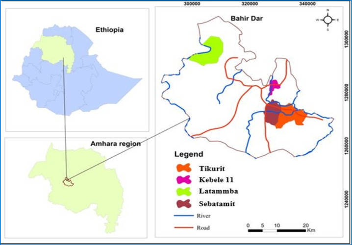 Figure 1
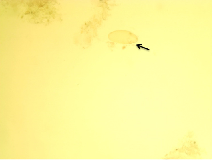 Figure 2
| Risk factor and category | Examined (n=384) | Positive (n=189), % | Chi-square ( | P value |
|---|---|---|---|---|
| Origin | 26.31 (3) | <.001 | ||
| Tikurit | 118 | 59 (50) | ||
| Sebatamit | 76 | 47 (61.84) | ||
| Latammba | 94 | 26 (27.65) | ||
| Kebele 11 | 96 | 57 (59.37) | ||
| Body condition | 28.60 (2) | <.001 | ||
| Poor | 125 | 80 (64) | ||
| Medium | 170 | 85 (50) | ||
| Fat | 89 | 24 (26.96) | ||
| Age | 0.839 (2) | .35 | ||
| Young | 129 | 65 (50.38) | ||
| Adult | 150 | 71 (47.33) | ||
| Old | 105 | 53 (50.47) | ||
| Sex | 0.844 (1) | .35 | ||
| Male | 187 | 93 (49.73) | ||
| Female | 197 | 96 (48.73) | ||
| Breed | 1.134 (1) | .29 | ||
| Local | 215 | 111 (51.62) | ||
| Crossbreed | 169 | 78 (46.15) |
Peer Reviews
No public reviews on file for this paper yet. If you reviewed it on a platform where reviews are public (OpenReview, ICLR, NeurIPS, ICML), you can paste yours below so the community can read it here.
Videos
No videos yet. Explain this paper in a talk, walkthrough, or lecture? Add one.
Taxonomy
TopicsHelminth infection and control · Parasites and Host Interactions · Parasitic infections in humans and animals
Introduction
Ethiopia hosts one of the largest livestock populations in Africa, with an estimated 55.03 million cattle, 27.32 million sheep, and 28.16 million goats as of 2019. Cattle, in particular, play a central role in the country’s agricultural economy, with the dairy sector contributing over 81% of total milk production. Despite this abundance, livestock productivity remains low due to constraints such as poor nutrition, inadequate management, and widespread infectious diseases. Among these, fasciolosis is one of the most impactful parasitic diseases, causing substantial economic losses through reduced growth, impaired fertility, decreased milk yield, and increased mortality [1].
Fasciolosis is caused by trematode parasites of the genus Fasciola, commonly known as liver flukes. Infection occurs when animals ingest metacercariae, the infective stage of the parasite, from contaminated pasture, water, or feed. Two species are primarily responsible: Fasciola hepatica, which predominates in temperate regions, and Fasciola gigantica, more common in tropical climates, including much of Africa and Ethiopia [2-4]. Transmission relies on the presence of aquatic snails, such as Lymnaea truncatula and Lymnaea natalensis, which serve as intermediate hosts under suitable conditions like stagnant water and moist environments [5].
Infected cattle experience liver tissue damage due to migrating immature flukes, resulting in inflammation, bile duct obstruction, hepatocellular necrosis, and fibrosis. Clinical signs often include weight loss, jaundice, poor body condition, and reduced productivity. Severe infections compromise liver function, predispose animals to secondary infections, and may lead to significant morbidity and mortality [6]. Economically, fasciolosis causes extensive liver condemnation at slaughterhouses, higher veterinary costs, and financial losses for farmers and the meat industry [7].
Epidemiology of fasciolosis is influenced by host and environmental factors, including age, sex, breed, management practices, and ecological conditions. Older animals are often more affected due to cumulative exposure, while some breeds show variable susceptibility. Differences in grazing behavior and reproductive cycles may contribute to higher prevalence in females in some cases [8-10].
Additionally, pasture type, access to contaminated water, and use of anthelmintics play critical roles in transmission. Although previous studies have provided insights into fasciolosis in Ethiopia, many were localized, leaving gaps in regional prevalence and risk factors. Given the ecological and management diversity across the country, region-specific studies are essential to inform targeted control strategies. In particular, the central and northern highlands, where cattle production is economically significant, may present unique environmental conditions that affect parasite dynamics [11-14]. Therefore, this study aimed to determine the prevalence and associated risk factors of bovine fasciolosis in Bahir Dar, Ethiopia.
Methods
Study Area
The study was conducted in Bahir Dar, Ethiopia, from November 2021 to April 2022 (Figure 1). Bahir Dar is located approximately 575 km northwest of Addis Ababa, at an elevation of 1500‐2600 meters above sea level, with geographic coordinates of 12°29’ N latitude and 37°29’ E longitude. The area receives an average annual rainfall of 1200‐1600 mm, and temperatures range from 8 °C to 31 °C. The landscape is predominantly plain plateaus, covering roughly 70% of the region, and the vegetation includes shrub formations, low woodlands, evergreen areas, and semi-humid highland vegetation. Agriculture is a key livelihood, with major crops including teff (Eragrostis tef), wheat (Triticum aestivum), maize (Zea mays), and various pulses [1].
Map of the study area
Study Animal and Sampling Method
The study animals consisted of cattle from four selected sites in Bahir Dar: Kebele 11, Sebatamit, Tikurit, and Latammba. Both indigenous and crossbred Holstein Friesian cattle reared under local management conditions were included. A total of 384 cattle were randomly selected, representing both sexes and multiple age groups. Animal age was assessed based on dentition and categorized as young or adult [15]. Body condition score was also evaluated for each animal following established guidelines to estimate nutritional and health status [16].
Study Design and Sample Size
A cross-sectional study was carried out in Bahir Dar, Ethiopia, from November 2021 to April 2022 to determine the prevalence and associated risk factors of bovine fasciolosis in Bahir Dar, Ethiopia. The risk factors considered included origin (location), breed, age, sex, and body condition of the cattle. The required sample size for fecal sample collection was calculated using a 95% confidence level, 5% absolute precision, and an expected prevalence of 50% in the absence of prior data for the study area [17].
where: n=required sample size; Pexp=expected prevalence (0.5); d=desired absolute precision (0.05); and Z=Z-value for confidence level (1.96). Based on this formula, a total of 384 cattle were included in the study.
Fecal Examination and Identification of Fasciola Eggs
Fresh fecal samples were collected directly from the rectum of each animal using disposable gloves and placed into universal bottles containing 10% formalin as a preservative. Samples were transported under controlled conditions to the Bahir Dar Regional Veterinary Laboratory for parasitological examination. The sedimentation technique was used to detect Fasciola eggs, following standard procedures [1819]. To differentiate Fasciola eggs from those of other trematodes, such as Paramphistomum species, the sediment was stained with methylene blue. Fasciola eggs are typically yellowish, large, and operculated, whereas Paramphistomum eggs stain blue [2021].
Data Management and Analysis
The raw data collected from the study were organized and entered into a Microsoft Excel spreadsheet for initial management. Subsequently, the data were exported to STATA (version 16.0; StataCorp) for statistical analysis. A χ² test was used to evaluate the correlation between infection rates and risk factors such as age, sex, breed, and location. The test evaluated infection rates based on these parameters, with a significance level of P<.05.
Ethical Considerations
Ethical clearance was obtained from the Ethics Research Review Committee of the University of Gondar, College of Veterinary Medicine and Animal Sciences (Ref. No. CVMASc/UoG/RERC/10/11/2021; November 7, 2021). Cattle were handled according to animal welfare guidelines, and owner consent was obtained prior to data collection.
Results
Prevalence of Bovine Fasciolosis
A total of 384 cattle were examined in this study, and the overall prevalence of bovine fasciolosis in the study area was 49.21% (n=189). Analysis by origin showed the highest prevalence in Sebatamit (n=47, 61.84%), followed by Kebele 11 (n=57, 59.37%), Tikurit (50%), and Latammba (n=26, 27.65%), with a statistically significant difference among sites (χ²=26.31, P<.001). Prevalence also varied according to body condition, with poor-conditioned cattle exhibiting the highest prevalence (n=80, 64%), followed by medium condition (n=85, 50%) and fat cattle (n=24, 26.96%), showing a significant difference (χ²=28.6, P<.001). When grouped by age, the prevalence was 50.38% (n=65) in young cattle, 47.33% (n=71) in adults, and 50.47% (n=53) in older animals; however, differences were not statistically significant (χ²=0.84, P=.35). Similarly, sex did not significantly influence prevalence: males had 49.73% (n=93), and females had 48.73% (n=96, χ²=0.844; P=.35). Finally, breed showed no significant effect on infection rates, although local cattle had a slightly higher prevalence (n=111, 51.62%) compared to crossbreeds (n=78, 46.15%) (χ²=0.287, P=.29) (Table 1).
Identification of Fasciola Eggs
The Fasciola eggs detected in cattle fecal samples are shown in Figure 2. Identification was based on morphological characteristics [2021]. The eggs are ovoid, possess a thick yellowish-brown shell, and typically have an operculum at one end.
Egg of Fasciola species: yellowish egg (arrow).
Discussion
Principal Findings
Bovine fasciolosis remains one of the most significant parasitic diseases affecting cattle in Ethiopia, causing substantial economic losses through reduced growth, poor milk production, liver condemnation, and increased susceptibility to secondary infections. The present study recorded an overall prevalence of bovine fasciolosis of 49.21% in the study area, which aligns with previous reports from different regions of Ethiopia. For instance, prevalence rates of 41.41% and 54.5% have been reported in Woreta [22] and Jimma [23], respectively. Similar rates were observed in North-East Amhara (47.10%) [13], and Eastern Shoa, Kuyu District (54.2%) [19] likely associated with common agro-ecological factors such as the presence of Lymnaea snails and practices like communal water use and irrigation that facilitate pasture contamination [24]. Conversely, lower prevalence rates have been reported in some regions. Investigations in Soddo (4.9%) [25], Nekemte (15.9%) [7], and Southern Ethiopia (15.9%) [8] demonstrated lower frequencies, while Zenzelma, Bahir Dar (26%) [1], and Bahir Dar (32.3%) [26] reported slightly lower prevalence. These variations are likely influenced by differences in altitude, geography, climate, snail host abundance, management practices, and anthelmintic use [27].
Geographical and Management Factors
Prevalence varied significantly among study sites in the current work, with Sebatamit recording the highest prevalence (61.84%), followed by Kebele 11 (59.37%), Tikurit (50%), and Latammba (27.65%). This observation is consistent with previous studies indicating substantial geographical variation in fasciolosis prevalence [28-31], which may be influenced by climatic conditions, animal management practices, and access to veterinary services [32].
Influence of Animal Factors
Cattle in poor body condition exhibited the highest prevalence (64%), compared to medium (50%) and fat cattle (26.96%). This pattern corresponds with several Ethiopian reports that found significantly higher infection rates in animals with poor body condition, likely reflecting increased susceptibility due to malnutrition, compromised immunity, and concurrent infections that weaken host defenses [33-36]. However, some studies reported no significant differences in prevalence among body condition categories, indicating that body condition may not always predict Fasciola infection [2337].
In this study, no significant differences were observed across age groups, with young cattle (50.38%), adults (47.33%), and older cattle (50.47%) showing similar prevalence. Several studies in Ethiopia have likewise reported no significant association between age and Fasciola infection [3839], although other reports documented age-related variation, likely reflecting differences in exposure or management practices [4041].
Similarly, sex did not significantly influence prevalence, with males at 49.73% and females at 48.73%. This finding aligns with previous studies reporting no significant differences between male and female cattle [35], although one study did observe sex-related variation, possibly due to differences in management practices or environmental exposure [25].
Finally, breed had no significant effect on infection rates, with local cattle and crossbreeds showing prevalence of 51.62% and 46.15%, respectively. This finding agrees with earlier research [2434], suggesting similar susceptibility between breeds, although a few studies reported breed-related differences, potentially attributable to genetic factors or breed-specific traits.
Limitations
The cross-sectional design and use of a single diagnostic method provide an accurate snapshot of prevalence and associated risk factors within the study population. While longitudinal or multimethod studies may provide additional detail, the current methodology is sufficient to address the study objectives and offers valuable insights for local disease control strategies.
Conclusion
This study revealed a high overall prevalence of bovine fasciolosis (49.21%) in the Bahir Dar area, confirming its significance as a major parasitic disease affecting cattle in the region. Prevalence varied notably across localities, with Sebatamit exhibiting the highest rate (61.84%) and Latammba the lowest (27.65%). Analysis of risk factors indicated a significant association between body condition and fasciolosis, with poorly conditioned cattle being more susceptible to infection. In contrast, no statistically significant differences were observed based on age, sex, or breed, suggesting that cattle across all demographic groups are at risk. These findings underscore the need for targeted, location-specific control strategies and highlight the importance of improved nutritional and health management practices to reduce the burden of fasciolosis in cattle populations.
The reference list from the paper itself. Each links out to its DOI / PubMed record.
- 1Legesse S Tsegaye S Lamesgen S Wolelaw Y Garikipati D Wondimagegn W Coprological prevalence and associated risk factors of bovine fasciolosis in and around Zenzelma, Bahir Dar, Ethiopia Eur J Exp Bio 201707534 doi 10.21767/2248-9215.100034 · doi ↗
- 2Ardo MB Aliyara YH Lawal H Prevalence of bovine fasciolosis in major abattoirs of Adamawa State, Nigeria Bayero Journal of Pure and Applied Sciences 201461216 doi 10.4314/bajopas.v 6i 1.3 · doi ↗
- 3Figtree M Beaman MH Lee R et al Fascioliasis in Australian travellers to Bali Med J Aust Aug 1720152034186188 doi 10.5694/mja 15.00010 Medline 26268290 · doi ↗ · pubmed ↗
- 4Pfukenyi DM Mukaratirwa S Willingham AL Monrad J Epidemiological studies of Fasciola gigantica infections in cattle in the highveld and lowveld communal grazing areas of Zimbabwe Onderstepoort J Vet Res Mar 20067313751 Medline 16715877 · pubmed ↗
- 5Mungube EO Bauni SM Tenhagen BA Wamae LW Nginyi JM Mugambi JM The prevalence and economic significance of Fasciola gigantica and Stilesia hepatica in slaughtered animals in the semi-arid coastal Kenya Trop Anim Health Prod 2006386475483 doi 10.1007/s 11250-006-4394-4Medline 17243475 · doi ↗ · pubmed ↗
- 6Mas-Coma S Valero MA Bargues MD Chapter 2. Fasciola, lymnaeids and human fascioliasis, with a global overview on disease transmission, epidemiology, evolutionary genetics, molecular epidemiology and control Adv Parasitol 20096941146 doi 10.1016/S 0065-308X(09)69002-3Medline 19622408 · doi ↗ · pubmed ↗
- 7Petros A Kebede A Wolde A Prevalence and economic significance of bovine fasciolosis in cattle slaughtered at Nekemte Municipal Abattoir, Western Ethiopia Journal of Veterinary Medicine and Animal Health 20135202205 UR Lhttps://academicjournals.org/article/article 1379686620_Petros%20et%20al.pdf Accessed 15-03-2022
- 8Asrese,NM Ali MG Bovine fasciolosis: prevalence and economic significance in Southern Ethiopia Acta Parasitologica Globalis 201457682 UR Lhttps://idosi.org/apg/5(2)14/1.pdf Accessed 15-03-2022