miR-7847-3p Serves as a Prognostic Biomarker and Suppresses Colorectal Cancer Progression
Haiqin Chen, Yusen Wang, Ming Lu

TL;DR
This study finds that miR-7847-3p is a potential biomarker for predicting colorectal cancer prognosis and may help suppress cancer progression by targeting AP1S1.
Contribution
The study identifies miR-7847-3p as a novel prognostic biomarker and therapeutic target in colorectal cancer through its regulation of AP1S1.
Findings
miR-7847-3p is downregulated in colorectal cancer tissues and linked to poor survival outcomes.
Overexpression of miR-7847-3p inhibits cancer cell proliferation, migration, and invasion.
AP1S1 is confirmed as a direct target of miR-7847-3p using dual-luciferase reporter assays.
Abstract
MiR-7847-3p is aberrantly expressed in multiple cancer types; however, its biological function and molecular mechanism in colorectal cancer (CRC) remain unclear. This investigation aims to explore the function of miR-7847-3p in CRC development and its potential regulatory mechanisms. Tumor tissues and paracancerous normal tissues from 122 CRC patients were collected and miR-7847-3p expression was detected by quantitative reverse transcription polymerase chain reaction. The predictive capacity of miR-7847-3p was evaluated through Kaplan–Meier survival analysis and subsequently validated via multivariable Cox proportional hazards modeling. The effects of miR-7847-3p on HCT116 and SW480 cells were evaluated through Cell Counting Kit-8 and Transwell assays. The miR-7847-3p target genes were predicted using TargetScan, miRDB, and miRWalk. Dual-luciferase reporter assay confirmed the…
Genes, proteins, chemicals, diseases, species, mutations and cell lines named across the full text — each resolved to its canonical identifier and authoritative record.
Click any figure to enlarge with its caption.
 Figure 1
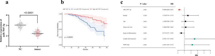 Figure 2
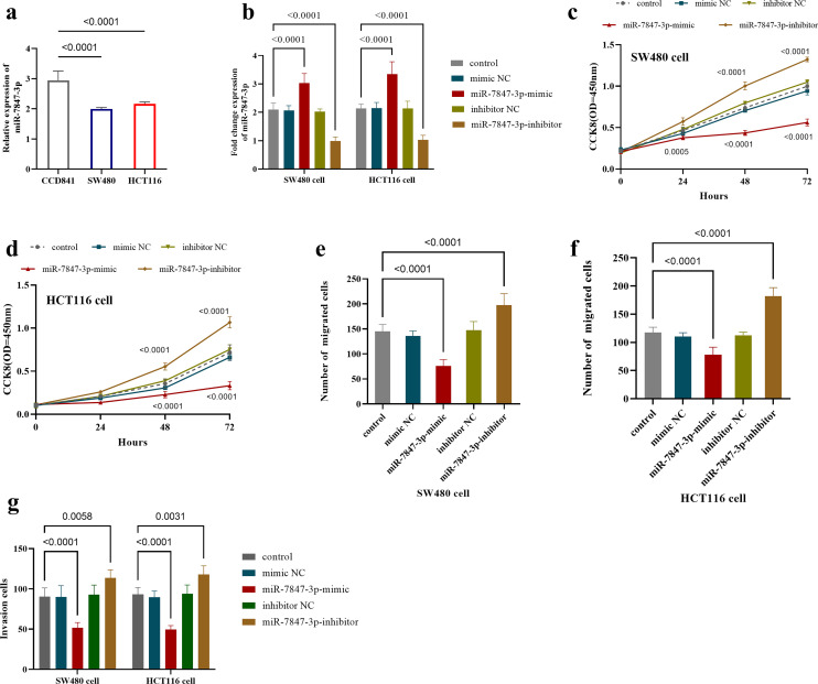 Figure 3
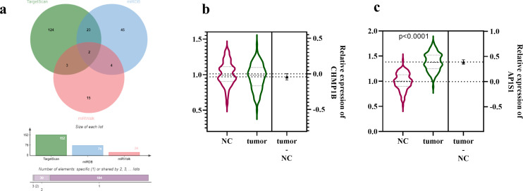 Figure 4
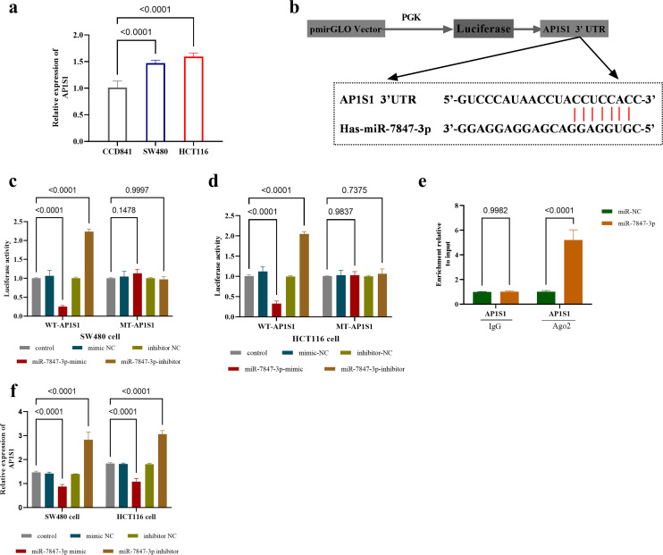 Figure 5
Peer Reviews
No public reviews on file for this paper yet. If you reviewed it on a platform where reviews are public (OpenReview, ICLR, NeurIPS, ICML), you can paste yours below so the community can read it here.
Videos
No videos yet. Explain this paper in a talk, walkthrough, or lecture? Add one.
Taxonomy
TopicsMicroRNA in disease regulation · Circular RNAs in diseases · Cancer-related molecular mechanisms research
Introduction
Colorectal cancer (CRC) is a common malignant tumor, with a worldwide incidence of 1 926 000 and mortality of 904 000 in 2022. This malignancy accounted for nearly one-tenth of all cancer cases (9.6%) and deaths (9.3%).1 Despite the current clinical use of colonoscopy combined with imaging assessment as the primary diagnostic tool, there is a high rate of underdiagnosis of early lesions2 and a lack of efficient molecular markers for prognostic stratification. Conventional treatment methods (such as surgical resection and chemotherapy) have limited efficacy for metastatic CRC, resulting in significantly reduced 5-year survival rates (less than 15%), increased risk of tumor recurrence, and severe treatment side effects (such as bone marrow suppression and intestinal dysfunction).3^,^4 The current clinical landscape demands rapid advancement in biomarker discovery to enable precise diagnosis and personalized treatment, thereby improving patient outcomes.
MicroRNAs (miRNAs) are involved in tumor development by regulating gene expression networks, and these molecular aberrations are pathologically implicated in driving tumor proliferation, metastatic spread, and chemoresistance development in CRC.5 The exceptional stability of miRNAs in bodily fluids and their tissue-specific expression patterns have positioned them as promising diagnostic biomarkers.6^-^8 Although some miRNAs have shown diagnostic potential in clinical studies on CRC,9^-^12 the number of miRNA markers translated into clinical applications remains limited. This underscores the urgent need for deeper research to discover and validate miRNA-based diagnostic markers and therapeutic targets. With the advancement of miRNA detection technology13 and the establishment of standardized sample processing processes,14 the reliability of miRNA marker research has been significantly improved. Based on the above technical advantages and clinical needs, screening miRNAs abnormally expressed in CRC is of great value for improving the disease diagnosis and treatment system.15
The research of miR-7847-3p in the field of oncology has gradually received attention. One study reported that miR-7847-3p was clearly downregulated in serum exosomes of patients with sepsis,16 and its expression was lower in bladder cancer tissues and shows an inverse relationship with tumor stage.^17^ Regarding CRC, only 1 microarray analysis study based on miRNA from CRC patient serum exosomes has reported downregulation of miR-7847-3p.18 However, the expression characteristics of miR-7847-3p in CRC, its clinical pathological relevance, and its regulatory mechanisms remain to be elucidated.
Given the key regulatory function of miR-7847-3p in cancers, investigating the clinical significance of miR-7847-3p in CRC will help deepen the understanding of CRC pathogenesis. This research endeavors to uncover the function and mechanisms of miR-7847-3p in CRC, thereby providing new potential molecular markers and a theoretical basis for the treatment of CRC.
Materials And Methods
Patients and Specimens
Tumor specimens and matched histologically normal adjacent tissues (sampled at a distance of ≥5 cm from the neoplastic edge19 were obtained from 122 CRC patients treated between January 2018 and December 2020. The specimens were independently verified by 2 pathologists. The study protocol was approved by the Ethics Committee at the First Affiliated Hospital of Xinjiang Medical University (approval number: 2018003, date: January 10, 2018), and this study adhere to the tenets of the Declaration of Helsinki. All enrolled subjects providing documented consent. Cases involving preoperative radiation therapy or concurrent additional malignancies were excluded from the analysis. The outcomes of all patients were monitored by telephone follow-up after surgery for 5 years. The postoperative treatment plans for the patients were all meticulously documented. For patients with stage II and III CRC, adjuvant chemotherapy regimens such as capecitabine combined with oxaliplatin (CapeOX) or fluorouracil, leucovorin, and oxaliplatin are employed. For metastatic CRC patients with RAS wild-type tumors, targeted therapies such as cetuximab or bevacizumab are used for treatment. For CRC patients with mismatch repair deficiency or high microsatellite instability, the use of immune checkpoint inhibitors such as pembrolizumab for immunotherapy will be explored. The specific treatment plan will be comprehensively evaluated based on factors such as tumor stage and the patient’s overall condition and will be implemented under the guidance of a multidisciplinary team.
Cell Culture
Colon cancer cells (SW480 and HCT116 cells) and normal control cells (CCD841) were sourced from the American Type Culture Collection. These cells were cultured in RPMI-1640 medium with 10% fetal bovine serum at 37°C and 5% CO_2_.
Cell Transfection
The transfection complexes were prepared by incubating either miR-7847-3p mimic or inhibitor (Reebok Bio) with Lipofectamine 3000 at a 1 : 10 (v/v) ratio in SW480 and HCT116 cell lines and cultured with Opti-MEM medium. Both cell lines were transfected with miR-7847-3p mimic complexes to induce miR-7847-3p overexpression, while loss-of-function experiments utilized inhibitor complexes for* miR-7847-3p* knockdown. Both mimic NC and inhibitor NC were parallelly transfected as the negative controls. Cells were harvested at the 48-hour timepoint following transfection for downstream analyses.
Reverse Transcription Quantitative Polymerase Chain Reaction Detection
Total RNA was extracted with Trizol (Invitrogen, USA). Total RNA (1 μg) was reverse transcribed into miRNA (Mir-X™ miRNA First-Strand Synthesis Kit, Takara) and mRNA (PrimeScript™ RT Reagent Kit, Takara). The cDNA was then subjected to quantitative polymerase chain reaction (qPCR) for amplification and detection. Primer sequences: miR-7847-3p (forward: 5′-CGTGACTGTCCCTCTGTGTC-3′, reverse: universal primer in Mir-X Kit); U6 (forward: 5′-CTCGCTTCGGCAGCACA-3′, reverse: 5′-TGCGTGTCATCCTTGCGCAG-3′); GAPDH (forward: 5′-GAAGGTGAAGGTCGGGAGTC-3′, reverse: 5′-GAAGATGGTGATGGGATTTC-3′); AP1S1 (forward: 5′-GCTGGAGGAGGAGGTGGAAGAGA-3′, reverse: 5′-CAGGTAGGCGTTGTCCTTGT-3′). Expression levels were calculated using the 2^-ΔΔCT^ method, with normalization to U6 (miRNA analyses) and GAPDH (mRNA analyses) as internal controls. In this study, U6 and GAPDH were selected as reference genes for miRNA and mRNA analyses, respectively. The stability of these reference genes was validated using geNorm and NormFinder software, which showed good stability (M value < 0.5) across different sample groups. Additionally, preliminary screening of reference gene expression levels ensured stable expression in all samples.
Cell Counting Kit-8
To assess cellular proliferation, a 96-well plate (5 × 10^3^ cells per well) was inoculated with 5 × 10^3^ cells per well and cultured for varying durations (0, 24, 48, and 72 hours). At each designated interval, 10 μL of Cell Counting Kit-8 (CCK-8) reagent (Nippon Kohin Chemical) was introduced, followed by a 2-hour incubation at 37°C. Optical density measurements were subsequently obtained at 450 nm to quantify viable cells. This experimental procedure was conducted in triplicate to ensure reproducibility.
Transwell
Cell migration: transfected cells (5 × 10^4^ cells/chamber) were resuspended in a serum-free medium and inoculated into the upper chamber of Transwell. Complete medium containing 10% fetal bovine serum (FBS) (600 μL) was added to the lower chamber as a chemokine. Following 24 hours incubation at 37°C, non-migrated cells on the membrane’s upper surface were gently removed. Cells were fixed, stained with crystal violet, and counted in 5 random fields.
Cell invasion: Firstly, Matrigel (BD Biosciences, NY, USA) was diluted with serum-free medium in the ratio of 1 : 8, evenly encapsulated on the membrane surface of the upper chamber of Transwell (Corning, NY, USA), and incubated at 37°C for 4 hours to form a gel layer. Transfectants (1 × 10^5^ cells/chamber) were suspended in serum-deprived medium and plated in the upper compartment. The lower compartment contained 400 μL of basal medium with 10% FBS as a chemoattractant. After 48 hours of incubation at 37°C, uninvaded cells were removed as above, fixed, stained, and counted for invaded cells crossing the stromal gel layer.
RNA Immunoprecipitation
RNA immunoprecipitation experiments were performed using the Magna RIP™ system (Millipore). The anti-Ago2 antibody (Abcam) and homologous IgG were incubated with cell lysate at 4°C overnight. The RNA was extracted after the Protein A/G magnetic bead capture complex, and the miR-7847-3p enrichment was detected by qPCR. The IgG group was used as a negative control.
Bioinformatics Analysis
The targets of miR-7847-3p were predicted using bioinformatics analysis through the TargetScan, miRDB, and miRWalk databases. Consensus targets identified by the Venn diagram from all 3 databases were selected for further analysis.
Dual-luciferase Assay
The 3′UTR of AP1S1 (WT) was amplified and cloned into the pmirGLO vector (Promega). A mutant (MT) version of the AP1S1 3′UTR, with predicted miR-7847-3p binding sites disrupted, was created via site-directed mutagenesis (Stratagene, La Jolla, CA, USA) and similarly inserted into pmirGLO. SW480 and HCT116 cells were co-transfected with either the WT or MT reporter plasmid, along with miR-7847-3p mimic, inhibitor, or corresponding negative controls. Luciferase activity was measured following a 48-hour transfection period.
Statistical Analysis
The study was statistically analyzed using SPSS 23.0 (IBM SPSS Corp.; Armonk, NY, USA) and GraphPad Prism 9.0 (GraphPad Software; San Diego, CA, USA). Measurements are shown as mean ± standard deviation. This study utilized G*Power 3.1.9.7 software (Heinrich Heine University Düsseldorf; Düsseldorf, Germany) for sample size and power analysis. The a priori analysis revealed that a total sample size of 172 is required with α = 0.05, power (1-β) of 0.90, and effect size d = 0.5, with the actual power exceeding 0.90. Post hoc analysis showed that with sample sizes of 105 and 122 and an effect size of d = 0.88, the power exceeded 0.99. The sample size is sufficient to reliably detect differences between the 2 groups. The Kaplan–Meier approach was employed to generate survival probability curves, with intergroup comparisons performed using the nonparametric log-rank test; prognostic determinants were evaluated using Cox proportional hazards modeling. Clinicopathological correlations were analyzed by chi-square test. Intergroup comparisons were performed employing Student’s t-test for pairwise analyses and 1-way ANOVA for multiple group comparisons. The predetermined significance criterion was P < .05 for all analyses.
Results
Analysis of the miR-7847-3p Expression in Colorectal Cancer and Its Correlation with Clinical Features
Tumor tissues showed a significant reduction in miR-7847-3p expression (P < .0001, Figure 1a). The miR-7847-3p expression showed a significant association with Tumor-node-metastasis (TNM) stage and lymph node metastasis (LNM) of CRC subjects. The proportion of participants in the low-expression group who were in TNM stage III and had LNM was higher than that in the high-expression group. No significant associations were observed between* miR-7847-3p* and participants’ gender, age, tumor size, or tumor differentiation (Table 1).
Association Between miR-7847-3p Expression Levels and Clinical Prognosis of Colorectal Cancer
Kaplan–Meier curves demonstrated markedly reduced overall survival in low miR-7847-3p expressers relative to high expressers (Figure 1b). In addition, miR-7847-3p (HR = 0.200, 95% CI: 0.089-0.451, P = .001), LNM (HR = 2.559, 95% CI: 1.153-5.676, P = .021) and TNM stage (HR = 2.204, 95% CI:1.018-4.772, P = .045) were considered as independent prognostic factors for CRC (Figure 1c).
Effect of miR-7847-3p on the Function of Colorectal Cancer Cells
Polymerase chain reaction analysis indicated that miR-7847-3p expression was significantly reduced (P < .0001) in CRC cell lines (SW480 and HCT116 cells, Figure 2a). In both cell lines, transfection with miR-7847-3p mimics significantly upregulated its expression levels (P < .0001), while transfection with inhibitors effectively downregulated its expression (P < .0001, Figure 2b).
Functional assessment using CCK-8 showed that overexpressing miR-7847-3p reduced SW480 cell proliferation, whereas its depletion enhanced proliferative capacity (Figure 2c). This regulatory pattern was consistently observed in HCT116 cells (Figure 2d).
Transwell tests gauged miR-7847-3p’s impact on SW480 and HCT116 cell migration and invasion. Findings revealed that* miR-7847-3p* overexpression significantly reduced the migration of SW480 cells (P < .0001), while inhibition of its expression raised migrating cell numbers (P < .0001, Figure 2E). A similar phenomenon was observed in HCT116 cells, where miR-7847-3p overexpression suppressed cell migration (P < .0001), while miR-7847-3p underexpression enhanced migration (P < .0001, Figure 2F). In both cell lines, it was also observed that overexpression of miR-7847-3p reduced cell invasion capacity (P < .0001), while inhibition of its expression increased invasion capacity (Figure 2g).
AP1S1 is a downstream Target Gene of miR-7847-3p
Bioinformatics analysis identified 2 potential target genes of miR-7847-3p, CHMPIB, and AP1S1 (Figure 3a). Subsequent expression analysis revealed that the expression level of CHMPIB did not show a significant difference between tumor tissues and adjacent non-tumor tissues (Figure 3b). In contrast, AP1S1 exhibited markedly elevated expression in tumor tissues than in adjacent non-tumor tissues (Figure 3c).
Validation of the Interaction Between miR-7847-3p and AP1S1
Quantitative analysis of AP1S1 expression in CRC cell lines (SW480 and HCT116) revealed that its expression level was significantly upregulated compared with normal colonic epithelial cells (P < .0001, Figure 4a). The binding site between miR-7847-3p and AP1S1 was identified by the TargetScan online database (Figure 4b). Further dual-luciferase reporter assays showed that overexpression of miR-7847-3p inhibited the luciferase activity of the WT-AP1S1 and knockdown of miR-7847-3p significantly promoted the luciferase activity of the WT-AP1S1 in both SW480 and HCT116 cells, while miR-7847-3p had no significant effect on the luciferase activity of the mutant type (Figure 4c, d).
The RIP experiments further indicated that miR-7847-3p was significantly enriched in the Ago2 protein complex (P < .0001 compared to IgG control) and overexpression of miR-7847-3p increased the association between AP1S1 and Ago2 (Figure 4e). The qPCR analysis confirmed that enforced miR-7847-3p expression strongly attenuated AP1S1 levels (P < .0001), while knockdown of miR-7847-3p significantly upregulated AP1S1 expression (P < .0001, Figure 4f).
Discussion
In recent years, miRNAs have emerged as important biomarkers for CRC due to their stable presence in bodily fluids and their role in tumor regulation.20 Numerous studies have demonstrated that alterations in miRNA expression profiles such as miR-130b-3p and miR-129-5p are closely associated with the onset and progression of CRC.21^,^22 In addition, miRNAs such as miR-135b and miR-223 have been widely investigated as CRC diagnostic markers.23^,^24 However, their expression differences between CRC tissues and normal tissues are relatively small, which limits their application in early diagnosis. In this study, systematic analysis revealed that* miR-7847-3p* was significantly downregulated in CRC tissues, consistent with previous reports of its low expression in serum exosomes from CRC patients,18 and provided a new candidate biomarker for the molecular diagnosis of CRC.
Although miR-135b and miR-223 play a certain role in the diagnosis of CRC, their prognostic value is relatively limited, and there is a scarcity of related research.25 Clinical data analysis has revealed the significant clinical value of miR-7847-3p in CRC. The levels of miR-7847-3p in CRC tumor tissues are significantly associated with TNM stage and LNM, which are important clinical indicators for assessing the progression and prognosis of CRC. Specifically, the TNM stage remains the gold standard for therapeutic decision-making, while LNM status (N-stage) is the strongest predictor of recurrence risk and a key determinant for adjuvant chemotherapy recommendations in stage II/III cancers.1^,^26 Kaplan–Meier analysis linked low miR-7847-3p expression to worse 5-year survival in CRC patients, and Cox regression confirmed it as an independent poor prognostic marker. These findings suggest its potential utility in risk stratification, though prospective validation is required before considering clinical translation. Notably, this study also found that LNM and TNM stages are equally independent prognostic factors, consistent with the results of many clinical studies.26 Based on the findings of this study, miR-7847-3p may serve as an effective prognostic marker for identifying CRC patients with potential adverse outcomes.
To investigate the tumor-regulatory function and molecular mechanisms of miR-7847-3p in CRC, a comprehensive suite of functional characterization experiments was conducted. Cell proliferation, migration, and invasion capabilities are key indicators for assessing the malignancy of tumors: abnormal proliferation driven by dysregulation of cell cycle checkpoints is the basis of tumor development, migration capabilities mediated by cytoskeleton remodeling determine the degree of local tumor infiltration, and invasion capabilities involving degradation of extracellular matrix are directly related to the occurrence of distant tumor metastasis.27 The experimental results showed that overexpression of miR-7847-3p significantly inhibited the proliferation, migration, and invasion capabilities of CRC cells. These functional assays collectively establish miR-7847-3p as a tumor suppressor in CRC development.
AP1S1, as the core subunit of the adaptor protein complex AP-1, plays a crucial regulatory role in the endocytosis and metastasis processes of tumor cells.28 Studies have shown that AP1S1 participates in tumorigenesis and progression by influencing the internalization process of receptor tyrosine kinases such as estimated glomerular filtration rate,29 and its abnormal expression is closely associated with the invasion and metastasis of various malignant tumors.30 The findings of this study further confirm that miR-7847-3p may exert its anti-cancer effects by targeting and suppressing the expression of AP1S1.
This study still has the following areas that require improvement: firstly, the limited number of clinical samples may have affected the analysis of the association between miR-7847-3p expression and clinical pathological features related to tumor malignancy (such as tumor size and differentiation). Secondly, due to some unforeseen factors, including inconsistent tumor engraftment rates and health issues in mice unrelated to tumor xenografts, the experimental conditions were complicated, resulting in a high failure rate in animal model construction. Thirdly, while it has been confirmed that miR-7847-3p exerts its anticancer effects by targeting AP1S1, the specific molecular mechanisms of AP1S1 in CRC require further exploration. Fourth, although the study design has attempted to control for confounding factors, there may still be other unidentified or unmeasured confounders that could influence the expression of miR-7847-3p and its relationship with the clinicopathological characteristics of CRC. Finally, although the variation in the severity of patients’ conditions leads to the inability to standardize postoperative treatment plans, postoperative treatment may influence the prognosis, a factor that was not taken into consideration in this study. These limitations will be the focus of future research.
This study reveals the significant role of miR-7847-3p in CRC, with its low expression being associated with poor prognosis in CRC patients.* miR-7847-3p* plays an antitumor effect in CRC by inhibiting cell proliferation, migration, and invasion of CRC cells. Furthermore, this study also confirmed that miR-7847-3p can predict patient prognosis and regulate cellular processes by targeting AP1S1. This discovery provides a novel biomarker for the molecular diagnosis and prognostic assessment of CRC, with potential clinical applications such as assisting in risk stratification and guiding the formulation of personalized treatment plans. Furthermore, the elucidation of the miR-7847-3p-AP1S1 axis provides direction for in-depth exploration of the pathogenesis of CRC and the development of novel therapeutic targets. Future research should expand the sample size to validate the prognostic efficacy of miR-7847-3p, delve deeper into its molecular mechanisms with* AP1S1*, and promote its clinical translation and application.
The reference list from the paper itself. Each links out to its DOI / PubMed record.
- 1Bray F Laversanne M Sung H Global cancer statistics 2022: GLOBOCAN estimates of incidence and mortality worldwide for 36 cancers in 185 countries. CA Cancer J Clin. 2024;74(3):229 263. (doi: 10.3322/caac.21834) 38572751 · doi ↗ · pubmed ↗
- 2Pickhardt PJ Hassan C Halligan S Marmo R. Colorectal cancer: CT colonography and colonoscopy for detection—systematic review and meta-analysis. Radiology. 2011;259(2):393 405. (doi: 10.1148/radiol.11101887) 21415247 PMC 3079122 · doi ↗ · pubmed ↗
- 3Fontana E Eason K Cervantes A Salazar R Sadanandam A. Context matters-consensus molecular subtypes of colorectal cancer as biomarkers for clinical trials. Ann Oncol. 2019;30(4):520 527. (doi: 10.1093/annonc/mdz 052) 30796810 PMC 6503627 · doi ↗ · pubmed ↗
- 4Ciardiello F Ciardiello D Martini G Napolitano S Tabernero J Cervantes A. Clinical management of metastatic colorectal cancer in the era of precision medicine. CA Cancer J Clin. 2022;72(4):372 401. (doi: 10.3322/caac.21728) 35472088 · doi ↗ · pubmed ↗
- 5To KK Tong CW Wu M Cho WC. Micro RN As in the prognosis and therapy of colorectal cancer: from bench to bedside. World J Gastroenterol. 2018;24(27):2949 2973. (doi: 10.3748/wjg.v 24.i 27.2949) 30038463 PMC 6054943 · doi ↗ · pubmed ↗
- 6Weber JA Baxter DH Zhang S The micro RNA spectrum in 12 body fluids. Clin Chem. 2010;56(11):1733 1741. (doi: 10.1373/clinchem.2010.147405) 20847327 PMC 4846276 · doi ↗ · pubmed ↗
- 7Lu J Getz G Miska EA Micro RNA expression profiles classify human cancers. Nature. 2005;435(7043):834 838. (doi: 10.1038/nature 03702) 15944708 · doi ↗ · pubmed ↗
- 8Schwarzenbach H Hoon DS Pantel K. Cell-free nucleic acids as biomarkers in cancer patients. Nat Rev Cancer. 2011;11(6):426 437. (doi: 10.1038/nrc 3066) 21562580 · doi ↗ · pubmed ↗