Dual interference with host neuropeptide signaling allows parasitoid wasp to hijack host sugar metabolism
Zhi-Zhi Wang, Ruo-Fei Ma, Li-Cheng Gu, Li-Zhi Wang, Ting Chen, Pei Yang, Jia-Ni Zou, Jiang-Yan Zhu, Zhi-Wei Wu, Yue-Nan Zhou, Min Shi, Xing-Xing Shen, Jian-Hua Huang, Xue-Xin Chen

TL;DR
A parasitoid wasp manipulates its host's sugar metabolism using neuropeptides to increase trehalose levels, affecting the host's physiology.
Contribution
The study reveals how parasitoid wasps use dual interference with host neuropeptide signaling to control sugar metabolism.
Findings
Parasitoid wasp sNPF promotes glycogenolysis in the host fat body via the sNPFR.
A symbiotic virus activates host sNPF expression, stimulating glycolysis in the midgut.
Host and parasite sNPFs activate distinct downstream signaling pathways via different receptor affinities.
Abstract
Changes in host carbohydrate metabolism determine the outcome of host–parasite relationships, but the underlying mechanistic basis remains elusive. Here, we show that the parasitoid wasp Cotesia vestalis induces trehalose accumulation in its host, the moth Plutella xylostella, largely independently of insulin/adipokinetic hormone signalling and food intake. Instead, parasitoids rewire host carbohydrate metabolism via two pathways activated by the evolutionarily conserved short neuropeptide F (sNPF), a functional analogue of mammalian neuropeptide Y. Parasitoid-derived teratocytes secrete sNPF that interacts with the sNPF receptor (sNPFR) on host cells, and contributes to host hypertrehalosemia by promoting glycogenolysis in the fat body. We further find that a parasitoid-symbiotic virus induces expression of host-encoded sNPF, which stimulates glycolysis in the host midgut. Furthermore,…
Genes, proteins, chemicals, diseases, species, mutations and cell lines named across the full text — each resolved to its canonical identifier and authoritative record.
Click any figure to enlarge with its caption.
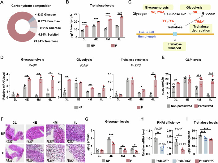 Figure 1
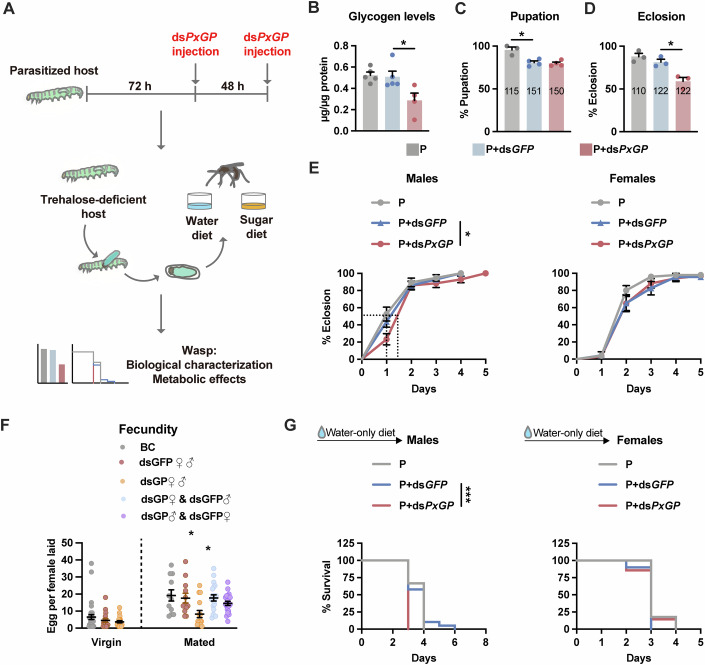 Figure 2
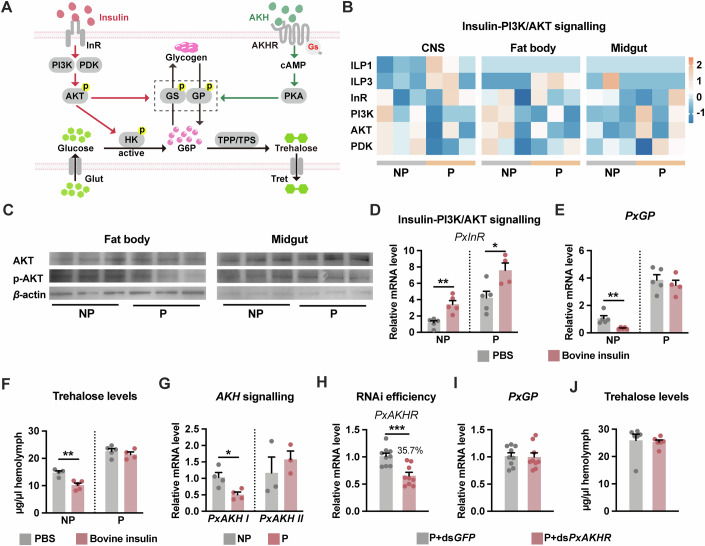 Figure 3
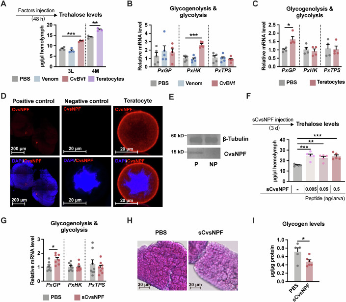 Figure 4
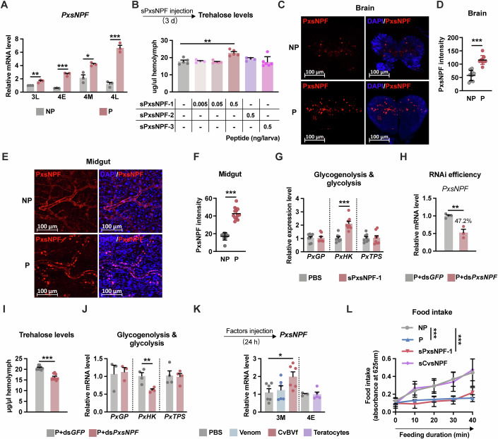 Figure 5
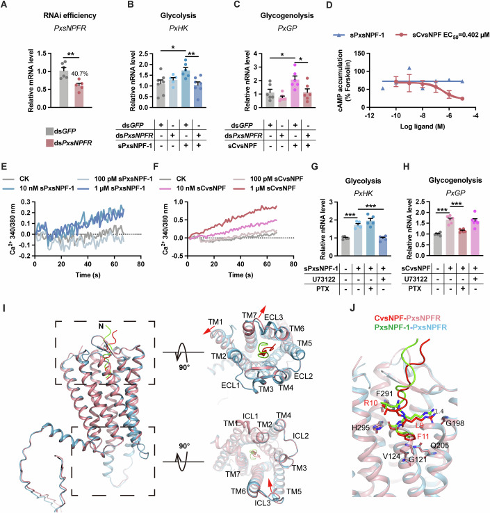 Figure 6
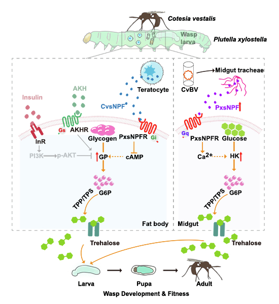 Figure 7
 Figure 8
- —http://dx.doi.org/10.13039/100017054MOST | NSFC | National Natural Science Foundation of China-Zhejiang Joint Fund for the Integration of Industrialization and Informatization (NSFC-Zhejiang Joint Fun
- —http://dx.doi.org/10.13039/501100001809MOST | National Natural Science Foundation of China (NSFC)
- —http://dx.doi.org/10.13039/501100012166MOST | National Key Research and Development Program of China (NKPs)
- —http://dx.doi.org/10.13039/501100012226MOE | Fundamental Research Funds for the Central Universities (Fundamental Research Fund for the Central Universities)
Peer Reviews
No public reviews on file for this paper yet. If you reviewed it on a platform where reviews are public (OpenReview, ICLR, NeurIPS, ICML), you can paste yours below so the community can read it here.
Videos
No videos yet. Explain this paper in a talk, walkthrough, or lecture? Add one.
Taxonomy
TopicsNeurobiology and Insect Physiology Research · Invertebrate Immune Response Mechanisms · Neuroendocrine regulation and behavior
Introduction
Carbohydrates serve as the primary energy source for animals (Matsuda et al, 2015). Metabolic homeostasis enables living organisms to adapt to environmental changes. To maintain a balance between carbohydrate catabolism and synthesis, organisms coordinate systemic nutrient homeostasis through humoral factors. Insulin and counterregulatory hormones, such as glucagon, are key humoral factors regulating circulating carbohydrate homeostasis (Bansal and Wang, 2008; Ojha et al, 2019). Glucagon stimulates hepatic glycogenolysis and gluconeogenesis in response to low levels of circulating glucose, and it is used clinically for the treatment of episodic hypoglycaemia (Sloop et al, 2005). In contrast, insulin inhibits hepatic glucose production and promotes circulating carbohydrate storage (Barthel and Schmoll, 2003). Impaired insulin and glucagon activities lead to dysregulated carbohydrate metabolism. Similar to mammals, carbohydrate homeostasis in invertebrates is also substantially influenced by humoral factors. The identification and characterisation of Drosophila melanogaster insulin-like peptides and their glucagon equivalent, adipokinetic hormone (AKH), revealed functional conservation in metabolic homeostasis and the underlying signalling mechanisms (Kim and Rulifson, 2004; Oh et al, 2019; Rulifson et al, 2002). In addition, other humoral factors linked to carbohydrate homeostasis include GABA, NPY and or its insect functional homologue short neuropeptide F (sNPF), etc. (Grayson et al, 2013; Marks and Waite, 1997; Meng et al, 2016; Oh et al, 2019) Together, the modulation of carbohydrate homeostasis is comparatively conserved between vertebrates and invertebrates and involves a complex interplay of multiple hormonal and neural signals.
Parasitism is an ecological behaviour that is common in nature and involves interactions between two or more species (Poulin and Morand, 2000). The survival of parasites primarily relies on the nutrient supply provided by their hosts. To this end, parasites sense the host’s nutrition status and evolve to adapt through genetic, epigenetic and metabolic adjustments (Mancio-Silva et al, 2017; Schneider et al, 2023; Zuzarte-Luís and Mota, 2018). In addition to directly consuming host nutrients, parasites usually modulate host physiology and metabolism to optimise their growth and development (Pennacchio et al, 2014). Changes in the content and composition of carbohydrates have been widely observed in the parasitised hosts. Notably, many parasitoid wasps, which are of agricultural and ecological importance, significantly increase the blood sugar level of their hosts during the parasitic process (Dahlman and Bradleighvinson, 1977; Dahlman and Vinson, 1975; Gade, 1991; Huang et al, 2008; Thompson, 1986; Thompson et al, 1990). Further studies revealed that the parasitisation-induced increase in host blood sugar was primarily attributed to parasitoid-derived factors, including venom, polydnavirus and teratocytes (Kryukova et al, 2021; Nakamatsu et al, 2001). Polydnavirus (PDV) is a type of specialist symbiotic virus, including Bracovirus associated with braconid wasps and Ichnovirus associated with ichneumonoid wasps, and displays multiple functions, including host immune regulation and development arrest during the host-parasitoid wasp interactions (Strand and Burke, 2014). Teratocytes are cells derived from the serosa surrounding the wasp embryo, and are released into the host’s haemocoel when eggs hatch (Strand, 2014). However, little is known about the molecular mechanism that controls carbohydrate homeostasis in the host following parasitisation.
Cotesia vestalis (Hymenoptera: Braconidae) is a solitary endoparasitoid wasp of the diamondback moth Plutella xylostella, one of the most widespread lepidopteran pest species of cruciferous vegetable crops (Wang et al, 2018; You et al, 2013). Here, we discovered two sources of peripheral sNPFs that regulate sugar metabolism through distinct G protein-mediated pathways in the C. vestalis–P. xylostella system, shedding light on host–parasite metabolic modulation.
Results
Parasitism affects both host glycogenolysis and glycolysis
To determine whether C. vestalis parasitisation impacts the sugar metabolism of the host P. xylostella larvae, we analysed the concentration of carbohydrates in the haemolymph of the host larvae. Ten common sugar components of the P. xylostella haemolymph were detected by HPLC (Fig. 1A; Appendix Table S1). Among them, trehalose, a glucose disaccharide, was the main carbohydrate, accounting for 79.94% of the total sugars, followed by fructose (8.7%), sorbitol (5.95%), glucose (4.43%) and sucrose (0.91%). The remaining five sugars are barely detectable. We next detected changes in haemolymph sugar contents in parasitised and non-parasitised P. xylostella larvae at different stages, including the middle 3rd instar (3M), late 3rd instar (3L), early 4th instar (4E), middle 4th instar (4M) and late 4th instar (4L) stages (Appendix Fig. S1A). The trehalose concentration in the parasitised P. xylostella larvae continually increased during the parasitism process and showed increases of ~20%, 27%, 70% and 38% at 3L, 4E, 4M and 4L, respectively (Fig. 1B). The concentrations of sorbitol and fructose were increased at the early stage of parasitism and decreased at the later stage (Appendix Fig. S1B,C), whereas the levels of glucose and sucrose remained unaffected (Appendix Fig. S1D,E). Therefore, we focused on the underlying mechanism of parasitism-induced hypertrehalosemia in subsequent research.Figure 1. Both host glycogenolysis and glycolysis are affected by parasitism.(A) Carbohydrate composition in the haemolymph of P. xylostella. (B) Circulative trehalose levels in non-parasitised (NP) or C. vestalis-parasitised (P) host larvae (n = 4). (C) Schematic diagram of the procedures for trehalose metabolism. (D) Relative mRNA levels of PxGP, PxHK and PxTPS (n = 3) from non-parasitised and C. vestalis-parasitised host larvae (whole body) at different instars. (E) G6P content in the fat body (n = 9). (F, G) PAS staining of the fat body (F) and its glycogen content (G, n = 4–5). Scale bars, 30 μm. (H) RNAi efficiency for the experiments in (I) was determined by relative mRNA levels of PxGP and PxHK in C. vestalis parasitised host larvae (whole body) at 4M instar (n = 4). (I) Trehalose concentration in the haemolymph of parasitised host larvae treated with dsPxGFP, dsPxGP or dsPxHK (n = 6–7). 3L, late 3rd instar; 4E, early 4th instar; 4M, middle 4th instar; 4L, late 4th instar. Data are represented as mean ± SEM. *P < 0.05, **P <0.01, ***P<0.005, not shown P > 0.05. The exact P values are provided in Appendix Table S5. Two-tailed unpaired Student’s t test, except for G, One-way ANOVA and Tukey’s multiple comparison tests. Source data are available online for this figure.
The circulating trehalose levels result from a well-controlled balance between enzymatic synthesis and degradation (Shukla et al, 2015). We measured the mRNA expression profiles of genes involved in trehalose metabolism (Fig. 1C). Compared with non-parasitised P. xylostella larvae, parasitised larvae showed increased expression of trehalose synthesis-related genes, including glycogen phosphorylase (PxGP), hexokinase (PxHK) and trehalose-6-phosphate synthase (PxTPS), but not phosphoglucose mutase (PxPGM) (Fig. 1D; Appendix Fig. S1F). The transcription and activity of trehalase (PxTREH), which catalyses the rate-limiting step of trehalose degradation, were not affected by C. vestalis parasitism (Appendix Fig. S1F,G).
GP and HK act as key rate-limiting enzymes for glycogenolysis and glycolysis, respectively, catalysing the production of glucose 6-phosphate (G6P), which is the upstream metabolite of trehalose (Tellis et al, 2023). The tissue expression pattern showed that PxGP and PxTPS were highly expressed in the fat body, whereas PxHK was highly expressed in the midgut; both of these tissues are important tissues for the storage, digestion, and utilisation of carbohydrates (Appendix Fig. S1H). G6P accumulation was greater in the fat body of parasitised P. xylostella larvae, and this increased accumulation was accompanied by a significant reduction in glycogen storage (Fig. 1E–G). We next performed RNA interference (RNAi) in the parasitised P. xylostella larvae to knock down the expression of PxGP and PxHK. As shown in Fig. 1H, the mRNA expression levels of PxGP and PxHK were significantly reduced by 61.3% and 71.1%, respectively, in the dsRNA-treated larvae compared with those in the control group. Strikingly, the trehalose levels in PxGP and PxHK knockdown hosts were markedly decreased by 20% and 17%, respectively (Fig. 1I). Taken together, our results suggest that C. vestalis parasitism might promote both glycogenolysis and glycolysis in host larvae, which would lead to an increase in the circulating trehalose levels.
Host hypertrehalosemia is crucial for wasp offspring fitness
To clarify the significance of high levels of circulating trehalose in the host haemolymph during parasitism by C. vestalis, we injected dsPxGP into parasitised host larvae to reduce the host trehalose levels, and then evaluated the biological traits of C. vestalis offspring (Fig. 2A). Strikingly, we detected a significant decrease in the glycogen content but not the glucose or trehalose content of C. vestalis offspring pupated from dsPxGP- treated hosts (Fig. 2B; Appendix Fig. S2A,B), suggesting that circulating trehalose from the host haemolymph is a key source of glycogen storage for the parasitoid offspring.Figure 2. Lower host trehalose concentration impaired the fitness of C. vestalis progenies.(A) Schematic diagram of the experiment designed for (B–G) and Appendix Fig. S2. Parasitised hosts were injected with dsPxGP twice consecutively to generate trehalose-deficient hosts. Newly emerged wasp offspring were collected and fed with sugar (orange) or water-only (blue) and were then subjected to metabolic and biological traits measurements. (B–D) Glycogen content (B, n = 4–5), pupation rate (C, n = 3–4) and eclosion rate (D, n = 3) of wasp offspring. The total number of wasps for each treatment in (C, D) was shown on each bar graph. (E) Eclosion time of C. vestalis males (left, n = 38 for grey, n = 57 for blue, n = 42 for red) and females (right, n = 49 for grey, n = 22 for blue, n = 33 for red). The dot lines mark time to an eclosion ratio of 50%. (F) Number of eggs laid per hour by each virgin (left) or mated (right) female offspring. Adult parasitoid wasps (both mated and virgin) used for parasitisation were fed a sugar diet for ~2 days prior to the experiment. (G) Survival curves of C. vestalis males (left, n = 30 for grey, n = 38 for blue, n = 44 for red) and females (right, n = 34 for grey, n = 40 for blue, n = 35 for red). Data are represented as mean ± SEM. *P< 0.05, **P < 0.01, not shown P > 0.05. The exact P values are provided in Appendix Table S5. One-way ANOVA and Tukey’s multiple comparison tests, except for (E, G), Kaplan–Meier log-rank test. Source data are available online for this figure.
The level of glycogen stored in feeding larvae always gradually decreases during metamorphosis to provide energy for pupae(Matsuda et al, 2015). Although there was no effect on the pupation rate, head width and pupal stage duration of parasitoid offspring (Fig. 2C; Appendix Fig. S2C,D), we found that reduced trehalose concentrations had negative effects on the eclosion, longevity and fecundity. First, approximately 30% of the pupae emerging from the dsPxGP-treated hosts failed to eclose (Fig. 2D). Male wasps from the dsPxGP-treated hosts exhibited an approximately 0.5 days delay before reaching an eclosion ratio of 50% compared with the controls (Fig. 2E). Second, the number of eggs laid per hour by a single mated female wasp decreased from 18 to 11 when host trehalose levels were reduced, whereas the fecundity of virgin wasps was not affected (Fig. 2F). To investigate the differential effects of host glycogen reduction on male and female offspring, two additional experimental groups were designed: females emerging from dsGP-treated hosts (dsGP♀) mated with males from dsGFP-treated hosts (dsGFP♂), denoted as the dsGP♀ × dsGFP♂ group, and the reciprocal cross, dsGP♂ × dsGFP♀. The dsGP♀ × dsGFP♂ group exhibited fecundity levels comparable to those of the control group. In contrast, the dsGP♂ × dsGFP♀ group showed a slight—though statistically insignificant—reduction in fecundity. Third, all of the male adult wasps emerging from the dsPxGP-treated hosts died at 3 days post eclosion on a water-only diet, whereas more than 50% of the individuals in the control groups survived for one or two additional days (Fig. 2G). This negative effect on the longevity of male wasps can be compensated for by providing sugar meals (Appendix Fig. S2E). Overall, the trehalose level in host larvae had a great impact on the fitness of parasitoid offspring, especially the male offspring.
Parasitism-induced hypertrehalosemia is largely independent of insulin-PI3K/AKT and AKH signalling
Insulin and AKH are the two crucial hormones involved in regulating carbohydrate metabolism. Insulin-phosphatidylinositol 3-kinase (PI3K) /Akt signalling regulates the storage and production of glucose, whereas AKH is involved in glycogen mobilisation (Gáliková and Klepsatel, 2023; Hopkins et al, 2020). We questioned whether parasitism impacts the insulin and AKH signalling pathways in the host (Fig. 3A). Transcriptome analysis revealed that wasp parasitisation did not affect the transcript levels of genes involved in insulin signalling in the central nervous system (CNS), fat body, or midgut of the host, except for insulin-like peptide 1 (ILP1), ILP3 and insulin receptor (InR) (Fig. 3B). As one of the most critical factors of the insulin-PI3K/Akt signalling axis, the activated AKT phosphorylates multiple targets and is involved in metabolic homeostasis (Hopkins et al, 2020). Immunoblot analysis further revealed that the levels of total AKT and phosphorylated AKT were comparable between the fat body and midgut of parasitised and non-parasitised larvae (Fig. 3C; Appendix Fig. S3A). Previous reports have shown that the exogenous addition of bovine insulin reduces haemolymph trehalose levels in insects (Kim and Hong, 2015). We next asked whether bovine insulin could correct parasitism-induced hypertrehalosemia. The expression level of PxInR was upregulated in both non-parasitised and parasitised hosts by bovine insulin treatment (Fig. 3D). However, haemolymph trehalose levels and GP expression levels were not reduced by bovine insulin in the parasitised host as in the non-parasitised group (Fig. 3E,F; Appendix Fig. S3B). Thus, hypertrehalosemia in parasitised hosts cannot be rescued by forced exogenous insulin supplementation, which is a typical phenotype of insulin resistance.Figure 3. Trehalose levels are not affected by insulin and AKH signalling after parasitism.(A) Schematic diagram of the regulation of glycogenolysis and glycolysis by insulin and AKH signalling. (B) The expression level of insulin-PI3K/AKT signalling-related genes in the CNS, fat body and midgut of 3L instar hosts. Orange and blue colours represent high and low expression levels based on the lgFPKM values, respectively. ILP1 insulin-like peptide 1, ILP3 insulin-like peptide 3, InR insulin receptor, PI3K phosphatidylinositol 3-kinase, AKT serine/threonine protein kinase Akt, PDK 3-phosphoinositide-dependent protein kinase. GenBank Accession Numbers are listed in Appendix Table S2. (C) Immunoblot analysis of total AKT and p-AKT (40 μg/input) in the fat body and midgut. β-actin (10 μg /input) was used as a loading control. (D–F) Whole-body relative PxInR (D) and PxGP (E) mRNA expression, and haemolymph trehalose concentration (F) from 4M instar hosts treated with bovine insulin (n = 4–5). (G) Relative mRNA levels of PxAKH I and PxAKH II in 3L instar hosts (whole body, n = 3–4). (H) RNAi efficiency of dsPxAKHR in parasitised host larvae (whole body) at 3 L instar (n = 9). dsPxGFP was used as the control. (I, J) Relative mRNA levels of PxGP from fat body at 3L instar (I, n = 9) and haemolymph trehalose concentration at 4M instar (J, n = 6) after PxAKHR knockdown. Data are represented as mean ± SEM. Two-tailed unpaired Student’s t test. *P< 0.05, **P< 0.01, ***P< 0.005, not shown P > 0.05. The exact P values are provided in Appendix Table S5. Source data are available online for this figure.
There are two AKH genes in P. xylostella, PxAKH I and PxAKH II. Previous transcriptome analyses revealed that the transcription level of AKH I was downregulated in the host after parasitisation (Shi et al, 2015). Our qPCR results also confirmed that the transcription level of PxAKH I in the 3 L larvae was decreased after parasitisation, whereas that of PxAKH II was not affected (Fig. 3G). The transcription level of PxGP in the fat body was unaffected after the elimination of AKH signalling in parasitised P. xylostella with RNAi-based knockdown of the AKH receptor (AKHR) (Fig. 3H,I). Furthermore, no substantial variations were detected in the amounts of trehalose in the haemolymph or glycogen in the fat body (Fig. 3J; Appendix Fig. S3C,D), suggesting that AKH might not be a cause of parasitism-induced hypertrehalosemia. Collectively, our results demonstrate that although parasitism induces insulin resistance in the host, the consequent hypertrehalosemia is not mediated by changes in insulin or AKH signalling, implying the involvement of additional regulatory mechanisms.
CvsNPF from teratocytes is responsible for PxGP-mediated glycogenolysis
During C. vestalis parasitism, venom and Coteisa vestalis bracovirus (CvBV) are maternal factors that are injected into P. xylostella larvae (host 3M stage) along with the wasp egg, and teratocytes derived from the embryonic membrane are released into the host haemocoel upon egg hatching (host 4E stage) (Wang et al, 2018). Thus, we wondered which factor is responsible for the elevated trehalose concentration. To mimic the parasitism process, we injected venom or CvBV particles into the 3M P. xylostella larvae and the teratocyte content into 4E larvae, separately. As a result, we found that the concentration of circulating trehalose was increased by 40% at 3L after the injection of CvBV and by 25% at 4M after the treatment of the teratocyte content, whereas venom injection did not have such effects (Fig. 4A). The injection of CvBV and teratocyte content in non-parasitised larvae increases the transcript level of PxHK and PxGP, respectively (Fig. 4B,C). Thus, our observations suggest that CvBV and the teratocytes may employ different mechanisms to increase the trehalose level in the host haemolymph.Figure 4. CvsNPF from teratocytes contributes to the PxGP-mediated glycogenolysis.(A) Effects of venom, CvBV or teratocytes injection on circulating trehalose of P. xylostella (n = 3–4). (B, C) Relative mRNA levels of PxGP, PxHK and PxTPS from P. xylostella larvae (whole body) injected with venom, CvBVat 3 L instar (B, n = 5) or teratocytes at 4M instar (C, n = 4), respectively. (D) Immunostaining of CvsNPF prepropeptide (red) in 3-day-old teratocytes. Positive control shows the immunostaining of CvsNPF neurosecretory cells in the brain of late 2nd instar C. vestalis larvae. The negative control shows the results of teratocytes probed without the primary antibody. Nuclei were labelled by DAPI (blue). (E) Immunoblot for CvsNPF in parasitised host haemolymph (anti-CvsNPF antibody). β-tubulin was used as a control. (F) Circulating trehalose levels after sCvsNPF injection (*n *= 5). (G–I) Whole body relative PxGP, PxHK and PxTPS mRNA levels (G, n = 7–8), PAS staining of the fat body (H) and its glycogen content at 4L instar (I, n = 5–6) from sCvsNPF-treated (0.5 ng) P. xylostella larvae. Scale bars, 30 μm. Data are represented as mean ± SEM. *P< 0.05, **P< 0.01, ***P<0.005; ns no significance. The exact P values are provided in Appendix Table S5. (A, B, F) One-way ANOVA and Tukey’s multiple comparison tests; (C, G, I) two-tailed unpaired Student’s t test. Source data are available online for this figure.
Previous studies on the C. vestalis teratocyte transcriptome identified 461 unigenes with signal peptides, which were predominantly categorised into four functional groups: regulation of host development, regulation of host immunity, nutrient metabolism and cellular structure (Gao et al, 2016). We found that C. vestalis teratocytes express a short neuropeptide F homologue, named CvsNPF (Appendix Fig. S3E), which is an important modulator of sugar metabolism in Drosophila (Lee et al, 2008; Oh et al, 2019). Multiple sequence alignments suggested that the CvsNPF prepropeptide had a conserved C-terminal sequence similar to that of other insects (Appendix Fig. S3F). Only one mature peptide sequence was predicted from the CvsNPF prepropeptide, which was highly similar to sNPF from P. xylostella. To investigate whether CvsNPF was secreted into the parasitised P. xylostella larvae, we produced an antibody specific to the CvsNPF prepropeptide. Immunohistochemistry revealed the localisation of CvsNPF in teratocytes (Fig. 4D). Furthermore, the CvsNPF antibody provoked immunological responses in the haemolymph of the parasitised host but not in that of the non-parasitised host (Fig. 4E). A band was observed at a higher molecular weight than expected, which may be due to post-translational modifications, such as glycosylation. Furthermore, LC/MS analysis of the corresponding band identified multiple fragments of the Cv-sNPF propeptide (Appendix Table S3).
We then injected synthetic CvsNPF (sCvsNPF) to assess the function of CvsNPF in P. xylostella larvae. The results showed that sCvsNPF significantly increased the trehalose levels in the haemolymph of P. xylostella larvae. The trehalose concentration increased by 40% after injections of 0.005 ng, 0.05 ng, or 0.5 ng sCvsNPF, suggesting that this effect was independent of the dose (Fig. 4F). qPCR analysis further showed that sCvsNPF upregulated the expression of PxGP but did not affect the transcript levels of PxHK and PxTPS (Fig. 4G). Because PxGP was primarily expressed in the P. xylostella fat body, we also detected a considerable decrease in the stored glycogen level in the fat body following sCvsNPF injection (Fig. 4H,I), consistent with the findings in the parasitised hosts (Fig. 1F,G). Thus, we concluded that CvsNPF produced by C. vestalis teratocytes promoted glycogenolysis by upregulating PxGP expression in the fat body, which led to the accumulation of circulating trehalose.
CvBV-induced PxsNPF results in high trehalose levels via PxHK-mediated glycolysis
Because sNPF from C. vestalis is involved in regulating trehalose metabolism, we wondered whether sNPF from the host P. xylostella would have a similar effect on circulating trehalose. Thus, we cloned the full-length cDNA of the P. xylostella short neuropeptide F, PxsNPF, which has an open reading frame of 543 bp. The amino acid sequence of predicted PxsNPF showed three amidation signals flanked by monobasic or dibasic cleavage sites, suggesting that PxsNPF was cleaved into three putative PxsNPF peptides, PxsNPF-1, PxsNPF-2 and PxsNPF-3. Sequence alignment analysis revealed that the putative PxsNPF-1, PxsNPF-2 and PxsNPF-3 had a conserved C-terminal sequence similar to that of other insects (Appendix Fig. S3F).
We analysed the mRNA expression profiles of PxsNPF in the parasitised host larvae. The results showed that the mRNA levels of PxsNPF were significantly upregulated in the parasitised larvae compared with those in the non-parasitised larvae at all four stages (3L, 4E, 4M and 4L) (Fig. 5A). To investigate the function of PxsNPF in P. xylostella, we separately injected the three synthetic PxsNPFs (sPxsNPFs) into the 3M larvae and detected the trehalose level of host larvae (4M). The results showed that a high dosage (0.5 ng per larva) of sPxsNPF-1 increased the haemolymph trehalose level, whereas sPxsNPF-2 and sPxsNPF-3 did not have this effect (Fig. 5B), suggesting that the trehalose-increasing effect of PxsNPF-1 was dependent on the concentration.Figure 5. CvBV-induced PxsNPF results in hypertrhalosmia via PxHK-mediated glycolysis.(A) Relative mRNA levels of PxsNPF (whole body, n = 3). (B) Circulating trehalose levels after injections of sPxsNPF-1, sPxsNPF-2 or sPxsNPF-3 (n = 4–6). (C and D) Immunostaining (C) and fluorescent intensity (D) of PxsNPF (red) in the brain of non-parasitised and parasitised P. xylostella larvae at 3L instar (n = 9). (E, F) Immunostaining and fluorescent intensity of PxsNPF (red) in the midgut of non-parasitised and parasitised P. xylostella larvae at 3L instar, quantified in (F) (n = 10). Nuclei were labelled by DAPI (blue). Magnified views (E) were generated from the areas in the white dashed box of Appendix Fig. S4C. Scale bar, 100 μm. (G) Relative mRNA levels of PxGP, PxHK and PxTPS (n = 8) from sPxsNPF-1-treated (0.5 ng) P. xylostella larvae (whole body) at 4L instar. (H) RNAi efficiency of dsPxsNPF in parasitised host larvae at 4M instar (whole body, n = 3–4). dsPxGFP was used as the control. (I and J) Circulating trehalose concentration (I, n = 9) and relative mRNA levels of PxGP, PxHK and PxTPS (J, n = 3–4) from C. vestalis parasitised host larvae (whole body) treated with dsPxGFP or dsPxsNPF. (K) Relative mRNA levels of PxsNPF from P. xylostella larvae (whole body) injected with venom, CvBV or teratocytes, respectively (n = 3–6). (L) Colourimetric quantification of food consumption in non-parasitised, parasitised, and sPxsNPF-1-injected and sCvsNPF-injected P. xylostella larvae at the middle 4th instar in a 40-min feeding assay (*n *= 10). Each synthetic peptide was injected at a dose of 0.5 ng per larva. Data are represented as mean ± SEM. *P<0.05, **P<0.01, ***P<0.005, not shown P > 0.05. The exact P values are provided in Appendix Table S5. A, D, F-J, Two-tailed unpaired Student’s t test; (B, K) one-way ANOVA and Tukey’s multiple comparison tests; (L) two-way ANOVA and Tukey’s multiple comparison tests. Source data are available online for this figure.
Next, we examined the source of PxsNPF signalling involved in the regulation of systemic trehalose homeostasis. We found that PxsNPF mRNA was primarily expressed in the CNS and midgut, and the transcript levels were significantly increased in these tissues after parasitism (Appendix Fig. S4A). Immunofluorescence assays revealed that PxsNPF is localised to the brain of P. xylostella and that parasitism led to a massive increase in PxsNPF peptide production (Fig. 5C,D). Strikingly, we found that midgut-derived PxsNPF was expressed by tracheal cells instead of intestinal cells (Appendix Fig. S4B). The PxsNPF-producing tracheal cells surrounded the anterior, middle, and posterior regions of the midgut (Appendix Fig. S4C). Parasitism significantly increased the PxsNPF signal in terminal trachea cells (Fig. 5E,F).
Further analysis revealed that the transcription of PxHK was upregulated after sPxsNPF-1 injection, whereas that of PxGP and PxTPS remained unaffected (Fig. 5G). Moreover, the dsPxsNPF injection significantly decreased the expression of PxsNPF in parasitised hosts compared with the negative control (Fig. 5H). The PxsNPF-knockdown hosts showed decreased trehalose levels in the haemolymph along with downregulation of PxHK expression, which is primarily expressed in the P. xylostella midgut (Fig. 5I,J). In contrast, the transcript levels of PxGP and PxTPS were not affected by dsPxsNPF injection (Fig. 5J). Thus, our results suggest that PxsNPF probably promotes trehalose synthesis by increasing the PxHK transcript levels in the midgut.
To identify which parasitic factor was responsible for the upregulation of PxsNPF, we injected C. vestalis venom, CvBV and teratocytes into the P. xylostella larvae separately. We detected an increase in PxsNPF expression 24 h after CvBV injection (Fig. 5K), which explained our previous finding that CvBV is responsible for the increase in the trehalose content in parasitised host larvae, and the increase of PxHK by CvBV corresponds to the upregulation of PxsNPF-1 (Fig. 4A,B). Overall, the results indicate that CvBV increases the host trehalose levels in the host by increasing the PxsNPF transcript levels.
PxsNPF inhibits feeding behaviour in host larvae
The intricate involvement of sNPF in feeding and metabolism has been well documented in many insects, and yet, depending on the species, sNPF can act as either a stimulatory or an inhibitory factor (Fadda et al, 2019). We wondered whether such consistency for the changes in the carbohydrate level is dependent on the feeding regulation. The food intake of the parasitised hosts decreased significantly with increases in the feeding interval (Fig. 5L; Appendix Fig. S4D). Consistently, we observed a similar inhibitory effect on feeding after injection of sPxsNPF-1 but not sCvsNPF (Fig. 5L; Appendix Fig. S4D). Compared with that of the control group, the feed interval time was likewise longer after the injection of sPxsNPF-1, but the feed time was unaffected. These findings imply that PxsNPF exhibits an inhibitory role in the control of feeding and hypertrehalosemia caused by parasitoid wasps may not depend on feeding behaviour regulation.
CvsNPF and PxsNPF activate the PxsNPF receptor via distinct G protein-mediated pathways
Neuropeptides exert their biological functions by binding to specific membrane receptors, most of which are G protein-coupled receptors (GPCRs) (Cui and Zhao, 2020). Bioinformatics and genome analysis revealed that P. xylostella possesses a single sNPF receptor homologue, PxsNPFR. Sequence and phylogenetic analyses confirmed that PxsNPFR is a member of the GPCR family (Appendix Fig. S5A,B). In P. xylostella, PxsNPFR was most highly expressed in the midgut, followed by the fat body and the CNS, which were CvsNPF/PxsNPF-targeted tissues (Appendix Fig. S6A). Immunofluorescence results further confirmed that PxsNPFR in the midgut was localised in both terminal tracheal cells and intestinal cells (Appendix Fig. S6B). By combining the PxsNPFR interference and sNPF treatment (Fig. 6A–C), we found that sCvsNPF-induced PxGP expression and sPxsNPF-1-induced PxHK expression were abolished following dsPxsNPFR treatment in non-parasitised host larvae, suggesting that both CvsNPF and PxsNPF exert their effects through PxsNPFR. Similar to the RNAi-mediated knockdown of sNPF, knockdown of sNPFR in parasitised host larvae also led to a significant reduction in trehalose levels (Appendix Fig. S6C,D). Despite the unchanged pupation rate and eclosion duration, other parameters—including the eclosion, the fecundity and longevity of male wasps fed with water—were comparable between wasps emerging from dssNPFR-treated hosts and those from dsGP-treated hosts (Appendix Fig. S6E–K). These results suggest that the sNPF/sNPFR signalling facilitates the larval development of parasitic wasps by modulating host trehalose levels.Figure 6. CvsNPF and PxsNPF activate the PxsNPF receptor with distinct downstream pathways.(A) RNAi efficiency of dsPxsNPFR in host larvae (whole body) at 3 L instar (n = 5–6). dsPxGFP was used as the control. (B, C) Relative mRNA levels of PxHK (B) and PxGP (C) in non-parasitised host larvae (whole body) at 3 L instar upon synthetic sNPF (0.5 ng) or dsPxsNPFR treatment (n = 6–7). (D) Dose-dependent inhibition of forskolin-induced cAMP accumulation in PxsNPFR–expressing HEK293 cells in response to sPxsNPF-1 or sCvsNPF. Cells were treated with 10 μM forskolin and various concentrations of the indicated peptide for 15 min at 37 °C (n = 3). (E, F) Intracellular Ca^2+^ mobilisation mediated by sPxsNPF-1 (E) and sCvsNPF (F) in PxsNPFR–expressing HEK293 cells. (G, H) Relative mRNA levels of PxHK in the midgut (G, n = 5) and PxGP in the fat body (H, n = 6) of host larvae incubated with the indicated sNPF peptide (20 µM) mixed with G_i_ inhibitor PTX (0.4 µM), PLCβ inhibitor U73122 (10 µM), or DMSO. (I) Structural alignment between CvsNPF (red)- PxsNPFR (pink) and PxsNPF-1 (green)-PxsNPFR (light blue) complex. Views from the extracellular (right-upper panel) and cytoplasmic (right-lower panel) sides are shown on the right. The red arrow represents the movement of the TM helix, extracellular loop (ECL) or intracellular loop (ICL) in the CvsNPF-PxsNPFR complex compared to the PxsNPF-1-bound PxsNPFR. (J) Structure of the hydrophobic region at the bottom of the binding pocket upon CvsNPF and PxsNPF-1 binding. The PxsNPFR residues (pink and light blue), C-terminal tail of CvsNPF (red) and PxsNPF1 (green) that participate in sNPF binding are presented as sticks. Data are represented as mean ± SEM. *P<0.05, **P< 0.01, ***P< 0.005, not shown P > 0.05. The exact P values are provided in Appendix Table S5. (A) Two-tailed unpaired Student’s t test; (B–H) one-way ANOVA and Tukey’s multiple comparison tests. Source data are available online for this figure.
To examine the PxsNPFR-mediated signalling, we constructed a pcDNA 3.1^(+)^/PxsNPFR plasmid and transfected it into HEK293 cells. The staining results showed that the PxsNPFR signal colocalised with the cell membrane (Appendix Fig. S7A,B). We next assayed cAMP production and Ca^2+^ mobilisation in the cells to confirm whether the cloned PxsNPFR was the functional receptor for sPxsNPF-1 and sCvsNPF. As revealed in Fig. 6D, only the sCvsNPF inhibited forskolin-stimulated cAMP accumulation in a concentration-dependent manner with an EC_50_ of ∼0.402 μM, whereas an inhibitory effect was not observed in the transfected cells treated with sPxsNPF. Moreover, both sPxsNPF-1 and sCvsNPF elicited a rapid and transient increase in intracellular Ca^2+^ mobilisation (Fig. 6E,F). Interestingly, the sCvsNPF showed stronger activity in evoking intracellular Ca^2+^, suggesting that PxsNPFR exhibits a stronger affinity for CvsNPF than for PxsNPF. These results suggest that the CvsNPF-PxsNPFR complex interacts with the G_i/o_ protein, leading to a clear inhibition of cAMP accumulation and that the PxsNPF-PxsNPFR interaction regulates G_q_ protein-mediated signalling.
To further investigate the pathways downstream of PxsNPF and CvsNPF activation, we separated the fat body and midgut from the host and stimulated the tissue with synthetic peptides and specific inhibitors of G_q_ and G_i_ signalling ex vivo. The inhibition of PLCβ, the key downstream effector of G_q_, by treatment with U73122, abolished the facilitatory effect of sPxsNPF-1 on PxHK (Fig. 6G). The incubation of the fat body with pertussis toxin (PTX), which prevents receptor coupling to G_i/o_, decreased the sCvsNPF activation of PxGP (Fig. 6H), indicating that PxsNPFR selectively stimulates G_i_ protein and G_q_ protein coupling.
To investigate signalling signatures associated with PxsNPF or CvsNPF, we performed a transcriptome analysis of the P. xylostella fat body and midgut following injection of sPxsNPF-1 or sCvsNPF. The intersection of these two sets of DEGs revealed that only 3.6% (157 transcripts) were found in both sets, suggesting the existence of significant differences in the downstream transcript profiles modulated by sCvsNPF and sPxsNPF-1 signalling, as corroborated by the KEGG enrichment analysis results (Appendix Fig. S8A–E and Datasets EV1 and 2). Activated G_i_ proteins subsequently block the cyclic adenosine 3,5-monophosphate (cAMP) response, whereas G_q_ proteins mobilise calcium and further modulate downstream effectors, such as phospholipase C (PLC) (Ritter and Hall, 2009). We observed that genes in the PKA pathway were downregulated by sCvsNPF treatment (Appendix Fig. S8F). In addition, genes involved in the G_q_ activation, such as PLC and inositol-1,4,5-trisphosphate (IP_3_), were significantly upregulated after sPxsNPF-1 injection (Appendix Fig. S8G), thereby stimulating the release of Ca^2+^ from the endoplasmic reticulum (Seyedabadi et al, 2019). This finding is consistent with the observed increase in the intracellular calcium levels in HEK293/PxsNPFR cells after sPxsNPF-1 stimulation (Fig. 6E). Collectively, these RNA-seq data further illustrate the ability of PxsNPFR to regulate sugar homeostasis by signalling through multiple G protein pathways.
Molecular docking predicts the interaction of CvsNPF and PxsNPF-1 with PxsNPFR
To compare the critical interactions of the CvsNPF-PxsNPFR and PxsNPF1-PxsNPFR complexes, we performed molecular modelling and docking based on AlphaFold2-predicted protein structures. Comparison of the two peptide-bound receptor complex structures revealed distinct conformational changes in the extracellular region, TM core and intracellular region of the receptor domains. The extracellular tip of TM1 in the CvsNPF-bound receptor structure is moved outwards by 1.2 Å compared with that in the PxsNPF-bound structure (Fig. 6I). Moreover, the α-helix on extracellular loop (ECL) 3 of the PxsNPF-1-bound receptor is not found in the structure of the CvsNPF-bound receptor. In the intracellular region, the intracellular loop (ICL) 3 of the CvsNPF-bound receptor constricts inwards, which plays a key role in the selectivity of GPCR for G protein coupling (Seyedabadi et al, 2019). The C-terminal sequence of sNPF is highly conserved and important for sNPF-bound receptor activation (Nassel and Wegener, 2011). In both the PxsNPF1 and CvsNPF peptide-bound PxsNPFR structures, the C-terminus of the peptide is located at the bottom of the ligand-binding pocket and inserts into a cavity shaped by helices II to VII (Fig. 6I). The side chains of L9, R10 and F11 in CvsNPF overlay well with those in PxsNPF1 (Fig. 6J). Compared with those of the CvsNPF-bound receptor structure, the R10 and F11 residues of PxsNPF1 form three additional hydrogen bonds with residues E186 and T97 in the receptor.
Previous studies have shown that the N-terminal of the NPY plays a role in mediating receptor-peptide recognition (Tang et al, 2022). The amount of buried surface area at the residue reveals the binding affinity to the receptor (Chen et al, 2013). For the N-terminal residues (S1-R3) of CvsNPF, the mean percentage of the buried surface area was ~57%, and the mean percentage of the buried surface area of the N-terminal residues in PxsNPF-1 was ~67.7%, which revealed that the interaction of the N-terminus of PxsNPF-1 with the receptor was stronger than that of CvsNPF (Appendix Fig. S9). These variations in the sNPF-PxsNPFR binding pocket support the specific binding of PxsNPFR to PxsNPF-1 or CvsNPF, which also partially explains the differences in downstream signalling.
Discussion
Parasites adopt various strategies to manipulate the physiological system of the host to ensure their survival (Pennacchio et al, 2014). In this study, we found that the host haemolymph carbohydrates, especially trehalose, the major circulatory sugar, changed after parasitisation. The increase in host trehalose throughout the parasitism process is crucial for the fitness of wasp offspring after emergence. Importantly, we characterised two sources of sNPF, CvBV-induced PxsNPF and teratocyte-derived CvsNPF, which act as modulators of carbohydrate anabolism and catabolism to increase the circulatory trehalose levels in the C. vestalis parasitised hosts.
The development and survival of parasites largely depend on host nutrition. It is well known that metamorphosis requires reprogramming of the carbohydrate metabolism (Nishimura, 2020). Although the carbohydrate compositions have not been fully characterised, our results strongly suggest that low trehalose levels in the host attenuate wasp offspring carbohydrate metabolism and development in wasp offspring (Fig. 2B–D). Similar pupation delays and eclosion deficiencies have been observed in Drosophila with low trehalose levels, which led to Tps1 mutant lethality during the late pupal stage (Matsuda et al, 2015; Nishimura, 2020). Adult parasitoid wasps are especially sensitive to nutrient deprivation, and the lifespan of many parasitoid species significantly decreases in the absence of sugar (Olson et al, 2000). In adult wasps, low glycogen levels are not crucial for physical fitness or lifespan under feeding conditions but are critical under low-carbohydrate conditions. This impact is notably stronger in male offspring than in female offspring. Meanwhile, low glycogen levels in the wasp larvae decreased oviposition ability in mated female wasps—an effect largely attributable to glycogen reduction in male offspring. Indeed, females and males show differences in their mechanisms for allocating nutritional resources to survival and reproduction, and their longevity also differs when exposed to nutritional stress. For instance, female Pachycrepoideus vindemmiae are more resistant to water and honey deficiency than males (Da Silva et al, 2019). Our findings suggest that these wasps exhibit greater sensitivity to sugar deficiency, potentially leading to an adverse impact on female reproduction. As an important biocontrol agent, it will be intriguing to explore sexual dimorphism in the nutritional requirements of this wasp to facilitate mass-rearing and application.
Alteration of the host physiology and behaviour by parasites is a widespread phenomenon, and the underlying mechanism is just beginning to be deciphered. It has been suggested that diverse parasites, ranging from viruses to parasitic worms and also some parasitic insects, manipulate their host through the central and peripheral nervous system (Hughes and Libersat, 2018). For instance, the jewel wasp Ampulex compressa injects venom directly into the CNS of the cockroach Periplaneta americana, achieving a local central paralysis to facilitate subsequent stings (Haspel et al, 2003). The effect of the venom appears to be mediated by two neuromodulators: GABA and dopamine (Hughes and Libersat, 2018). In some host-parasitoid wasp systems, host neuroendocrine alterations are generally caused by three powerful weapons (venom, PDV and teratocyte) of parasitoid wasps (Hughes and Libersat, 2018; Shi et al, 2015). During parasitism, teratocytes, which are larger in size and exhibit increased ploidy but barely divide, express a cocktail of genes and exhibit multiple functions to regulate host homeostasis (Gao et al, 2016; Wu et al, 2023). In this case, we discovered that sNPF secreted by teratocytes act as a humoral factor to modulate the host physiology. The concept of molecular mimicry, in which parasites share certain antigens with their hosts, has been known for a long time (Inal, 2004). The highly conserved CvsNPF expressed by teratocytes is like a neurohormone mimicry of PxsNPF, which targets distinct tissues but exhibits comparable biological functions, representing novel evidence of hormone crosstalk in host–parasite relationships.
CvBV circles are integrated into the genome of P. xylostella and express hundreds of functional genes, which may be involved in host neuroendocrine changes (Shi et al, 2015; Wang et al, 2021). Although our study does not yet provide direct mechanistic evidence, previous findings demonstrated that insect tracheal epithelial cells serve as a major conduit for systemic viral dissemination (Engelhard et al, 1994). The enlarged tracheal cell nuclei and the upregulation of sNPF in these cells collectively suggest that they are a direct target tissue of CvBV. It is well established that tracheal and intestinal tissues communicate extensively (Perochon et al, 2021; Tamamouna et al, 2021). Terminal tracheal cells play a role in regulating the metabolic state of gut epithelial cells by mediating both systemic and local neuronal signals (Linneweber et al, 2014). It would be interesting to investigate how tracheal sNPF/sNPFR signalling regulates gut metabolism and the mechanism by which CvBV gene expression increases sNPF levels.
The insulin/ILP function in metabolism, growth, reproduction, and ageing has been thoroughly investigated due to its evolutionarily conserved function in the animal kingdom. In normal P. xylostella larvae, insulin treatment reduces the blood trehalose level, possibly through preventing the breakdown of glycogen as it does in mammals and other insects (Broughton et al, 2008; Sato-Miyata et al, 2014). Unexpectedly, we observed insulin resistance in parasitised hosts. Except for the activation of InR, the cascade of downstream signalling molecules (AKT and GP) appears to be unaffected. Similar results were found in the AKH-AKHR pathway. These results support the notion that systemic metabolism modulation under parasitism conditions is distinct from that under homoeostatic conditions (Medzhitov, 2021). Given the complex roles of the three parasitic factors, we propose that certain factors from the parasitoid wasp may block signal transduction of these two pathways.
sNPF is a multifunctional hormone involved in the regulation of various physiological processes in insects, such as feeding, moulting, courtship, development and so on (Cholewinski et al, 2024). Depending on the species, sNPF either promotes feeding or inhibits food intake. In D. melanogaster, overexpression of sNPF increases food intake in larvae and adults, resulting in obesity or larger body size (Lee et al, 2004). Conversely, feeding inhibition by sNPF is also observed in mosquitoes and locusts (Christ et al, 2018; Dillen et al, 2014; Liesch et al, 2013). Though loss-of-function to detect the role of sNPF in P. xylostella larvae is absent, the consistent inhibitory effect on food uptake by parasitism, which induces the expression of PxsNPF, and injection of PxsNPF-1, suggests that PxsNPF exerts a negative role in food uptake, while CvsNPF does not affect host feeding. It is well known that blood sugar level usually positively correlates with feeding behaviour (Nagata, 2019). Based on our findings, we propose that parasitism-induced hypertrehalosemia may not be driven by PxsNPF-mediated feeding behaviour regulation.
Besides feeding regulation, sNPF interacts with insulin/ILP signalling to regulate energy metabolism and growth (Hong et al, 2012; Kapan et al, 2012; Lee et al, 2008). In D. melanogaster, the circulating levels of glucose and trehalose in the haemolymph are elevated in sNPF mutant Drosophila due to suppressed insulin signalling (Kapan et al, 2012; Lee et al, 2008). A recent study revealed that CN neurons expressing sNPF reciprocally regulate the insulin and AKH to maintain carbohydrate homeostasis, i.e., sNPF activates insulin expression in insulin-producing cells but inhibits AKH activity in the corpus cardiacum (Oh et al, 2019). sNPF therefore seems to function as a signal-integration hub by regulating insulin and AKH release. As the transcript data show, the upregulation of ILP1 and the downregulation of AKH I in the CNS of parasitised larvae suggest a possible interplay between PxsNPF and ILP or AKH. However, results regarding the downstream signalling molecules in target tissues reveal that ILP and AKH are not major contributors, suggesting a direct manipulation of sugar homeostasis by sNPF. A few reports indicate that blocking peripheral Y1 receptor (Y1R) signalling improves insulin secretion from pancreatic β-cells and enhances insulin-stimulated glucose uptake in skeletal muscle (Yang et al, 2022). Selective peripheral antagonism of Y1R leads to measurable improvements in glucose tolerance under conditions of diet-induced obesity (Yan et al, 2021). These findings indicate that sNPF/NPY signalling is likely to be an alternative pathway involved in sugar homeostasis regulation.
Most neuropeptides elicit their biological effects through the GPCRs that are present on the cell surface (Cui and Zhao, 2020). GPCRs activated by ligands interact with different effector proteins, including G_q_, G_s_ and G_i_, triggering intracellular signalling pathways (Ritter and Hall, 2009; Seyedabadi et al, 2019). The interaction between sNPF and its receptor in different cells leads to opposing outcomes in the Drosophila brain due to the different G protein signalling pathways coupled by sNPFR (Oh et al, 2019). In our studies, the difference in cAMP or Ca^2+^ in HEK293/PxsNPFR cells after treatment with CvsNPF or PxsNPF suggested that CvsNPF has a stronger affinity for PxsNPFR than for PxsNPF, and the effects of the two sNPF may be mediated by G_i_ proteins in the fat body and by G_q_ protein in the midgut, consistent with the results of the inhibitor injection experiments (Fig. 6G,H). Likewise, the inhibition of cAMP/PKA cascade is much more like peripheral NPY-Y1R interaction, which plays a major role in the respiratory system, physiology and endocrine system (Shende and Desai, 2020). This functional specificity is largely supported by structural prediction and receptor docking analysis (Fig. 6I). Regarding the differential effects of sPxsNPFs (sPxsNPF-1, -2 and -3) on increasing circulating trehalose level or reducing food intake, the variation may be attributed to differences in the binding affinities between the ligands and their respective receptors. A similar phenomenon has been observed in Bombyx mori. Despite the co-expression of multiple sNPF isoforms, only sNPF-2 significantly enhanced feeding behaviour, while sNPF-1 and sNPF-3 were ineffective (Matsumoto et al, 2019). Despite the high similarity of amino acid sequences between PxsNPF and CvsNPF, the structural difference in the sNPF-PxsNPFR binding pocket further supports the specific binding of PxsNPFR to PxsNPF or CvsNPF.
Disorders of carbohydrate metabolism are common features in parasitology. A few studies report examples of parasite-induced host carbohydrate metabolism regulation, which has been suggested that the flow of carbohydrates is usually undirected and mediated by multiple factors, implying that the exact regulatory mechanism may be much more sophisticated in parasitised organisms (Ramos et al, 2022; Shah-Simpson et al, 2017; Vandermosten et al, 2018). For example, infection with malaria and other protozoan parasites often leads to hypoglycaemia, and the survival and reproduction of these intracellular pathogens are usually sensitive to exogenous glucose (Ramos et al, 2022; Shah-Simpson et al, 2017). When the blood glucose level decreases to below a threshold that would lead to the death of the host, adrenal hormones prevent severe hypoglycaemia by exhausting hepatic glycogen (Vandermosten et al, 2018). Intriguingly, the regulation of glycaemia during malaria infection is independent of insulin. Combining our findings, these results support the notion that metabolism modulation under parasitism conditions is distinct from that under homeostatic conditions (Medzhitov, 2021).
In conclusion, by characterising the roles of two sNPF homologues in the parasitoid wasp-host system, our study discovers a cross-species hormone regulation of the host’s metabolism, thereby addressing a crucial gap in the comprehension of how parasites modulate host carbohydrate metabolism. Once parasitism occurs, CvBV-induced PxsNPF, which is mainly expressed in the P. xylostella brain and midgut tracheal, promotes glycolysis through PxsNPF- PxsNPFR signalling in the midgut. In addition to the PxsNPF, parasitoid wasp teratocyte-derived CvsNPF has an even stronger affinity to PxsNPFR and activates a distinct downstream pathway that reprograms carbohydrate metabolism in the host through GP-mediated glycogenesis in the fat body. Our findings provide a paradigm of molecular mimicry by demonstrating sNPF-sNPFR coevolution as a driver of parasitic metabolic hijacking and thus reveal the complexity of metabolism modulation in the host–parasite interplay.
Methods
Reagents and tools tableReagent/resourceReference or sourceIdentifier or catalogue number Experimental models
Cotesia vestalis Wang et al, 2018N/A Plutella xylostella Wang et al, 2018RRID: NCBITaxon_51655Human embryonic kidney 293 (HEK293T)CellosaurusRRID: CVCL_0063 Recombinant DNA Plasmid: pcDNA 3.1(+)/PxsNPFRThis paperN/A Antibodies Mouse anti-β-ActinABclonalAC004HRP Goat Anti-Rabbit IgGABclonalAS014;HRP Goat Anti-Mouse IgGABclonalAS003;Rabbit anti-phospho-AKT (Ser473)AbmartCat# T0067Rabbit anti-AKTHuabioCat# ET1609-51Alexa Fluor 488InvitrogenA32731Alexa Fluor 594InvitrogenA32740Rabbit polyclonal anti-CvsNPF prepropeptide: AENYLDYGEENADRNIThis paperN/ARabbit polyclonal anti-PxsNPF prepropeptide: QALSQYDSVAQSAQEAAThis paperN/ARabbit polyclonal anti-PxsNPFR (1-42 AA)This paperN/A Oligonucleotides and other sequence-based reagents PrimersThis paperSee Appendix Table S4 Chemicals, enzymes and other reagents Pertussis toxin (PTX)Tocris Bioscience3097U73122MedChemExpressHY-13419Dulbecco’s PBS (D-PBS)Sigma-AldrichD8537TNM-FH mediumHyCloneSH30280.03RNA-easy Isolation ReagentVazymeR701-01KOD OneTM PCR master MixTOYOBOKMM-201RNA isolater Total RNA Extraction ReagentVazymeR401-01THUNDERBIRD® SYBR qPCR MixToyoboQPS-201Gold Antifade MountantInvitrogenS36937ForskolinBeyotimeS1612IBMXSigma-AldrichI7018Fura-2/AMDojindoCat# 108964-32-5Bradford ReagentThermo FisherCat# 23200AmyloglucosidaseSigma-AldrichA7420Blue food dyeSigma861146sCvsNPF: SQRSPSLRLRFamideThis paperN/AsPxsNPF-1: SVRSPSRRLRFamideThis paperN/AsPxsNPF-2: DTRQPVRLRFamideThis paperN/AsPxsNPF-3: SVRAPSMRLRFamideThis paperN/ACarbohydrates KitSigma-AldrichCAR10D-(+)-Trehalose dihydrateSigma-AldrichT5251; CAS: 6138-23-4D-SorbitolSigma-Aldrich85529; CAS: 50-70-4DMSOSigma-AldrichD2650 Software DNASTAR 5.02 https://www.dnastar.com/software/ N/AZEN 2.3 imaging software https://www.zeiss.com/microscopy/en/products/software/zeiss-zen.html RRID:SCR_013672GraphPad Prism 9 https://www.graphpad.com/features RRID:SCR_002798PRED COUPLE http://bioinformatics.biol.uoa.gr/PRED-COUPLE RRID:SCR_006193CCTOP https://cctop.ttk.hu/ RRID:SCR_016963Multi-state modelling of the GPCR and kinase using AlphaFold2 https://github.com/huhlim/alphafold-multistate N/ASIGNALP 4.0 http://www.cbs.dtu.dk/services/SignalP/ RRID:SCR_015644AlphaFold2-Multimerhttps://github.com/sokrypton/ColabFold (Evans et al, 2022)N/ASnapGene 6.0.2 www.snapgene.com RRID:SCR_015052Ligplot 2.2.8https://www.ebi.ac.uk/thornton-srv/software/LigPlus/download.html (Laskowski and Swindells, 2011)RRID:SCR_018249PyMOL 2.1 http://www.pymol.org/ RRID:SCR_000305PDBePISA https://www.ebi.ac.uk/pdbe/pisa/ RRID:SCR_015749MEGA 11https://www.megasoftware.net (Tamura et al, 2021)RRID:SCR_023017 Critical commercial assays SuperScript III First-Strand Synthesis SystemInvitrogen180800515 min TA/Blunt-Zero Cloning KitVazymeC601cAMP-Glo™ AssayPromegaCat# V1501Glucose (HK) Assay kitSigma-AldrichCat# GAHK20-1KTGlucose (GO) Assay KitSigma-AldrichCat# GAGO20Glucose-6-Phosphate Assay KitSigma-AldrichCat# MAK014-1KTGlycogen periodic acid schiff (PAS/Hematoxylin) stain kitSolarbioG1281FastPure Gel DNA Extration Mini KitVazymeDC301T7 High Yield RNA Transcription KitVazymeTR101-01Minute™ Total Protein Extraction Kit for InsectsInvent BiotechnologiesSA-07-ISMinute™ Plasma Membrane/Protein Isolation and Cell Fractionation KitInvent BiotechnologiesSM-005
Insect rearing and parasitisation
C. vestalis and P. xylostella used in this study have been continuously maintained for decades in our lab (Wang et al, 2018), and both insects were reared at 25 °C, 65% relative humidity, and 14 h light: 10 h dark photoperiod. P. xylostella larvae were maintained on an artificial diet*. C. vestalis* larvae were bred on P. xylostella larvae, and adult wasps were fed with 20% (v/v) honey solution.
For parasitisation experiments, middle 3rd instar P. xylostella larvae were individually exposed to a single mated female wasp until oviposition was observed.
Cell line
The human embryonic kidney cell 293 (HEK293) cells were maintained in DMEM supplemented with 10% foetal bovine serum (Gibco, USA) at 37 °C with 5% CO_2_. Cell culture medium was added with 1000 units/L penicillin and 1000 mg/L streptomycin.
Sequence and phylogenetic analysis
The amino acid sequences of CvsNPF, CvsNPFR, PxsNPF and PxsNPFR were obtained from the genomes of C. vestalis and P. xylostella, respectively (Shi et al, 2019; You et al, 2013). Deduced amino acid sequences of CvsNPF, PxsNPF and PxsNPFR were analysed using DNASTAR (Version 5.02, DNASTAR Inc., USA). Database searches were performed with BLASTP (http://www.ncbi.nlm.nih.gov/). The signal sequence was predicted by SIGNALP 4.0 (http://www.cbs.dtu.dk/services/SignalP/). Transmembrane segments were predicted using the topology prediction server CCTOP (https://cctop.ttk.hu/). The web server programme PRED COUPLE (http://bioinformatics.biol.uoa.gr/PRED-COUPLE) was used to predict GPCR coupling specificity. The protein sequence alignment was created by SnapGene 6.0.2 software (www.snapgene.com) using the algorithm Clustal Omega. To construct an sNPFR phylogenetic tree, selected protein sequences were aligned by ClustalW. A phylogenetic tree was constructed using the neighbour-joining method, followed by 1000 bootstrap tests in MEGA version 11 (Tamura et al, 2021).
Three-dimensional structure prediction and molecular docking
Prediction and building of the activation state structure of PxsNPFR was based on methods reported previously (Heo and Feig, 2022). PxsNPF-bound and CvsNPF-bound PxsNPFR complexes were modelled using a modified AlphaFold2-Multimer following the instructions on the website https://github.com/sokrypton/ColabFold (Evans et al, 2022). The predicted activation state of the receptor model was input as a custom template. The 2D and 3D peptide-receptor interaction models were visualised by Ligplot 2.2.8 (Laskowski and Swindells, 2011) and PyMOL 2.1, respectively. The protein interface analyses were performed by PDBePISA (https://www.ebi.ac.uk/pdbe/pisa/).
cDNAs cloning
Genes of P. xylostella were identified by analysing the expressed sequence tags (ESTs) in the DBM transcriptome apparatus and genome (Shi et al, 2015; You et al, 2013). CvsNPF was identified by analysing the ESTs in the transcriptome data of C. vestalis teratocytes(Gao et al, 2016). cDNA library was constructed by the SuperScript III First-Strand Synthesis System (Invitrogen) from total RNA of 2nd to 4th instar P. xylostella larvae or 1st to 3rd instar C. vestalis larvae, which was extracted with the RNA-easy Isolation Reagent (Vazyme) following the manufacturer’s instructions. The entire coding regions (CDS) of genes were amplified from the cDNA library by KOD One^TM^ PCR master Mix (Toyobo) with specific primers (Appendix Table S4) and the following conditions: initial denaturation for 5 min at 95 °C, then amplification for 1 min at 95 °C 1 min at 56–60, 1 min at 72 °C for 30 cycles, followed by a 10 min 72 °C incubation. All PCR-amplified DNA products were purified by FastPure Gel DNA Extraction Mini Kit (Vazyme) and inserted into pCE2 TA/Blunt-zero Vector by 5 min TA/Blunt-Zero Cloning Kit (Vazyme) and sequenced by Tsingke Biological Technology (Hangzhou, China).
Peptide synthesis
The following peptides (sCvsNPF: SQRSPSLRLRFamide; sPxsNPF-1: SVRSPSRRLRFamide; sPxsNPF-2: DTRQPVRLRFamide; sPxsNPF-3: SVRAPSMRLRFamide) were chemically synthesised by Sangon Biotech with a purity of >90%. The peptide was dissolved in double-distilled water at 5 mg/ml and stored at −80 °C. A fresh working solution needs to be prepared by diluting the storage solution with PBS.
RNA sequencing and data analysis
The fat body and midgut of the 4 M instar P. xylostella larvae were dissected under the microscope in PBS on ice. Total RNA was isolated using an RNA isolater Total RNA Extraction Reagent (Vazyme) according to the manufacturer’s instructions. The quality and the concentration of the isolated total RNA were estimated by electrophoresis and a NanoDrop 2000. The transcriptome sequencing was performed at Annoroad Gene Technology. cDNA libraries for transcriptome sequencing made from the total RNA were prepared using the NEBNext Ultra RNA Library Prep Kit for Illumina in conjunction with the NEBNext Poly (A) mRNA Magnetic Isolation Module. Libraries were validated and quantified before being pooled and sequenced on an Illumina HiSeq 2000 (Illumina) sequencer with a 150 bp paired-end protocol. The raw reads were filtered by removing adaptor sequences, empty reads and low-quality sequences (reads with unknown sequences ‘N’). Sequences were de novo assembled using Trinity on a Galaxy Portal, and both ends were sequenced. The fragments per kilobase of transcript per million mapped reads (FPKM) method was used to analyse the expression profiles of all unigenes in different samples. Read counts were input into DESeq2 to calculate differential gene expression and statistical significance. Differentially expressed genes (DEGs) were screened using two criteria: (1) Log_2_ (fold change) >1 and (2) a corresponding adjusted P value less than 0.05. OmicShare (https://www.omicshare.com/tools/home/report/koenrich.html) was used to analyse the KEGG enrichment pathways.
Quantitative PCR
To analyse gene expression in different tissues, P. xylostella larvae were dissected under the microscope, followed by the collection of the silk gland, fat body, midgut, haemocytes and central nervous system (CNS, brain and ventral nerve cord). Haemocytes were collected on clean Parafilm membranes with capillaries by bleeding the larvae from a cut proleg. To explore gene expression at different instars of parasitised or non-parasitised larvae, non-parasitised larva was isolated individually using RNA-easy Isolation Reagent (Vazyme). Parasitoid larvae were removed before parasitised hosts were isolated. As for the measurement of other experiments, per larva with different treatments was isolated individually using RNA-easy Isolation Reagent according to the manufacturer’s instructions.
The quality and concentrations of total RNAs were estimated by electrophoresis and NanoDrop 2000 spectrophotometer (Thermo Fisher Scientific). First-strand cDNAs were synthesised using the ReverTra Ace qPCR RT kit (Toyobo) according to the manufacturer’s instructions. Both β-actin (GenBank Acc. No.: AB282645) and β-tubulin (GenBank Acc. No.: EU127912) of P. xylostella were used as internal controls. Primer sequences used for qPCR analysis are shown in Appendix Table S4. qPCR was performed by using THUNDERBIRD^®^ SYBR qPCR Mix (Toyobo) on the CFX Connect real-time system (Bio-Rad). All qPCR assays were performed using four internal replicates and at least three biological replicates under the following conditions: 95 °C for 60 s and 40 cycles of 95 °C for 15 s and 60 °C for 30 s. Relative gene expression levels were calculated using the 2^−ΔΔCt^ method.
Collection of venom and CvBV particles
About 2-day-old female adults were used for the collection of CvBV and venom, as previously described (Wang et al, 2018; Yu et al, 2007). Wasp individuals were swabbed with 95% ethanol (v/v), and then the venom reservoirs were carefully removed from the abdomen of female wasps and placed into 40 μl prechilled PBS in a 0.5 ml Eppendorf tube on ice. The reservoirs were then torn open with forceps to release the venom and then centrifuged at 12,000×g for 10 min at 4 °C. The venom supernatant was transferred to a clean Eppendorf tube and stored frozen at −80 °C. One venom reservoir equivalent (VRE) was defined as the supernatant from one torn venom reservoir in 1 μl PBS. To obtain CvBV particles, ovaries were dissected into prechilled PBS, and the calyx was punctured individually. The calyx fluid with PBS was filtered using a 0.22 μm filter to remove cellular debris, and centrifuged at 20,000×g for 1 h. The viral particle pellet was resuspended in PBS and stored frozen at −80 °C. The viral particles collected from one single adult female were defined as one female equivalent (FE).
Teratocyte collection
Teratocytes from C. vestalis were collected using previously established methods (Wang et al, 2018). Briefly, parasitised P. xylostella larvae were dissected in culture dishes containing TNM-FH medium (HyClone) plus antibiotics (ampicillin and kanamycin, each at 100 μg/l). The larvae were dissected to release the C. vestalis teratocytes into the medium. Then, 5-day-old teratocytes were collected and transferred to another dish containing medium.
Teratocytes were further washed five times in the medium, transferred to a microfuge tube and then gently centrifuged at 500×g for 5 min. The resulting pellet consisted of only teratocytes whose abundance was estimated using a haemocytometer. The teratocytes collected from one single parasitised larva were defined as one teratocyte equivalent (TE). To obtain the teratocytes’ contents, ~100 × 200 teratocytes were collected from parasitised P. xylostella larvae and transferred to 10 μl PBS. Teratocytes were broken using ceramic beads and centrifuged at 16,100×g for 10 min at 4 °C to remove the debris. The supernatant was stored at −80 °C.
Microinjection
Microinjections were performed under a Stemi 2000C microscope (Zeiss) using glass capillary injection needles and an Eppendorf Femtojet (Eppendorf) with a microcontroller (Narishige). To confirm the function of bovine insulin on hosts, 1 μg bovine insulin (Sigma-Aldrich, I0305000) was injected into the middle 3rd instar P. xylostella larvae. To identify the effect of each parasitic factor on hosts, 0.05 FE CvBV or 0.05 VRE venom was injected into the abdomen of the middle 3rd instar P. xylostella larva (Wang et al, 2018; Yu et al, 2007). 1 TE teratocytes’ content was microinjected into the early 4th instar P. xylostella larvae. To confirm the function of each synthetic sNPF peptide, 0.005 ng, 0.05 ng, or 0.5 ng of each peptide was injected into the middle 3rd instar P. xylostella larvae in a volume of 0.1 μl/per larva once per day for 3 consecutive days.
In vitro sNPF peptide and inhibitor treatment
The in vitro incubations of fat body or midgut were performed as previously described with minor modifications (Cheng et al, 2022). The fat bodies and midguts from non-parasitised middle 4th instar P. xylostella larvae were collected in 0.5 mL of PBS. The collected tissues were then preincubated in culture dishes containing TNM-FH medium (HyClone) for 30 min. sNPF peptides or inhibitors were administered to isolated fat body and midgut in vitro using a modified method from a previous study (Oh et al, 2019). 1 mM stock solution of pertussis toxin (PTX, Tocris Bioscience) or U73122 (MedChemExpress) was dissolved in DMSO (Sigma-Aldrich) and kept at −80 °C. 0.1 ng/µl PTX or 10 µM U73122 were then mixed with 20 µM sNPF-contained TNM-FH medium. Preincubated tissues were incubated in these mixtures at 25 °C for 1 h, followed by total RNA isolation and qPCR.
RNA interference
DNA fragments of ∼500 bp in size were amplified by PCR. Forward and reverse primers containing T7 promoter sequences at their 5’end were listed in Appendix Table S4. T7 High Yield RNA Transcription Kit (Vazyme) was used for the production and purification of double-stranded RNA (dsRNA) according to the manufacturer’s instructions. A 328 bp coding sequence from green fluorescent protein (GFP, GenBank Acc. No.: AAB02574.1) was used as a control dsRNA (dsGFP). Synthetic dsRNA was confirmed by an agarose gel, and the concentration was determined by NanoDrop2000 spectrophotometer (Thermo Scientific). About 500 ng of dsRNA was injected into P. xylostella larvae 72 h post-parasitisation, and the RNAi efficiency was detected 48 h post-injection by qPCR. At least three biological replicates were performed.
Western blotting and LC-MS/MS Analysis
For immunoblotting of CvsNPF, cell-free plasma of haemolymph from non-parasitised or parasitised 4L instar P. xylostella was collected after 4-fold dilution with cold PBS and centrifugation at 10,000×g for 10 min. For immunoblotting of AKT and phospho-AKT, the fat body and midgut of non-parasitised or parasitised 3 L instar P. xylostella were collected. For immunoblotting of PxsNPF and PxsNPFR, the midguts of non-parasitised 4 M instar P. xylostella were collected. Total protein from samples was extracted by Minute™ Total Protein Extraction Kit for Insects or Minute™ Plasma Membrane/Protein Isolation and Cell Fractionation Kit (Invent Biotechnologies) according to the manufacturer’s protocol. Proteins were diluted in 5× Protein Sodium Dodecyl Sulfate Polyacrylamide Gel Electrophoresis Loading Buffer (Sangon Biotech), then boiled for 10 min. Proteins were separated by SDS-polyacrylamide gel electrophoresis (PAGE) and then transferred to a polyvinylidene difluoride (PVDF) membrane (Bio-Rad) using the Mini-ProTEAN Tetra system (Bio-Rad) at 12 V for 10 min.
PVDF membrane was incubated in a blocking solution (Tris-buffered saline containing 0.1% Tween 20, 5% BSA) for 1 h. Rabbit polyclonal anti-CvsNPF prepropeptide (generated against the peptides AENYLDYGEENADRNI by ABclonal; 1:500 dilution), rabbit polyclonal anti-PxsNPF prepropeptide (generated against the peptides QALSQYDSVAQSAQEAA, ABclonal, 1:500 dilution), rabbit polyclonal anti-PxsNPFR generated against the recombinant protein PxsNPFR (amino acids 1-42; ABclonal; 1:500 dilution), rabbit anti-phospho-AKT (Abmart, Cat# T40067, Ser473; 1:500 dilution) or rabbit anti-AKT (Huabio, Cat# ET1609-51, 1:500 dilution), mouse anti-β-Actin (ABclonal, 1:2000 dilution) and mouse anti-β-Tubulin (Fudebio, 1:1000 dilution) were used as primary antibodies. The HRP Goat Anti-Rabbit IgG (ABclonal, 1:2000 dilution) or HRP Goat Anti-Mouse IgG (ABclonal, 1:2000 dilution) were used as the secondary antibody. After five washes with PBST, membranes were incubated with an ECL Chemiluminescence Kit (Vazyme) and imaged by Chemi Doc-It Imaging System (Bio-Rad).
To validate the specificity of polyclonal antibodies, the target band from SDS-PAGE was excised and subject to LC-MS/MS analysis for protein identification after proteolytic digestion by trypsin. Briefly, the treated sample was then subjected to a home-made C18 Nano-Trap column (Thermo Fisher, 164535, 5 cm × 75 μm, 3 μm). The separated peptides were analysed by Orbitrap Eclipse matched with FAIMS (Thermo Fisher). All resulting spectra were searched against target protein sequence by the search engines: Proteome Discoverer 3.1 (PD 3.1, Thermo). A maximum of 2 missed cleavage sites were allowed. In order to improve the quality of analysis results, the software PD 3.1 further filtered the retrieval results: Peptide Spectrum Matches (PSMs) with a credibility of more than 99% were identified PSMs. The identified PSMs and proteins were retained and performed with FDR no more than 1.0%. To validate the antibody specificity of anti-PxsNPF and anti-PxsNPFR, a band around 60 kDa was excised from the SDS-PAGE gel based on the western blot results (Appendix Fig. S10) and subjected to LC-MS/MS analysis. The detection of PxsNPF and PxsNPFR fragments were shown in Appendix Table S3, comfirmed the specificity of anti-PxsNPF and anti-PxsNPFR.
Measurement of haemolymph carbohydrate levels by HPLC
For the determination of carbohydrate levels in the haemolymph of non-parasitised or parasitised P. xylostella larvae, high-performance liquid chromatography (HPLC) analysis was used as previously described with minor modifications (Mayack et al, 2020). 1 μL of haemolymph was collected, diluted 1000 times with double-distilled water and then heated at 95 °C for 5 min. Samples filtered through a 0.22-μm filter were used for HPLC analysis.
In all, 10 μl of supernatant was injected per sample. Carbohydrates were separated using a Dionex ICS-3000 Ion Chromatograph equipped with a Carbo PA1 Carbohydrate Column (Dionex) at 30 °C with a flow rate set at 1 ml/min. The mobile phase was NaOH at a concentration of 200 mM. Sample concentrations were calculated from the standard curve obtained from a serial dilution of the standard solution. Different sugar standards (Sigma-Aldrich), including sucrose, glucose, fructose, trehalose, sorbitol, arabinose, maltose, mannose, xylose and lactose, were mixed for HPLC analysis to identify each carbohydrate peak according to the typical retention times (Appendix Fig. S1A). Standard curves for each run were generated from the peak areas of the five standards (sucrose, glucose, fructose, trehalose, and sorbitol) with known sugar concentrations.
Sugar measurement
For P. xylostella larvae, 1 μl of haemolymph from the moth was diluted 60-fold in cold PBS and subjected to trehalose determination. The fat body of P. xylostella larvae was homogenised in 200 μl of cold PBS. Half of the homogenate was centrifuged at 13,000 rpm for 15 min, and the supernatant was used for protein content determination by Bradford Reagent (Invitrogen). The rest was heated at 70 °C for 5 min followed by centrifuging at 13,000 rpm for 15 min, and the supernatant was used for glycogen determination.
For C. vestalis larvae, ~20 wasp larvae were collected from parasitised 4 L instar P. xylostella and homogenised with 400 μL PBS. Half of the homogenate was centrifuged at 13,000 rpm for 15 min, and the supernatant was quantified for protein content using the Bradford assay described above. The rest was heated at 70 °C for 5 min followed by centrifuging at 13,000 rpm for 15 min, and the supernatant was used for trehalose, glycogen and glucose determination, respectively.
Trehalose content determination was performed by the anthrone sulfate method. 15 μL of the sample was incubated with 15 μL of 1% H_2_SO_4_ at 90 °C for 10 min, then 15 μL of 30% KOH was added and incubated at 90 °C for 10 min. Finally, 300 μl of 0.2% anthrone (Sigma-Aldrich) solution in 96% H_2_SO_4_ was added and incubated at 90 °C for 10 min. All reactions were stopped by immersion in ice. To create a trehalose standard curve, 15 μl of trehalose standards (Sigma-Aldrich) were treated similarly as a sample. Finally, 100 μl of each sample was added to a 96-well plate. The absorbance of samples was then measured at 630 nm on a SpectraMax® iD5 Multi-Mode Microplate Reader (Molecular Devices), and the trehalose levels were calculated based on the standard curve and normalised by protein content or sample volume.
For glycogen measurement, 50 μL of heat-treated homogenate was incubated with either 50 μL of amyloglucosidase (Sigma-Aldrich) or 50 μl of PBS. To create a glycogen standard curve, 50 μL of glycogen standards (Sangon Biotech) were treated with either 50 μL of amyloglucosidase or 50 μL of PBS. All samples were incubated at 37 °C for 1 h. Then, 30 μL of each sample was added to a 96-well plate. Next, samples were determined by using the GAHK20 Glucose Assay kit (Sigma-Aldrich), according to the manufacturer’s directions and normalised by protein content. The absorbance of samples was then measured at 340 nm and normalised by subtracting the absorbance of the free glucose of untreated samples from the absorbance of the total amount of glucose present in samples treated with amyloglucosidase. Glycogen content was then calculated based on the glycogen standard curve. Glucose levels were determined by the GAHK20 Glucose Assay kit (GAHK20) according to the manufacturer’s directions and normalised by protein content.
Measurement of trehalase activity
Trehalase activity was detected as previously described with minor modifications (Shi et al, 2016). P. xylostella larvae at different instars were homogenised in 200 μL of cold PBS. The homogenate was then centrifuged at 12,000×g for 10 min at 4 °C. The supernatant was directly used to measure the soluble trehalase activity. The reaction mixture (250 μL) consisted of 62.5 μL of 0.04 M trehalose (Sigma-Aldrich), 50 μL of the supernatant, and 137.5 μL of PBS. The mixture was incubated at 37 °C for 30 min, and the reaction was stopped by heating at 95 °C for 5 min. The trehalase activity was based on the rate of glucose released from trehalose, measured using a Glucose (GO) Assay Kit (Sigma-Aldrich). The protein amount in each sample was estimated as described above. Trehalase activity was expressed as μg (glucose) mg^-1^ (protein) min^−1^.
Glucose 6-phosphate determination
Glucose 6-phosphate (G6P) was determined using a G6P kit (Sigma-Aldrich) following the manufacturer’s instructions. The fat body of P. xylostella larvae was homogenised in 200 μL of cold PBS. The homogenate was centrifuged at 13,000×g for 15 min to remove insoluble material, and then the supernatant was collected. In all, 15 μL of the supernatant was quantified for protein content determination using the Bradford assay described above. The remaining supernatant was deproteinized with a 10 kDa MWCO spin filter (Merck). Then, 30 μL of sample and 20 μL of G6P assay buffer were added to duplicate wells of a 96-well plate, and 50 ul of reaction mixture containing 46 μL of G6P assay buffer, 2 μL of G6P enzyme, and 2 μL of G6P substrate was applied to each well and carefully mixed. Triplicate assays were set up for each sample. In addition, a blank sample was set up by omitting the G6P enzyme mix to remove the effect of NADH or NADPH background. The blank readings were then subtracted from the sample readings. Finally, the plate was protected from light and incubated at room temperature for 30 min. Absorbance was measured at 450 nm. The amount of G6P (nmole) was calculated based on the standard curve. The concentration of G6P was determined using the following equation and normalised by protein content: amount of G6P/sample volume.
Glycogen staining
Staining glycogen was performed using the glycogen periodic acid schiff (PAS/Hematoxylin) stain kit (Solarbio) according to the manufacturer’s instructions. In brief, the fat bodies of P. xylostella larvae were collected and fixed with 4% paraformaldehyde for 30 min, washed twice with PBS, incubated with buffer A (periodic acid solution) for 5 min, and washed twice with PBS. Samples were stained with buffer B (Schiff Reagent) for 10 min, and washed with PBS for 10 min. For staining nuclei, buffer C (Hematoxylin solution) was applied for 1 min and transferred with buffer D (Eosin solution). Then samples were washed with PBS for 15 min and mounted in gold antifade mountant (Invitrogen). Images were acquired with a Zeiss Primo Star microscope equipped with a Zeiss AxioCam ERc camera, except for dsAKHR- and dsGFP-treated samples (KEYENCE VHX-2000C).
Cyclic AMP and intracellular Ca2+Assay
To establish a stable HEK293/PxsNPFR cell line, the pcDNA 3.1^(+)^/PxsNPFR plasmid was transfected into the HEK293 cells using Lipofectamine 3000 (Invitrogen). Two days later, 800 μg/ml G418 (Sangon Biotech) was added to the culture medium to select cells that stably expressed the receptor. After three weeks, resistant clones were trypsinised in cloning cylinders and transferred to 12-well plastic plates for expansion.
Measurement of cyclic AMP in the HEK293/PxsNPFR cell line was accomplished with the cAMP-Glo™ Assay (Promega) according to the manufacturer’s instructions. Cells were seeded into the 96-well tissue culture plate (Thermo) overnight to grow at a recommended cell density (2500-10,000 cells/well). For the assay, cells were washed three times with PBS and then stimulated with 10 μM forskolin (Beyotime, added with 500 μM IBMX, Sigma-Aldrich) alone or 10 μM forskolin (added with 500 μM IBMX) with the indicated concentrations of synthetic sNPF for 15 min at room temperature. To generate a cAMP standard curve, cAMP standards supplied with the kit were prepared in a separate 96-well. Chemiluminescent signals were measured for 1 s/well on a SpectraMax iD5 Multi-Mode Microplate Reader (Molecular Devices).
To measure the intracellular calcium flux, the transfected cells HEK293/PxsNPFR were harvested with PBS solution containing 0.02% EDTA, rinsed twice with PBS, and resuspended in Hanks’ balanced salt solution containing 0.025% BSA. Cells were then loaded with 2.5 μM Fura-2/AM (Dojindo) for 30 min at 37 °C and washed twice in Hanks’ balanced saline solution. These cells were stimulated with the indicated concentrations of synthetic sNPF, and Hanks’ solution was used as a negative control. The calcium flux was measured using excitation at 340 nm and 380 nm on a SpectraMax iD5 Multi-Mode Microplate Reader (Molecular Devices) at 37 °C.
Immunohistochemistry
To localise PxsNPF prepropeptide and PxsNPFR, the midgut and brain of non-parasitised or parasitised P. xylostella at 3L instar and 4M instar were collected as described above. To localise CvsNPF prepropeptide, 3-day-old teratocytes and the brain of late 2nd instar C. vestalis larvae were collected. To localise PxsNPFR in the HEK293/PxsNPFR cell line, the cells were washed with PBS carefully and then plated into a chamber slide (Thermo).
Samples were fixed in a 4% paraformaldehyde solution for 30 min. After rinsing in PBST, tissues were blocked with 5% BSA in PBST for 2 h and incubated overnight at 4 °C with the primary antibody. The following primary antibodies were used: rabbit polyclonal anti-PxsNPF prepropeptide (generated against the peptides QALSQYDSVAQSAQEAA, ABclonal, 1:200 dilution), rabbit polyclonal anti-CvsNPF prepropeptide (generated against the peptides AENYLDYGEENADRNI, ABclonal, 1:200 dilution) or rabbit polyclonal anti-PxsNPFR generated against the recombinant protein PxsNPFR (amino acids 1-42; ABclonal; 1:200 dilution). Samples were then washed three times in PBST and incubated with the following secondary antibody for 2 h. Alexa Fluor 488 (Invitrogen, 1:1000 dilution) or Alexa Fluor 594 (Invitrogen, 1:1000 dilution) was used as the secondary antibody. CellMask™ Plasma Membrane Stains (Invitrogen) were used for the membrane staining by incubation for 30 min. The nuclei were stained with DAPI (Roche) for 5 min. The specimens were mounted in Gold Antifade Mountant (Invitrogen). Images were visualised using a Zeiss LSM 800 confocal microscope and analysed with ZEN imaging software v.2.3 Lite (ZEISS) or ImageJ 2.9.0 (National Institutes of Health). For fluorescence intensity quantification, corrected total cell fluorescence was calculated by subtracting the mean background fluorescence from the mean fluorescence intensity of each selected region.
Food intake assay
Quantification of food ingestion was performed as previously described (Zhan et al, 2016). Briefly, after fasting for 30 min, every 10* P. xylostella* larvae were allowed to transfer into a dish and fed on an artificial diet that contained 0.5% (w/v) blue food dye (Sigma-Aldrich). Feeding was interrupted at different time points by freezing the dishes at −80 °C. Larvae were homogenised in 300 μl PBS buffer with 1% Triton X-100 and centrifuged at 13,000 rpm for 30 min to clear the debris. The absorbance of the supernatant was measured at 625 nm. The background absorbance for supernatants was from larvae fed with regular food. The net absorbance reflected the amount of food ingested.
Biological characterisation of parasitoid offspring
Wasp larvae from the 4 M instar parasitised P. xylostella larvae with dsRNA injection were collected. A KEYENCE VHX-2000C microscope was used to photograph wasp larvae and measure the head capsule width. The head capsule (viewed dorsally) of per larva was measured transversely across the eyes at the broadest point of head width.
The pupation and eclosion rate were calculated as the percentage of wasps that successfully pupated and eclosed from the total of parasitised hosts. The pupation and eclosion time of wasps emerging from parasitised P. xylostella larvae were recorded at 24 h intervals.
Wasps from parasitised P. xylostella larvae injected with dsRNA were divided into two groups after eclosion. In the mated group, females and males were kept together for 48 h to mate sufficiently; in the virgin group, female and male wasps were kept separately to prevent the females from mating. Each box contained 40 3M instar P. xylostella and one mated or virgin female wasp. The females were removed after 1 h of parasitism. Wasp eggs of the hosts were collected 24 h after parasitism. The oviposition ability of each female wasp was assessed by the number of eggs laid in 1 h.
The wasp cocoon from the host injected with dsRNA was collected and reared separately. They were then divided into two groups according to diet. In the sugar-diet group, wasps were fed with 20% (v/v) honey solution. In the water-diet group, wasps were fed with water-only. The survival days of adult wasps from eclosion to death were recorded every 24 h.
Quantification and statistical analysis
ImageJ 2.9.0 was used for quantitative comparison of positive signals in immunofluorescent staining images. Data are presented as mean ± standard error of the mean (SEM). Statistical analyses were performed with GraphPad Prism 9.0. Comparisons between two groups were performed using two-tailed unpaired Student’s t tests; comparisons across multiple groups were assessed using one-way ANOVA and two-way ANOVA (used only if there were more than one variant). The post hoc test with Tukey’s correction was performed for multiple comparisons following ANOVA. The statistical methods of sequencing data were described in the figure legends. The sample numbers (n) are shown in the figure legends. P < 0.05 indicated the differences were statistically significant. The exact P values of each figure are provided in Appendix Table S5.
Supplementary information
Appendix Peer Review File Data Set EV1 Data Set EV2 Source data Fig. 1 Source data Fig. 2 Source data Fig. 3 Source data Fig. 4 Source data Fig. 5 Source data Fig. 6 Appendix Figure Source Data
The reference list from the paper itself. Each links out to its DOI / PubMed record.
- 1Lee KS, You KH, Choo JK, Han YM, Yu K (2004) Drosophila short neuropeptide F regulates food intake and body size. J Biol Chem 279:50781–5078910.1074/jbc.M 40784220015385546 · doi ↗ · pubmed ↗
- 2Liesch J, Bellani LL, Vosshall LB (2013) Functional and genetic characterization of neuropeptide Y-like receptors in. P Lo S Neglect Trop D 7:e 248610.1371/journal.pntd.0002486 PMC 379497124130914 · doi ↗ · pubmed ↗
- 3Perochon J, Yu YC, Aughey GN, Medina AB, Southall TD, Cordero JB (2021) Dynamic adult tracheal plasticity drives stem cell adaptation to changes in intestinal homeostasis in Drosophila. Nat Cell Biol 23:48510.1038/s 41556-021-00676-z PMC 761078833972729 · doi ↗ · pubmed ↗
- 4Shende P, Desai D (2020) Physiological and therapeutic roles of neuropeptide Y on biological functions. In: Turksen K (ed.) Cell biology and translational medicine. 7 Springer, Cham, pp 37–4710.1007/5584_2019_42731468359 · doi ↗ · pubmed ↗