A responsive Co(ii) 19F PARAShift probe: activation of Fermi contact interactions triggered by pH-dependent coordination changes
Kathleen M. Scott, Rahul T. Kadakia, Christopher D. Hastings, Georgia E. F. Barone, Jackson A. Reyna, Jin Xiong, Yisong Guo, Emily L. Que

TL;DR
A new 19F MRI probe shows a large chemical shift change in response to pH changes, useful for imaging.
Contribution
A novel 19F MR probe with a large pH-dependent chemical shift change is introduced.
Findings
The probe CoNO2ASF5 shows over 30 ppm 19F chemical shift change with pH.
Fermi contact interactions are identified as the mechanism behind the shift.
Abstract
We report a novel 19F MR imaging probe CoNO2ASF5 exhibiting a large 19F chemical shift change of over 30 ppm in response to physiological pH changes. Calculations identify Fermi contact interactions drastic as the mechanism of enhanced 19F MR shift. We report a novel 19F MR imaging probe CoNO2ASF5 exhibiting a large 19F chemical shift change of over 30 ppm in response to physiologically relevant pH changes.
Genes, proteins, chemicals, diseases, species, mutations and cell lines named across the full text — each resolved to its canonical identifier and authoritative record.
Click any figure to enlarge with its caption.
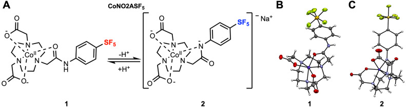 Figure 1
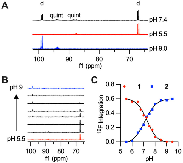 Figure 2
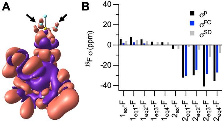 Figure 3
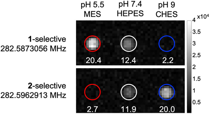 Figure 4
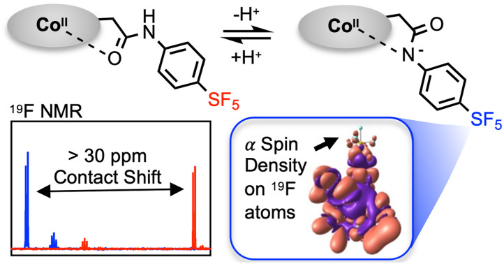 Figure 5
 Figure 6
- —National Institutes of Health10.13039/100000002
- —National Science Foundation10.13039/100000001
Peer Reviews
No public reviews on file for this paper yet. If you reviewed it on a platform where reviews are public (OpenReview, ICLR, NeurIPS, ICML), you can paste yours below so the community can read it here.
Videos
No videos yet. Explain this paper in a talk, walkthrough, or lecture? Add one.
Taxonomy
TopicsMagnetism in coordination complexes · Organometallic Compounds Synthesis and Characterization · Lanthanide and Transition Metal Complexes
Mammalian blood acts as a buffer to maintain pH homeostasis at 7.4 and aberrations from this pH are associated with several pathologies including metabolic and respiratory acidosis/alkalosis, chronic obstructive pulmonary disease, and pneumonia.^1^ In cancer, tumor microenvironments are often acidic, primarily due to increased anaerobic glycolysis causing higher concentrations of lactic acid.^2–4^ Thus, accurate pH mapping has the potential to be a powerful diagnostic tool. While sensors exist to monitor biological pH (colorimetric, optical imaging, positron emission tomography), magnetic resonance imaging (MRI) has the distinct benefit of being nonionizing with infinite penetration depth, making this a promising modality for developing non-invasive, in vivo quantitative pH sensors.^5–10^
MRI is a widely used clinical imaging technique characterized by high spatial resolution and contrast. Traditional MRI detects ^1^H; however, due to the high proton concentration in living organisms, ^1^H MRI contains unavoidably high background signal which lowers specificity and quantitative power. As an alternative nucleus, fluorine (^19^F) has favorable properties: 100% ^19^F natural isotopic abundance, a nuclear spin of ½, excellent receptivity (83% of proton), and a large chemical shift range (>350 ppm). Given the lack of MR active fluorine in the body, quantitative images of biological environments can be taken with higher specificity than ^1^H MRI.
Paramagnetic metals can modulate chemical shifts in ^19^F MR imaging agents via pseudocontact shift (PCS) and Fermi contact (FC) interactions. PCS derives from through-space magnetic dipole interactions in which a frequency shift is caused by the anisotropy of the paramagnetic species’ magnetic susceptibility. ^19^F PARAshift agents commonly employ PCS as the metal fluorine distance is often in the 5–10 Å range.^11–14^ Cobalt(ii) has a relatively large PCS range (26 Å) due to fast Orbach relaxations causing a short electronic relaxation time (∼10^−12^ s).^15^ We have reported redox responsive ^19^F PARAshift probes that use a spin change from paramagnetic (PCS-active) Co(ii) to diamagnetic Co(iii) using an amide-linked perfluoro-tert-butyl ^19^F tag.^13,16,17^ However, with PCS-based probes the shift range has been limited (∼3–10 ppm). Cobalt(ii) probes have been developed for ^1^H PARAshift and Chemical Exchange Saturation Transfer (CEST) that utilize PCS and FC to induce proton frequency shifts.^18–21^ FC interactions result from direct overlap of unpaired electron spin on the MR active nucleus and can result in much larger chemical shift changes compared to PCS.^22,23^ However, effects are generally limited to a couple bond lengths away from the paramagnetic center and have not been widely exploited in responsive ^19^F MR sensors.
We report CoNO2ASF_5_, a responsive ^19^F MR probe that exhibits a >30 ppm ^19^F chemical shift change between acidic and basic environments. This probe contains an anilide-linked pentafluorosulfanyl moiety (–SF_5_) as our ^19^F reporter. This SF_5_ group's ^19^F signal (∼+60 ppm) is highly shifted from CF_3_ groups (∼−70 ppm), and thus these tags could be used in tandem to monitor multiple analytes simultaneously. Further, despite inequivalent fluorines splitting the SF_5_ signal, studies successfully employ this tag as a sensitive, biocompatible ^19^F reporter group.^24,25^ Crucially, using an anilide moiety, as opposed to an alkyl amide, imbued new coordination properties to enable pH-responsiveness within a physiologically relevant range and activation of FC interactions several bonds away from the Co(ii) center. We characterized the probe's sensing properties and validated the pH-dependent shift change mechanism through theoretical calculations.
CoNO2ASF_5_ uses 1,4,7-triazacyclononane (TACN) as a macrocyclic base with two carboxylic acid arms (NO2A) and one anilide arm containing the aryl-SF_5_ moiety. This scaffold utilizes a hexa-coordinate structure for kinetic stability of the Co(ii).^26,27^ The anilide donor is key to biologically relevant pH sensitivity (compared to alkyl amide donors employed by our group) as the electron withdrawing nature of the aryl group lowers the pKa of the anilide NH to be within the physiological pH range. We propose that anilide deprotonation results in a CoNO2ASF_5_ coordination shift (Fig. 1a), converting from the protonated, neutral, oxygen-bound species (O-bound, 1) to the deprotonated, anionic, nitrogen-bound species (N-bound, 2).
Synthesis of CoNO2ASF_5_ was carried out from previously reported ^t^BuNO2A^13^ with chloroacetyl chloride and 4-(pentafluorothio)-aniline in two steps to afford ^t^BuNO2ASF_5_. Acid deprotection generated the final ligand NO2ASF_5_. Ligand NO2ASF_5_ was reacted with CoCl_2_·6H_2_O in anaerobic conditions to yield CoNO2ASF_5_. Intermediates were purified by reverse-phase chromatography and confirmed by HRMS and NMR.
Crystals of CoNO2ASF_5_ were grown as pink prisms in acidic (acetonitrile/water/HCl) and basic (methanol/water/NaOH) environments. Single crystal XRD structures demonstrate that the anilide oxygen coordinates to the cobalt in acidic conditions and the deprotonated anilide nitrogen coordinates with the cobalt in basic conditions, confirming the proposed coordination switch (Fig. 1). The coordination spheres consist of a plane of three TACN nitrogens and a plane of two carboxylate anions with the anilide. The twist angle of these planes shifts from a pseudo-octahedral angle of 41° for O-bound 1 to a less offset angle of 25° for N-bound 2. The added anionic donor in 2 is reflected by a lengthening of the Co–N_TACN_ (from 2.109 Å to 2.144 Å) and Co–O_carboxylate_ (from 2.039 Å to 2.102 Å) bond lengths after deprotonation. This is accompanied by a decrease in the deprotonated Co–N_anilide_ (2.07 Å) bond lengths compared to the protonated Co–O_anilide_ (2.123 Å) bond length. The cobalt-fluorine distance is also shortened in N-bound 2 (7.674 Å) compared to O-bound 1 (8.992 Å).
Magnetic susceptibility studies of CoNO2ASF_5_ in acidic and basic conditions confirm that both complexes are high spin S = 3/2 species with large orbital angular momentum contributions characteristic of high spin Co(ii), Table 1. This is reflected in the ^1^H NMR spectra of 1 and 2 which reveal the presence of highly shifted resonances ranging from −60 to +220 ppm (Fig. S2 and S3).
^19^F NMR characterization of 1 mM CoNO2ASF_5_ at pH 7.4 showed two doublets corresponding to the four-equivalent equatorial fluorines of the –SF_5_ moiety and two quintets corresponding to the axial fluorine. Each doublet and quintet pair could be detected in isolation by placing CoNO2ASF_5_ in acidic or basic buffers. At acidic pH (pH 5.5), the doublet and quintet peaks of complex 1 are found at +66.3 ppm and +88.0 ppm respectively. At basic pH (pH 9), the doublet and quintet peaks of deprotonated complex 2 are found at +98.0 ppm and +93.8 ppm respectively. There is an unexpectedly large >30 ppm chemical shift in the doublet peak when transitioning between the O-bound and N-bound complexes (Fig. 2A). Given the low intensity of the quintet, we will highlight the doublet peaks for all NMR samples. The doublets’ ^19^F relaxation times are significantly shortened due to the paramagnetic effect of the Co(ii), Table 1. Despite the high T1/T2 ratio and short T2, the doublet is well resolved with minimal broadening of the full-width at half maximum (FWHM) value.
A pH titration of CoNO2ASF_5_ was performed to characterize the pKa of the observed coordination change and to demonstrate that the conversion between the two ^19^F NMR doublets as pH is increased. The titration was performed using 1 mM CoNO2ASF_5_ dissolved in sulfonic acid buffers at different pH values (pH 5.0–6.5 MES, pH 7.0–8.0 HEPES, pH 8.5–9.5 CHES) and monitored by ^19^F NMR spectroscopy. Fig. 2B shows that as the pH increases, the peak at +66.3 ppm decreases and the peak at +98.0 increases. This data was fit to a pKa of 7.2 for the anilide proton, which is firmly within the biologically relevant range. Additional cyclability tests were performed and demonstrated reversible coordination and dynamic pH monitoring by ^19^F NMR (Fig. S4).
To better understand the large change in shift between O- bound 1 and N-bound 2, NMR chemical shift calculations were performed using a hybrid protocol combining density functional theory (DFT) and ab initio multireference approaches.^28,29^ DFT based geometric optimization showed coordination modes consistent with their crystal structures. After deprotonation, the anilide N atom replaces the carbonyl O atom as the coordinating atom. Complete active space self-consistent field (CASSCF) calculations with N-electron valence state perturbation theory (NEVPT2) treatment of dynamic correlation validate the triplet ground states for both complexes but reveal distinct zero field splitting (ZFS) characteristics. Complex 1 exhibits a positive axial parameter D of +51.1 cm^−1^ (D = 3Dzz/2) with E/D = 0.079 (E = (Dxx − Dyy)/2) and g = [2.026, 2.481, 2.536], whereas complex 2 shows a negative D of −55.0 cm^−1^ with E/D = 0.142 and g = [2.085, 2.213, 2.766]. The calculated g factors well reproduce the measured effective magnetic moments (1: exp. 4.61 ± 0.04μB, calcd 4.57μB; 2: exp. 4.72 ± 0.03μB, calcd 4.60μB).
Calculated NMR chemical shifts using variant DFT functionals are summarized in Tables S5 and S6 with atom labels shown in Fig. S6. For ^19^F nuclei, the calculated values reproduce the experimental trends, Table 2. Increasing fraction of Hartree–Fock exchange in the hybrid PBE functional, from 20% to 30% (PBE-20, 20%; PBE0, 25% and PBE-30, 30%), the calculated ^19^F chemical shifts for the equatorial ^19^F in complex 2 shows slight upfield shifts, from 110.2 ppm to 108.5 ppm, while the other F sites exhibit slight downfield shifts of similar magnitude. These opposite change in chemical shifts come from the distinct magnitude of paramagnetic contributions (σ^p^), thus leading to their different sensitivities to the functionals used in the calculations. With PBE0 functional, the equatorial F in complex 2 (denoted 2_eq_-F) display large σ^p^ of ca. −35 ppm, while the other F nuclei only show |σ^p^| < 8 ppm (Tables S7 and S8). All F sites in complex 1 have positive σ^p^, while those in complex 2 are negative. Due to the similar Mulliken charges on F atoms of the same type, this contrast in σ^p^ cannot arise from anionic-fragment charge bias.
Instead, Mulliken reduced orbital spin-density analysis reveals that in O-bound 1 all orbitals of all F atoms carry nearly zero spin density, whereas in N-bound 2 the axial F atom remains almost spin-neutral, while the p_x,y_ orbitals of the equatorial F atoms exhibit alpha-spin density, albeit small in magnitude (Table S9). The alpha-spin density stems from weak spin delocalization through the π conjugated pathway extending from the deprotonated amide N, which directly coordinates to Co, through the aromatic ring and S atom, and ultimately to the p_x,y_ orbitals of 2_eq_-F atoms, Fig. 3A. The alpha-spin density on p_x,y_ orbitals polarizes beta-spin density on the F 1s orbital, producing a positive hyperfine coupling through FC (Aiso of ca. 0.16 MHz) and a corresponding negative σ^p^ of ca. 30 ppm. This mechanism is consistent with the drastic 31.7 ppm upfield shift for the equatorial ^19^F signal after deprotonation.
The individual contributions of FC versus PCS were determined by extracting the FC term and the spin-dipole (SD) term of the hyperfine coupling tensor, Fig. 3B. PCS is based on the anisotropic component of the hyperfine tensor which is composed primarily of the SD term with small contributions from the gauge correction (diamagnetic) and spin-orbital coupling terms. The majority of the paramagnetic contributions to 1 come from PCS with minor to negligible FC contributions. We therefore hypothesize that the ∼4 ppm shift from the diamagnetic ligand to O-bound 1, is a result of PCS. This relatively small shift reflects the longer Co–F distance. The PCS effects in 2 are more pronounced but are insignificant compared to the large FC effects produced by the beta-spin density. Overall, these DFT results support a novel signal switching mechanism in which FC interactions are activated following deprotonation of the N-amide and coordination to Co^II^.
Given the distinct and well resolved ^19^F NMR signal, we continued with ^19^F MRI phantom studies. Selective pulse sequence imaging was utilized with optimized parameters based on the relaxation values and frequencies to image 5 mM CoNO2ASF_5_ at pH 5.5, 7.4 and 9. Pulse sequences tailored specifically to either species yielded SNR values 20 for both complexes in 30 minutes (Fig. 4). Each pulse sequence was selective for either the O- or N-bound complexes with the signal of the other complex below the limit of detection (SNR < 3). Both species were observed at physiological pH 7.4.
In conclusion, we have synthesized and characterized a novel pH-responsive cobalt complex for ^19^F MR applications. By utilizing an anilide linker, the pKa of the coordinating amide moiety is lowered to 7.2. This pH sensitivity is clearly observed by ^19^F MR imaging as the O- and N-bound species exhibit a 30 ppm chemical shift difference. The coordinating deprotonated anilide nitrogen allows for delocalization of spin density through a pi conjugated pathway to the fluorine atoms. This provides FC interactions to the equatorial fluorine in 2 whereas 1 has much weaker paramagnetic contributions mostly through PCS. As a new strategy for responsive PARAshift probes, this design stands out due to the exceptional shift magnitude despite a long Co–F distance. So, while many FC based paramagnetic probes exhibit strong signal quenching due to the close Co–F proximity, CoNO2ASF_5_ displays minimal signal broadening. Future work will evaluate the versatility of applying FC shifts through the extend pi system in developing sensing molecules to a variety of stimuli for MR sensing applications.
K. S. and R. K. performed experimental investigation, formal analysis, conceptualization, and writing original draft. C. H., G. E. F. B., and J. A. R. performed experimental investigation and analysis. J. X. preformed DFT and CASSCF/NEVPT2 calculations/interpretation and writing. Y. G. provided expertise, funding and editing. E. Q. provided conceptualization, formal analysis, writing, and project administration, resources and funding.
Conflicts of interest
There are no conflicts to declare.
Supplementary Material
CC-062-D6CC00957C-s001
CC-062-D6CC00957C-s002
The reference list from the paper itself. Each links out to its DOI / PubMed record.
- 1Hopkins E. , Sanvictores T. and Sharma S., Physiology, Acid Base Balance, Stat Pearls Publishing, Treasure Island (FL), 2022
- 2Kato Y. Ozawa S. Miyamoto C. Maehata Y. Suzuki A. Maeda T. Baba Y. Acidic Extracellular Microenvironment and Cancer Cancer Cell Int.20131318910.1186/1475-2867-13-8924004445 PMC 3849184 · doi ↗ · pubmed ↗
- 3Bogdanov A. Bogdanov A. Chubenko V. Volkov N. Moiseenko F. Moiseyenko V. Tumor Acidity: From Hallmark of Cancer to Target of Treatment Front. Oncol.20221297915410.3389/fonc.2022.97915436106097 PMC 9467452 · doi ↗ · pubmed ↗
- 4Ward C. Meehan J. Gray M. E. Murray A. F. Argyle D. J. Kunkler I. H. Langdon S. P. The Impact of Tumour p H on Cancer Progression: Strategies for Clinical Intervention Explor. Targeted Anti-Tumor Ther.2020127110010.37349/etat.2020.00005 · doi ↗
- 5Tantama M. Hung Y. P. Yellen G. Imaging Intracellular p H in Live Cells with a Genetically Encoded Red Fluorescent Protein Sensor J. Am. Chem. Soc.201113326100341003710.1021/ja 202902 d 21631110 PMC 3126897 · doi ↗ · pubmed ↗
- 6Hou H. Zhao Y. Li C. Wang M. Xu X. Jin Y. Single-Cell p H Imaging and Detection for p H Profiling and Label-Free Rapid Identification of Cancer-Cells Sci. Rep.201771175910.1038/s 41598-017-01956-128496209 PMC 5431805 · doi ↗ · pubmed ↗
- 7Steinegger A. Wolfbeis O. S. Borisov S. M. Optical Sensing and Imaging of p H Values: Spectroscopies, Materials, and Applications Chem. Rev.202012022123571248910.1021/acs.chemrev.0c 0045133147405 PMC 7705895 · doi ↗ · pubmed ↗
- 8Demoin D. W. Wyatt L. C. Edwards K. J. Abdel-Atti D. Sarparanta M. Pourat J. Longo V. A. Carlin S. D. Engelman D. M. Andreev O. A. Reshetnyak Y. K. Viola-Villegas N. Lewis J. S. PET Imaging of Extracellular p H in Tumors with 64 Cu- and 18 F-Labeled p HLIP Peptides: A Structure–Activity Optimization Study Bioconjugate Chem.20162792014202310.1021/acs.bioconjchem.6b 0030627396694 PMC 5034329 · doi ↗ · pubmed ↗