Visceral Leishmaniasis in Southern Brazil: an overview of current epidemiology
Bibiana Paula Dambrós, Amabilli de Souza Rosar, Patricia Hermes Stoco, Patricia Flávia Quaresma, Edmundo Carlos Grisard, Mario Steindel

TL;DR
This paper reviews the spread of visceral leishmaniasis in southern Brazil, focusing on the emergence of cases in regions where it was previously absent.
Contribution
The paper provides an updated overview of visceral leishmaniasis epidemiology in southern Brazil, highlighting recent geographic expansions and transmission dynamics.
Findings
Visceral leishmaniasis cases have emerged in multiple municipalities in Rio Grande do Sul and Santa Catarina since 2008.
Lutzomyia longipalpis, the main vector, has not been found in all endemic areas, suggesting other sandfly species may contribute to transmission.
Autochthonous human cases have been reported in Paraná and Santa Catarina, indicating disease spread beyond initial locations.
Abstract
Visceral leishmaniasis (VL), caused by the protozoan parasite Leishmania (Leishmania) infantum, is transmitted in Brazil through the bites of female phlebotomine sandflies. Until 2008, no cases of autochthonous VL transmission were reported in the southern Brazilian states of Paraná (PR), Santa Catarina (SC), and Rio Grande do Sul (RS). The first case of canine VL (CVL) in RS was reported in 2008, followed by that of human VL (HVL) in 2009. Since then, CVL and HVL cases have emerged in 12 and five additional municipalities in the RS, respectively. In SC, CVL was first reported in Santa Catarina Island in 2010 and is now present in 44 of 89 localities. By 2014, CVL had appeared in western SC, with new cases reported in the midwestern and southern areas in 2021and 2024, respectively. Between 2017 and 2022, five autochthonous HVL cases were diagnosed in Florianópolis. In the PR, CVL was…
Genes, proteins, chemicals, diseases, species, mutations and cell lines named across the full text — each resolved to its canonical identifier and authoritative record.
Click any figure to enlarge with its caption.
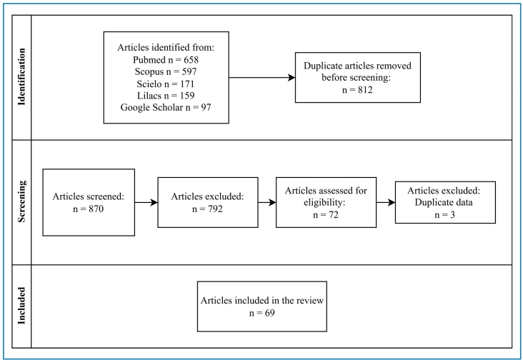 Figure 1
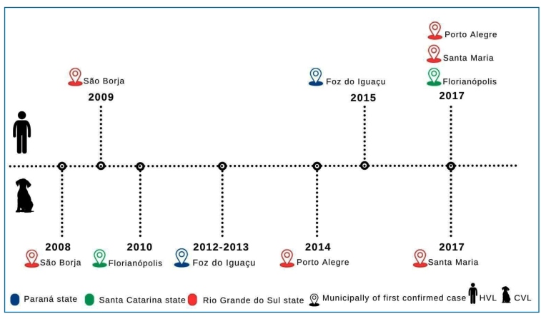 Figure 2
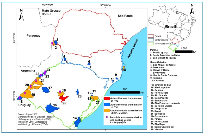 Figure 3
- —CNPq
- —CAPES
- —FAPESC
Peer Reviews
No public reviews on file for this paper yet. If you reviewed it on a platform where reviews are public (OpenReview, ICLR, NeurIPS, ICML), you can paste yours below so the community can read it here.
Videos
No videos yet. Explain this paper in a talk, walkthrough, or lecture? Add one.
Taxonomy
TopicsResearch on Leishmaniasis Studies · Trypanosoma species research and implications · Syphilis Diagnosis and Treatment
INTRODUCTION
Visceral leishmaniasis (VL) is a potentially fatal disease caused by the protozoan parasites Leishmania donovani and Leishmania infantum, affecting 83 countries and territories, resulting in 50,000-90,000 cases per year1 ^,^ 2. Transmitted through the bite of infected female sandflies of the genus Lutzomyia in the Americas, symptoms include fever, weight loss, fatigue, weakness, loss of appetite, enlarged liver and spleen, anemia, and swollen lymph nodes1. This neglected and socially determined tropical disease mainly affects vulnerable populations and is one of the most significant infectious diseases worldwide1.
Human VL (HVL) cases have been reported in Argentina, Bolivia, Brazil, Colombia, Costa Rica, El Salvador, Guatemala, Honduras, Mexico, Nicaragua, Paraguay, Uruguay, and Venezuela. These countries report approximately 3,500 cases per year, ranging from 7% to 9%. Over 90% of cases occur in Brazil3, which has one of the highest rates of canine VL (CVL) worldwide4. Thus, parasite circulation through humans, vectors, and mammals potentially allows distinct genotypes or adaptations to local vectors5 ^-^ 7.
Historically, in impoverished endemic rural areas, CVL and HVL have progressively spread to the mid- and southwestern regions of Brazil, establishing an urban transmission cycle in which CVL precedes human cases in 23 of 26 states, including the Federal District8 ^,^ 9. In this review, we examined the expansion of CVL and HVL in the southern Brazilian states of Paraná (PR), Santa Catarina (SC), and Rio Grande do Sul (RS), as well as their epidemiological aspects and prophylactic strategies.
METHODS
To evaluate the status of autochthonous canine and human VL, as well as the phlebotomine fauna and epidemiology of VL in southern Brazil, a comprehensive literature search was conducted using the following primary databases: PubMed, SciELO, and SCOPUS, covering the period from January 2008 to December 2024. The following terms were used to retrieve articles: “Canine OR Human Visceral Leishmaniasis AND Rio Grande do Sul OR Santa Catarina OR Paraná,” “Lutzomyia OR Phlebotomine OR Sandfly AND Rio Grande do Sul OR Santa Catarina OR Paraná,” “Leishmaniasis OR Epidemiology AND Rio Grande do Sul OR Santa Catarina OR Paraná,” and “Visceral Leishmaniasis OR Epidemiology AND Southern Brazil.”
This review adhered to the guidelines outlined in the Preferred Reporting Items for Systematic Reviews and Meta-Analyses statement (PRISMA), which can be accessed at http://www.prisma-statement.org. The inclusion criteria were as follows: (1) articles written in English or Portuguese, (2) free full access or availability through Periódicos CAPES, and (3) publication dates and data collection from 2008 to 2024. The exclusion criteria were as follows: (1) reviews, theses, dissertations, monographs, editorials, letters to the editor, forums, comments, author corrections, and books, (2) studies with overlapping or duplicate data, and (3) abstract-only papers.
Only full papers in English and Portuguese were reviewed, and only those containing detailed information were retrieved and analyzed according to the eligibility criteria. To minimize potential bias in the literature search, the titles and abstracts of the selected articles were examined by two independent reviewers based on predefined inclusion criteria (Figure 1).
FIGURE 1:Flowchart of article selection.
Additionally, data were retrieved from the World Health Organization (WHO), Pan American Health Organization (PAHO), Brazilian Health Authority databases (SINAN and DATASUS), and technical publications from the RS, SC, and PR state health authorities. Raw data were manually curated to include data exclusively related to southern Brazilian States and then organized in Excel spreadsheets to allow comparative analysis. Figure 1 and Figure 2 were generated using Canva (https://www.canva.com/pt_br/), and Figure 3 was generated using QGIS Desktop 3.40.10.
FIGURE 2:Timeline of autochthonous canine (CVL) and human (HVL) visceral leishmaniasis in southern Brazilian states of Paraná (PR), Santa Catarina (SC), and Rio Grande do Sul (RS).
FIGURE 3:Distribution of autochthonous cases of canine visceral leishmaniasis (CVL) and human visceral leishmaniasis (HVL) and the presence of Lutzomyia longipalpis in the states of Paraná (PR), Santa Catarina (SC), and Rio Grande do Sul (RS) between 2008 to 2024.
CANINE AND HUMAN VISCERAL LEISHMANIASIS
HVL has been documented in Brazil since 1913; however, L. infantum was likely introduced to Brazil by Portuguese or Spanish colonizers in the northeastern region, where susceptible vectors facilitated parasite transmission10. Since then, primary routes for L. infantum dispersion in central and southern Brazil have been proposed.
Retrospective analyses have pointed to the Bolivia-Brazil route of introduction, associated with population/animal displacement from Bolivia to the neighboring states of Mato Grosso and Mato Grosso do Sul in Brazil, and possibly São Paulo, during the construction of the Bolivia-Brazil gas pipeline (1998-2005)11.
Microsatellite analyses have indicated two possible introduction routes. The Paraguay-Brazil route, linked to the introduction of VL from urban transmission in Paraguay into the Brazilian states of Mato Grosso do Sul and Paraná, particularly around the triple border area (Argentina-Paraguay-Brazil) around 2012; and the introduction of a distinct genetic cluster of L. infantum strains in western Santa Catarina, closer to the Argentinian border, that expanded towards southern Paraná state around 201311.
As the introduction of VL into the RS was associated with CVL and HVL cases reported in Posadas, Argentina, in 200612 ^,^ 13, we cannot rule out the expansion of VL within the Brazilian territory, since the country has one of the highest prevalence rates in the Americas.
The emergence of VL in the southern states is a recent phenomenon, as only a few imported human cases were reported before 200714 ^-^ 17. Since then, a typical pattern of disease expansion has been observed, with increasing reports of autochthonous CVL, followed by HVL (Figure 2).
Autochthonous cases of CVL in RS were initially reported in São Borja in 2008, close to the Brazil-Argentina border, where Lu. longipalpis was identified as the primary vector, highlighting the vulnerability of these areas18 ^,^ 19. In the following year, autochthonous HVL cases were reported in the same municipality18. In 2010, autochthonous CVL cases were reported in the metropolitan area of Porto Alegre, the state capital, located approximately 585 km east of São Borja20 ^-^ 22. Between January and July 2014, the CVL seroprevalence was 4.0% in Porto Alegre, reaching 5.6% in Canoas and 4.6% in São Leopoldo neighboring cities20. Between 2016 and 2021, 766 CVL and 20 HVL autochthonous cases were reported in Porto Alegre21, where Lu. longipalpis has not been reported23. In 2010, an autochthonous case of CVL was reported for the first time in the non-endemic municipality of Santa Maria in the central region of the state24. Almost simultaneously, several autochthonous CVL and two cases of HVL were recorded between 2017-2021 in Santa Maria, in the central region of RS, where the presence of Lu. longipalpis was confirmed25 ^-^ 27.
Between 2009 and 2023, 52 autochthonous HVL cases were documented in RS, with seven fatal cases in Porto Alegre and one in Santa Maria26 ^,^ 28, indicating the spread of VL and its associated morbidity and mortality in the region. According to the RS State Health Authority, the active transmission of L. infantum was recorded in 12 municipalities (Figure 3), 67% of which shared borders with Argentina and Uruguay28.
By 2023, autochthonous HVL in southern Brazilian states had an annual average rate of 0.67 cases for SC, 1.0 for PR, and 4.67 for RS, encompassing low transmission risk areas29 (Table 1). However, a comparative analysis of data obtained from the Brazilian Ministry of Health, State Health authorities, and scientific literature revealed some discrepancies. For instance, municipalities such as Castro and Umuarama in PR, Imaruí and Joinville in SC, and Cachoeira do Sul, Capão da Canoa, Nova Petrópolis, Rio Grande, and São Sepé in RS were classified as areas of autochthonous HVL transmission despite a lack of evidence28 ^,^ 30 ^,^ 31. Conflicting epidemiological reports must be properly addressed, as the accuracy of such data depends primarily on specific diagnoses, directly impacting treatment, decision-making processes, and the effectiveness of educational campaigns, demanding collaboration between health authorities, researchers, and the public.
TABLE 1:Number of reported and confirmed cases of human visceral leishmaniasis in the states of Paraná, Santa Catarina, and Rio Grande do Sul between 2009 to 2023, according to DATASUS.State Origin (municipality of infection)Number of cases Deaths
ReportedConfirmed
Paraná Foz do Iguaçu24246 Maringá200 Santa Terezinha de Itaipú220 Alto Paraiso100 Castro100 Curitiba100 Ivate100 Londrina100 Terra Rica 101 Umuarama100 Santa Catarina Florianópolis550 Imaruí100 Joinville100 Rio Grande do Sul Porto Alegre25257 São Borja14140 Uruguaiana660 Itaqui440 Viamão400 Santa Maria331 Cachoeira do Sul100 Capão da Canoa100 Nova Petrópolis100 Rio Grande100 São Sepe100 Total
103
83 (80.6%)
15 (14.5%)
A retrospective analysis of the evolution of CVL in the state of PR illustrated the challenges associated with diagnosis, epidemiological assessment, and decision-making for the control of disease transmission. A seroepidemiological survey of CVL was conducted in the municipalities of Foz do Iguaçu and Santa Terezinha de Itaipu between 2014 and 2015, and 1,129 dogs were tested32. The seroprevalence of CVL was 23.8% in the rural areas of Foz do Iguaçu and 4.7% in the neighboring city of Santa Terezinha de Itaipu32. Between 2015 and 2020, a survey of CVL conducted on 12,205 dogs in Foz do Iguaçu revealed a seroprevalence of 37.94%, indicating a heightened risk for HVL33.
By 2015, the first autochthonous case of HVL was diagnosed34. Between 2015 and 2022, these municipalities, located on the triple border between Argentina and Paraguay, reported 26 autochthonous HVL cases, resulting in six fatalities29. This highlights the potential of VL to spread across neighboring municipalities and countries.
The emergence of CVL in SC began in 2010 in the neighborhoods of Canto dos Araçás and Lagoa da Conceição on Santa Catarina Island (Florianópolis), located on the eastern Atlantic border17 ^,^ 35. According to data from the Zoonosis Control Center in Florianópolis, the percentage of euthanasia cases was 57.35% between 2010 and 2019. In contrast, from 2020 to 2022, the percentage decreases to 28%. In 2013 and 2015, CVL surveys were conducted in São Miguel do Oeste and Descanso in the western region of the state36 and in Erval Velho and Herval d’Oeste in the midwestern region37. Based on serological and PCR testing, these surveys revealed prevalences of 19.0% and 7.14% in the western and midwestern regions, respectively. Following the canonical spreading pattern, three autochthonous HVL cases were reported in Florianópolis in 2017, with two additional cases each in 2020 and 202331 ^,^ 38.
An overall seroprevalence of 0.6% for CVL was estimated among 1,227 dogs sampled across six mesoregions of SC using DPP and ELISA tests39. Positive cases were primarily concentrated in the eastern region of the state, with 43% of these cases being asymptomatic and 57% exhibiting polysymptomatic characteristics. Later, a survey of L. infantum infection based on PCR analysis of blood samples from dogs sheltered at the Zoonosis Control Centers in the cities of Tubarão (77 dogs) and Criciúma (30 dogs), located in southeastern SC, revealed positivity rates of 16.9% and 23.5%, respectively40. Notably, 85% of the dogs were asymptomatic. Another study in Tubarão reported a PCR positivity rate of 4.2% for L. infantum in conjunctival swabs from 47 sheltered dogs41. Taken together, these findings confirm the transmission of L. infantum in SC, not only in Florianópolis, but also in the southern, midwestern, and western regions. This indicates that the seroprevalence rates may vary considerably owing to the different sampling and diagnostic methods employed. According to the SC Health Authority, between 2019 and 2020, 50 of 295 municipalities reported CVL cases, mostly non-autochthonous42, whereas Florianópolis recorded 1,058 CVL cases in 11 out of 18 districts by 202443, representing an increased risk of VL transmission to humans.
SANDFLY FAUNA
Phlebotomine sand flies are blood-feeding insects and vectors of Leishmania parasites, the causative agents of leishmaniasis. Sand flies are found in tropical and temperate regions worldwide, and the majority of medically important species belong to the genus Phlebotomus in the Old World and Lutzomyia in the New World44. Thus, the assessment of local sandfly fauna, the prevalence of Leishmania spp. infection, and the vectorial competence of these species is crucial for understanding the epidemiology of VL. Xenodiagnosis is a classical parasitological technique used to determine and quantify the infectiousness of a specific host to a blood-sucking insect species, and provides conclusive data to distinguish infectious from noninfectious45. However, this method is labor-intensive, complex, and time-consuming, and requires the establishment of sandfly colonies, trained personnel, and research facilities.
The complex interactions between mammalian-infective Leishmania species or genotypes and sand flies may influence transmission dynamics and require multivariate analysis46. In the context of VL, such studies are of major relevance to endemic or vulnerable transmission areas where the primary vector Lu. longipalpis and Lu. cruzi have not yet been reported. Other sandfly species, such as Lu gaminarai, Mi. migonei, Ny. neivai, Pi. fischeri, and Lu. evansi have also been implicated in the transmission of L. infantum in Brazil14 ^,^ 47 ^-^ 51.
The state of Paraná was considered free of VL transmission until 2012, with neither CVL, HVL, nor Lu. longipalpis reported there52 ^,^ 53. Lu. longipalpis was sampled for the first time in Foz do Iguaçu in 2012, when the city was declared an area at risk for the disease54. An entomological survey conducted in Foz do Iguaçu and Santa Terezinha de Itaipu between 2014 and 2015 revealed that Lu. longipalpis was the predominant species, accounting for 71.78% of the 3,543 sandflies captured55. Other species, such as Evandromyia edwardsi, Expapillata firmatoi, Micropygomyia ferreirana, and Pi. christenseni documented for the first time in this region, may also be involved in L. infantum transmission55.
A summary of the phlebotomine sandfly fauna in vulnerable and endemic areas of VL in southern Brazil is presented in Table 2.
TABLE 2:Records of sandfly species in endemic areas of visceral leishmaniasis in the states of Paraná, Santa Catarina, and Rio Grande do Sul.StateMunicipalitySandfly speciesReferences Paraná Foz do Iguaçu and Santa Terezinha de Itaipu Lu. longipalpis, Ny. whitmani, Ny. intermedia, Ny. neivai, Mi. quinquefer, Mi. migonei, Pi. pessoai, Pi. monticola, Pi. fischeri, Pi. christenseni, Brumptomyia brumpti, Martinsmyia alfabética, Ev. cortelezzii, Ev. edwardsi, Evandromyia sp., Pa. shannoni, Expapillata firmatoi, Micropygomyia ferreirana, Phlebotominae sp. 5 ^,^ 54 ^,^ 55
Santa Catarina Florianópolis Pi. fischeri, Mi. migonei, Ny. neivai, Brumptomyia sp. 14 ^,^ 42
Joaçaba, Herval d'Oeste, Ouro, Lacerdópolis and Erval Velho Brumptomyia cunhai, Br. guimaraesi, Br. mangabeirai, Brumptomyia sp*., Mi. migonei, Ny. neivai, Pi. fischeri, Ps. lanei, Ev. edwardsi, Martinsmyia alfabetica*
Tubarão Ny. neivai, Mi. migonei, Pi. fischeri, Ev. edwardsi, Brumptomyia sp*.*
Rio Grande do Sul São Borja, Barra do Quaraí, Garruchos, Pirapó, Porto Xavier, Uruguaiana, Itaqui Lu. longipalpis
Porto Alegre Ps. lanei, Brumptomyia sp., Mi. migonei, Pi. fischeri, Ny. neivai, Lutzomyia gaminarai
Estrela Brumptomyia cunhai, B. nitzulescui, Ev. edwardsi, Pi. fischeri, Ps. lanei, Mi. migonei, L. misionensis, Ny. neivai, L. pascalei
Santa Maria Lu. longipalpis, Mi. migonei, Pi. fischeri, Ps. lanei, Brumptomyia sp. 26 ^,^ 27
In 2006, the first case of VL was identified in Argentina’s province of Misiones, where several cases of CVL and one case of HVL were reported in Posadas along with the presence of Lu. longipalpis. This represents the southernmost transmission of L. infantum in Latin America12. Shortly after, Lu. longipalpis was also reported in Salto and Bella Unión-Cuarein in Uruguay56, where CVL cases were confirmed in 201557 ^,^ 58, indicating the ongoing cross-border spread of VL and underscoring the necessity for both national and international surveillance efforts55 ^,^ 56 ^,^ 58.
Since 1990, Santa Catarina has reported the autochthonous transmission of human cutaneous leishmaniasis in the western region. Entomological surveys conducted in the northeastern and midwestern regions of SC did not record the presence of Lu. longipalpis 59 ^-^ 61. Since 2011, entomological surveillance conducted by the Zoonosis Control Centre in Florianópolis has revealed the presence of Brumptomyia sp., Pi. fischeri, Mi. migonei, Ny. neivai, Ev. edwardsi, Lu. tupynambai, and Lu. firmatoi. Natural infection assessed by PCR confirmed the presence of L. infantum DNA in 1.6% of Ny. neivai, as well as Leishmania spp. in 0.6% of Pi. fischeri and 0.3% of Lu. migonei 14. In a more recent study, L. infantum was confirmed in 0.4% Ny. neivai in Tubarão, 150 km south of Florianópolis, accounting for 85.8% of the sandflies captured41. In this area, a CVL prevalence of 4.2% was also confirmed.
Approximately 50% of the 52 autochthonous HVL cases in RS occurred in areas where Lu. longipalpis has not been reported, including in the capital city of Porto Alegre, where sandfly fauna were investigated between 2014 and 2015 for L. infantum infection using PCR. In the Agronomia district, located 2.5 km from the Jardim Carvalho district, Pi. fischeri tested positive for L. infantum 23. Meanwhile, in the Morro Santana and Jardim Carvalho districts, which are 3.5 km apart, Lu. gaminarai, Pi. fischeri, and Mi. migonei also tested positive50. These findings in a restricted, vulnerable urban area bordering a forested region highlight the necessity for ongoing biological and epidemiological research to understand the dynamics of VL transmission, which may differ between endemic areas. Understanding these unique transmission patterns is crucial for developing targeted interventions and control strategies tailored to specific challenges faced in such environments.
The high prevalence of CVL and HVL in Porto Alegre (RS) and Florianópolis (SC), where Lu. longipalpis has not been reported, suggests that other species, such as Ny. neivai, Mi. migonei, Pi. fischeri, and Lu. gaminarai, which have been found naturally infected with L. infantum, may also play a role in L. infantum transmission in these regions.
DIAGNOSIS OF CVL
The clinical diagnosis of CVL relies on clinical signs and clinicopathological abnormalities; however, a definitive diagnosis requires serological, parasitological, and molecular laboratory tests62. The Brazilian Ministry of Health recommends two serological tests based on different methodologies: an initial immunochromatographic dual-path platform (TR-DPP) (Bio-Manguinhos, Fiocruz, Brazil) and an ELISA assay for confirmation63. An indirect immunofluorescence assay (IFA) using standardized antigens and serum dilutions of at least³ 1:160 represents an alternative diagnostic assay63.
The microscopic examination of stained smears from popliteal lymph nodes or bone marrow aspirates and in vitro culture of these aspirates are useful parasitological techniques; however, histopathological and immunohistochemical tests have limited specificity and sensitivity63.
Molecular diagnosis using PCR offers high specificity and sensitivity, even in the early stages of infection, and can detect asymptomatic infections64 ^,^ 65. PCR techniques targeting L. infantum sequences such as kinetoplast DNA minicircles (kDNA), 18S rRNA, HSP70, and hypothetical proteins (Linj31) facilitate accurate diagnosis, parasite load monitoring, treatment evaluation, and assessment of vector infectiousness66 ^-^ 69.
Diagnostic methods vary among the southern states. According to the state and municipal healthcare authorities of PR, SC, and RS, DPP and ELISA are currently used in CVL surveys in all states. In SC and RS, standard immunological techniques (DPP and ELISA) are used to diagnose HVL and CVL, as recommended by the Brazilian Ministry of Health. In PR, where the Instituto Carlos Chagas (ICC/Fiocruz) is the state reference laboratory, serological, molecular, and parasitological methods are employed as confirmatory/complementary diagnostic tools for both HVL and CVL.
PROPHYLATIC AND CONTROL MEASURES
For over five decades, the Brazilian Ministry of Health has recommended dog culling as the primary public health measure for controlling CVL4 ^,^ 62 ^,^ 70. However, this policy has led to societal conflict, and numerous studies have shown that dog culling is ineffective as a standalone strategy to control CVL transmission4 ^,^ 70 ^,^ 71, prompting calls for new and more effective approaches62. Combined strategies, such as the use of repellents, vaccination, and treatment, may work at the individual level. However, public policies with community engagement may provide more humane and effective methods for managing the impact of VL on human and animal populations.
Since 2016, the Brazilian government has authorized the use of miltefosine (Hexadecylphosphocholine, Milteforan®) for the treatment of CVL, representing the first therapeutic alternative to euthanasia for infected animals72. Treatment depends on the disease stage and can be achieved by the administration of miltefosine alone or in combination with domperidone or allopurinol. While miltefosine can initially improve symptoms and reduce the parasitic load, monotherapy yields unsatisfactory results because many animals experience relapse, which increases the risk of disease transmission73. To mitigate this, veterinarians have also used immunomodulators, including allopurinol, domperidone, and the Leish-Tec® vaccine when it was still available73 ^-^ 75, as recommended by Brasileish and Conselho Federal de Medicina Veterinária (CFMV)63 ^,^ 76. Therefore, the continuous development of comprehensive treatment protocols that incorporate immunotherapy, along with careful monitoring and supportive care, is essential for managing CVL and reducing the risk of transmission to humans73 ^,^ 74.
Although treatment with miltefosine and associated therapies can help reduce the parasite burden and improve the health of VL-infected dogs73, cases of parasitological cure have not been documented. Thus, a significant issue remains as to whether treated animals can act as reservoirs of L. infantum, which severely affects transmission control. Therefore, neither miltefosine therapy nor prophylactic or immunotherapeutic vaccination for CVL is a public health measure in Brazil77.
Consequently, the responsibility and costs associated with the vaccination or treatment of infected dogs fall solely on their owners, demanding that dog owners take responsibility for their pets’ health and highlighting the importance of individual accountability in managing CVL cases63 ^,^ 76.
It is well established that dogs play a pivotal role as reservoirs to maintain the urban transmission cycle of VL in Latin American and Mediterranean countries78 ^,^ 79. The proximity of L. infantum-infected dogs to humans, parasite genetic plasticity, and the variety of sandfly species significantly influence disease dynamics, requiring control and prevention strategies. Since 2020, the CFMV has implemented mandatory reporting of positive or suspected CVL cases in Brazil, recommending complementary preventive and control measures, such as vaccination and the use of insecticide-impregnated collars76. In southern Brazil, key control measures include the euthanasia of infected dogs, seroepidemiological surveys in transmission areas, establishment of municipal zoonosis control centers, and urban/peridomiciliary cleaning to eliminate potential breeding sites for sandflies. The municipality of Florianópolis (SC) has offered free treatment to VL-infected dogs from low-income owners since 202280, and the SC State Health Authority has implemented mandatory reporting of CVL since 202481.
Finally, it is worth noting that various sylvatic and domestic mammals of the orders Carnivora, Marsupialia, and Rodentia act as primary reservoirs of L. infantum, thereby sustaining the zoonotic transmission cycle82, which has been understudied across a wide geographical area.
FINAL REMARKS
The geographic expansion of VL in southern Brazil is an undeniable reality that emphasizes the need for continuous monitoring and improved intervention strategies towards risk management in both endemic and vulnerable areas. However, it is crucial to consider that inadequate entomological and medical surveillance, as well as non-standardized human or animal testing, may lead to underreporting of both CVL and HVL, which can significantly affect our understanding of the disease’s epidemiology in southern Brazil.
Additionally, the border effect, along with increased domestic and international migration and cargo transport in recent decades, raises concerns regarding the potential spread of VL. These factors necessitate the effective control of the movement of infected animals to mitigate the risk of transmission. Comprehensive strategies focusing on surveillance, public awareness, and strict regulations regarding animal transport are essential to address the challenges posed by VL in this region.
While Lu. longipalpis is a key player in the epidemiology of VL throughout Brazil, other sandfly species, often regarded as secondary vectors, may act as competent or primary vectors of L. infantum in areas where Lu. longipalpis has not yet been reported. This is supported by the high seroprevalence of CVL in vulnerable and formerly non-endemic areas. These findings suggest the need for further research on the roles of alternative vectors and their potential impact on the transmission dynamics of leishmaniasis, as well as the importance of adapting control measures to address the specific risks associated with these species.
The reference list from the paper itself. Each links out to its DOI / PubMed record.
- 1Jain S Agua J Ruiz-Postigo J Maia-Elkhoury N Valadas S Zamani G Global leishmaniasis surveillance updates 2024: consolidating gains and new initiatives World Health Organization 2024
- 2Alvar J Vélez ID Bern C Herrero M Desjeux P Cano J Leishmaniasis Worldwide and Global Estimates of Its Incidence P Lo S ONE 201275 e 356712269354810.1371/journal.pone.0035671 PMC 3365071 · doi ↗ · pubmed ↗
- 3Pan American Health Organization (PAHO) Leishmaniasis: Epidemiological Report for the Americas 20232024 May 5Available from: https://iris.paho.org/handle/10665.2/59155
- 4Bermudi PMM Costa DNCC Nunes CM Tolezano JE Hiramoto RM Rodas LAC Canine serological survey and dog culling and its relationship with human visceral leishmaniasis in an endemic urban area BMC Infect Dis 20202014014013250346110.1186/s 12879-020-05125-0PMC 7275440 · doi ↗ · pubmed ↗
- 5Thomaz-Soccol V Gonçalves AL Piechnik CA Baggio RA Boeger WA Buchman TL Hidden danger: Unexpected scenario in the vector-parasite dynamics of leishmaniases in the Brazil side of triple border (Argentina, Brazil and Paraguay)P Lo S Negl Trop Dis 2018124 e 00063362962458610.1371/journal.pntd.0006336 PMC 5906028 · doi ↗ · pubmed ↗
- 6Barroso PA Nevot MC Hoyos CL Locatelli FM Lauthier JJ Ruybal P Genetic and clinical characterization of canine leishmaniasis caused by Leishmania (Leishmania) infantum in northeastern Argentina Acta Tropica 20151502182232627706710.1016/j.actatropica.2015.08.007 · doi ↗ · pubmed ↗
- 7Valdivia HO Roatt BM Baptista RDP Ottino J Coqueiro-dos-Santos A Sanders MJ Replacement of Leishmania (Leishmania) infantum Populations in an Endemic Focus of Visceral Leishmaniasis in Brazil Front Cell Infect Microbiol 2022129000849000843581168210.3389/fcimb.2022.900084 PMC 9263273 · doi ↗ · pubmed ↗
- 8Bruhn FRP Werneck GL Barbosa DS Câmara DCP Simões TC Buzanovsky LP Spatio-temporal dynamics of visceral leishmaniasis in Brazil: A nonlinear regression analysis Zoonoses and Public Health 20247121441563798483710.1111/zph.13092 · doi ↗ · pubmed ↗