From Synthesis to Therapeutics: Bioactive‐Coated Zein Nanoparticles in Drug Delivery
Milena Ferreira de Lima, Andy Joel Taipe Huisa, Azael da Silva Neto, Clarice Beatriz Gonçalves Silva, Iago Dillion Lima Cavalcanti, Karolliny Barbosa de Araújo, Mariane Cajubá de Britto Lira‐Nogueira, Nereide Stela Santos Magalhães, Priscila Gubert

TL;DR
This review explores how zein nanoparticles coated with bioactive materials can improve drug delivery by enhancing stability and therapeutic effects.
Contribution
The paper systematically reviews synthesis methods, coating materials, and biological activities of bioactive-coated zein nanoparticles.
Findings
The antisolvent method is most commonly used for zein nanoparticle synthesis due to its simplicity and cost-effectiveness.
Chitosan is identified as the preferred coating material for improving nanoparticle stability and performance.
Bioactive-coated zein nanoparticles show antioxidant, anticancer, and other therapeutic properties in preclinical studies.
Abstract
Polymeric zein nanoparticles (ZNP), derived from corn protein, are biodegradable drug carriers with high stability and low synthesis costs. Their amphiphilic nature allows efficient encapsulation of both hydrophilic and lipophilic drugs, making them promising for drug delivery. However, their instability under physiological pH can limit therapeutic efficacy, necessitating protective coatings for improved absorption. This review discusses synthesis methods, coating materials, and biological activities of bioactive‐coated ZNP. We found that the antisolvent method is the most commonly used due to its simplicity and cost‐effectiveness, while chitosan is the preferred coating material. ZNP exhibit antioxidant, anticancer, anesthetic, antidiabetic, hypoglycemic, and immunogenic properties, as demonstrated in both in vitro and in vivo studies. Their ability to enhance bioavailability, reduce…
Genes, proteins, chemicals, diseases, species, mutations and cell lines named across the full text — each resolved to its canonical identifier and authoritative record.
Click any figure to enlarge with its caption.
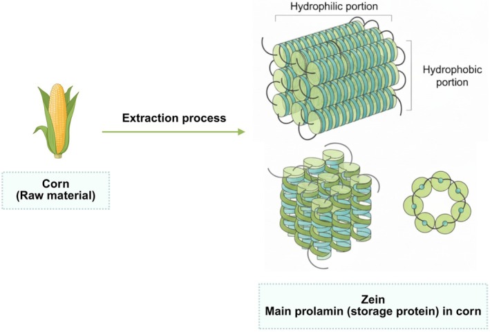 FIGURE 1
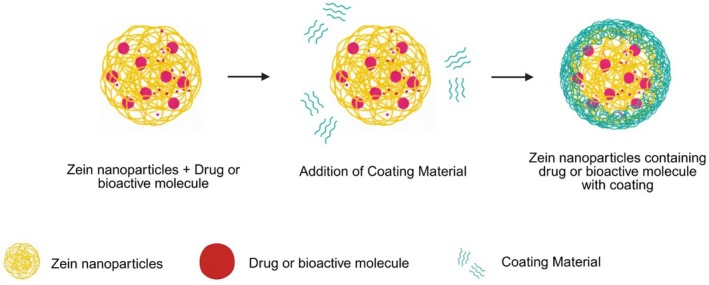 FIGURE 2
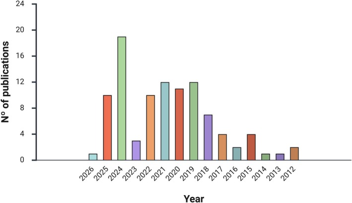 FIGURE 3
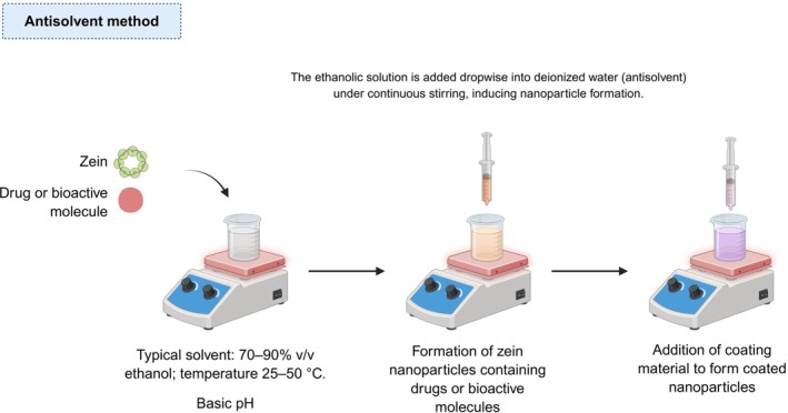 FIGURE 4
| Synthesis method | Drug | Coating | Particle size (nm) | Encapsulation efficacy (%) | References |
|---|---|---|---|---|---|
| Antisolvent | Benznidazole | Eudragit L100‐55 | 209 | 36–60 | Pilicita et al. [ |
| Antisolvent | Cinnamaldehyde and naringin | Lactoferrin and sodium caseinate | 268.5 | 73.61–79.54 | Mohamed et al. [ |
| Antisolvent | Colchicine | Eudragit S100 | 104.3 | 59.8 | Taymouri et al. [ |
| Antisolvent | Curcumin | Dextran sulphate | 180 | 96 | Albogamy et al. [ |
| Antisolvent | Curcumin | Alginate oligosaccharides | 116.5 | 89.7 | Jiang et al. [ |
| Antisolvent | Curcumin | Tremella polysaccharides | 164.0–225.2 | 93.34 | Li et al. [ |
| Antisolvent | Curcumin | Dextran sulfate | 135 | 85.37 | Yuan et al. [ |
| Antisolvent | Curcumin | Xanthan gum | 179 | 60–80 | Zhang et al. [ |
| Antisolvent | Curcumin | Ethanol‐soluble polysaccharide | 253–266 | 88.7%–80.4% | Zhang et al. [ |
| Antisolvent | Curcumin | Silk sericin | 330–400 | 80–90 | Zhu et al. [ |
| Antisolvent | Curcumin | Carboxylic curdlans | 183 | 66.7 | Yu et al. [ |
| Antisolvent | curcumin | Carboxymethyl cellulose | 277.5 | 70 | Öztekin et al. [ |
| Antisolvent | Curcumin | Chitosan and genipin | 185–190 | 82.3 | Sha et al. [ |
| Antisolvent | Curcumin | Gum Arabic | 192.5–216 | 87–92.4 | Ye et al. [ |
| Antisolvent | Curcumin and piperine | Chitosan, hialuronic acid | 599 | 90.4 | Chen et al. [ |
| Antisolvent | Docetaxel and cynomorium songaricum polysaccharide | Green tea polyphenols/iron coordination complex | 274 | 45.0 | Yu et al. [ |
| Antisolvent | Eugenol | Chitosan | 100 | — | Ferreira et al. [ |
| Antisolvent | Fucoxanthin | Chitosan; soybean polysaccharide | 230 | 91 | Zhao et al. [ |
| Antisolvent | Gambogenic acid | Polydopamine | 312 | 82.18 | Zha et al. [ |
| Antisolvent | Guavinoside B | Caseinate | 176.2–178.7 | 84.11–88.70 | Yang et al. [ |
| Antisolvent | Honokiol | Polysialic acid | 107.2 nm | 79.2 | Zhang et al. [ |
| Antisolvent | Hypericin | Polietilenoglicol (PEG)/caseinates | 92.7 ± 3.0 | 57.8 | Abdelsalam et al. [ |
| Antisolvent | Hyperoside | Pectin | 272–298 nm | 33.2–90.5 | Wang et al. [ |
| Antisolvent | Magnolol | Chondroitin sulfate | 142.27–164.36 nm | 65.34–80.71 | Wang et al. [ |
| Antisolvent | Naringin | Glutamine (Gln)‐modified chito‐oligosaccharides | 116 | 88.25 | Guo et al. [ |
| Antisolvent | Niclosamide | Bovine serum albumin | 200 | — | Rejinold et al. [ |
| Antisolvent | OMV‐F4 e OMV‐F18 | Gantrez AN–mannosamine polymer | 220–280 | — | Matías et al. [ |
| Antisolvent | Only zein | Chitosan | 181 | — | Lima et al. [ |
| Antisolvent | Proanthocyanidins | Pectin | 404.35 | — | Li et al. [ |
| Antisolvent | Propolis | Carboxymethyl chitosan | 197–220 | 83 | Zhang et al. [ |
| Antisolvent | Quercetin | Polysaccharides | 200 | 82.5 | Li et al. [ |
| Antisolvent | Quercetin | Fucoidan | 211.7 | 80.54–90.83 | Zhang et al. [ |
| Antisolvent | Quercetin | Sodium caseinate and chitosan | 188 | 82.78 | Zhou et al. [ |
| Antisolvent | Quercetin | Trimethylated chitosan | 200–250 | 90 | Dai et al. [ |
| Antisolvent | Quercetin | Gelatin and carboxymethyl starch | 189–219.1 | 95.2 | Zhang et al. [ |
| Antisolvent | Quercetin | Alginate‐pectin | 298.6–475.8 | 86 | Wan et al. [ |
| Antisolvent | Quercetin | Chitosan | 199.50–225.47 | 59.36–74.95 | Liu et al. [ |
| Antisolvent | Resveratrol | Caseinate–dextran conjugates | 224 | — | Davidov‐Pardo et al. [ |
| Antisolvent | Tannic acid | Pectin | 166 | 89 | Liang et al. [ |
| Antisolvent | Tannic acid and resveratrol | Pectin | 166.8 | 51.5–77.2 | Liang et al. [ |
| Antisolvent | Tanshinone | Pectin | 132 | 79.41 | Elmizadeh et al. [ |
| Antisolvent | Thymol | Caseinates | 200 | — | Kang‐Kang et al. [ |
| Antisolvent | β‐caroteno | Carboxymethyl chitosan | 243.9 | 92.7 | Wang et al. [ |
| Antisolvent co‐precipitation | Curcumin | Ethylcellulose | 167 | 63 | Hasankhan et al. [ |
| Antisolvent co‐precipitation | Insulin | Carboxy‐ methylated short‐chain amylose | 168 | 90.5 | Ji et al. [ |
| Antisolvent co‐precipitation | Insulin | Chitosan | 311.32 | 89.6 | Ji et al. [ |
| Antisolvent nanoprecipitation | Ellipticine | Sodium caseinate and polyethylene imine complex | 137.6–166.7 | 94.8 | Pourhossein et al. [ |
| Antisolvent precipitation and electrophoretic deposition | Tannic acid | Copper‐doped bioactive glasses and sodium carboxymethyl cellulose | 213–255 | 99 | Hadzhieva et al. [ |
| Antisolvent precipitation and electrostatic deposition | Fusidic acid | Lactoferrin | 80.78 | 60.5 | Marey et al. [ |
| Antisolvent with modifications | Curcumin | Oxidized dextran | 150 | 90 | Rodriguez et al. [ |
| Atomizing/antisolvent precipitation | Docetaxel | Soy lecithin and carboxymethyl chitosan | 206.9 | 79.22 | Wu et al. [ |
| Co‐extrusion method | Paclitaxel | B16 cancer cell membrane | 95.4–289.99 | — | Huang et al. [ |
| Complex coacervation | Doxorubicin hydrochloride | Complex of metal–tannic acid | 156.4–181.7 | 91 | Liang et al. [ |
| Desolvation | Insulin | Gantrez AN‐thiamine conjugate | 258–345 | — | Inchaurraga et al. [ |
| Desolvation | Insulin | Poly(anhydride)‐thiamine conjugate | 250 | 80 | Inchaurraga et al. [ |
| Desolvation | Insulin | Gantrez AN‐PEG conjugate | 248 | 88.7 | Martínez‐López et al. [ |
| Desolvation | Insulin | Poly(ethylene glycol) | 263 | 84.8 | Reboredo et al. [ |
| Desolvation | Only zein | Poly(ethylene glycol) | 200 | — | Reboredo et al. [ |
| Desolvation | Only zein | Poly(ethylene glycol) 35,000 | 200 | — | Reboredo et al. [ |
| Desolvation/deslocation |
| Gantrez–mannosamine conjugate | 211 | 70 | Berzosa et al. [ |
| Electrostatic deposition | Curcumin | Alginate; carrageenan; pectin; gum arabic; carboxymethyl cellulose | 160–210 | 80 | Chang et al. [ |
| Electrostatic deposition | Curcumin | Pectin | 112–201.5 | 61.6–91.7 | Chang et al. [ |
| Electrostatic deposition | Lutein | Sophorolipid | 200 | 90 | Yuan et al. [ |
| Electrostatic deposition | Zein/caseinate complex | Pectin | 200 | — | Chang et al. [ |
| Emulsification‐solvent evaporation | Carbamazepine | Chitosan | 221.46 | 62.28 | Alak et al. [ |
| Ethanol‐injection | Atorvastatin | lecithin | 191.95 | 70 | Elgendy et al. [ |
| Ionic complexation | Doxorubicin | Tannic acid/FeII; tannic acid/AlIII; tannic acid/EuIII | 135.9–145.9 | 78.4–83.1 | Liang et al. [ |
| Ionic complexation | Doxorubicin | (PEG)/tannin acid (TA) complex | 221.3–275.7 | 81.8–88.6 | Liang et al. [ |
| Liquid antisolvent precipitation | Resveratrol | Maillard conjugates of sodium caseinate and dextran | 180–198 | 83% | Davidov‐Pardo et al. [ |
| Liquid–liquid dispersion | Curcumin | Sodium caseinate and Sodium alginate complex | 185.63–191.10 | 36.10–76.06 | Liu et al. [ |
| Liquid–liquid dispersion | Resveratrol | Chitosan | 295 | 51 | Pauluk et al. [ |
| Liquid–liquid dispersion | Thymol | Caseinates | 398.90–521.80 | 80.51–82.89 | Zhang et al. [ |
| Liquid–liquid phase separation | indole‐3‐carbinol (I) or 3,30‐diindolylmethane (D) | Chitosan | 113.5–89.1 | 77.79–78.08 | Luo et al. [ |
| Liquid–liquid phase separation | Simvastatin | Caseinate | 131 | 89 | Ahmed et al. [ |
| Low‐energy phase separation | Curcumin | Water‐soluble chitosan | 66–170 | 94.9 | Liang et al. [ |
| Low‐energy phase separation | Vitamin D3 | Carboxymethyl chitosan | 200 | 87.9 | Luo et al. [ |
| Nanoprecipitation | — | Sodium deoxycholate | 100–200 | — | Gagliardi et al. [ |
| Nanoprecipitation | Genistein | Tretinoin | 154.5 | — | Kamel et al. [ |
| Nanoprecipitation | Outer membrane vesicles (OMV) obtained from | Gantrez‐mannosamine polymer conjugate | 132 | — | Matías et al. [ |
| Nanoprecipitation | Paclitaxel | Sodium deoxycholate | 103–158 | 40 | Gagliardi et al. [ |
| Nanoprecipitation | Resveratrol | Hydrophilic pectin and Eudragit S 100 | 100–200 | 94 | Contado et al. [ |
| Nanoprecipitation | Ceftazidime and tobramycin | Chitosan | 315–335 | 55 | Campos et al. [ |
| New method | Folic acid and caffeic acid | Chitosan | 176.3 | CA: 15.1–64.1|FA: 5.9–84.1 | Wusigale et al. [ |
| Oxidative self‐polymerization | Doxorubicin | Tannic acid | 155.8 | 68.6 | Liang et al. [ |
| pH‐driven | curcumin | Shellac | ≅30 | 96 | Lv et al. [ |
| pH‐driven | Curcumin | Quaternary ammonium chitosan | 218.2 | 89.3 | Liu et al. [ |
| pH‐driven | Quercetin | Auricularia cornea Ehrenb polysaccharides | 185.6 | 75.8 | Wang et al. [ |
| Phase separation | Exemestane and luteolin | PEGylated phospholipids, lactoferrin | 229.5–500.9 | 61.8–88.3 | El‐Lakany et al. [ |
| Phase separation | Resveratrol | Caseinates | 100 | 81.42 | Shaomin et al. [ |
| Phase separation | Curcumin | Polydopamine | 53.2–150 | 36–79 | Zhang et al. [ |
| Phase separation | Resveratrol | Chitosan | 94–132 | 91 | Khan et al. [ |
| Sacrificial template | Astaxanthin | Chitosan‐α‐lipoic acid copolymer and sodium alginate | 239.07 | 88.13 | Wang et al. [ |
| Sacrificial template | Quercetin/doxorubicin | Chitosan | 218.8 | 67.32 | Da Paz Do Nascimento et al. [ |
| Thin‐film hydration | Terbinafine | Dextran sulphate | 273.2 | 9.2%–93.1% | Al‐Sawahli et al. [ |
| Ultrasonic‐antisolvent method | Curcumin | Carboxymethylated corn fiber gum | 158.17 to 380.93 | 91.19 | Ma et al. [ |
| Test conducted | Drug/coating | Results | Applicability of study | Author |
|---|---|---|---|---|
|
DPPH | Curcumin/ethyl cellulose |
Nanoparticles maintained approximately 50% antioxidant activity after 6 months of formulation. Free drug showed antioxidant activity lower than 20%. |
Food formulations Drug delivery | Hasankhan et al. [ |
| Tanshinone/pectin |
Higher antioxidant in T/Z NPs: Tanshinone in lipophilic T/Z NPs showed stronger DPPH* scavenging (77.35%) than in T/Z/P NPs (72.04%). Hydrophilic pectin coating in T/Z/P NPs slowed DPPH* penetration, limiting immediate antioxidant activity. Z/P NPs without tanshinone still exhibited moderate antioxidant activity (~45%) due to zein's inherent properties. | Antioxidant | Elmizadeh et al. [ | |
|
Curcumin/Alginate and NaCas |
Nanoencapsulation significantly enhanced curcumin's antioxidant activity, as shown by SC50 values. Ethanol‐dissolved curcumin had the worst antioxidant activity (SC50 = 8.79 μg/mL), equivalent to 67% of vitamin C (SC50 = 5.90 μg/mL). Nanoencapsulated curcumin (SC50 = 2.65 μg/mL) was 3.32 times stronger than vitamin C. SA/SC/Z nanoparticles effectively improved curcumin's antioxidant capacity by enhancing hydrophilicity and radical scavenging. |
Drug delivery Antioxidant enhancement | Liu et al. [ | |
| Thymol/sodium caseinate |
Nanoparticles without thymol showed < 20% DPPH* scavenging activity. As the thymol‐to‐zein ratio increased from 0% to 40%, the DPPH* scavenging activity increased from 25% to 52%. |
Antioxidant preservation in packaging Drug delivery | Kang‐Kang et al. [ | |
| Curcumin/chitosan |
Cur/zein–HTCC nanoparticles showed enhanced antioxidant activity. At a 1:1 zein–HTCC ratio, DPPH scavenging increased by:
13.3% (pasteurization at low temperature) 26.9% (pasteurization at high temperature) 29.0% (UV exposure) Encapsulation improved stability against heat and UV degradation. |
Drug delivery Antioxidant therapy | Liang et al. [ | |
| Curcumin/dextran |
Antioxidant activity increased with concentration for both free and encapsulated curcumin. Encapsulated curcumin had higher DPPH radical scavenging efficiency than the free form. Dextran‐zein nanoparticles enhanced curcumin's antioxidant capacity by protecting its structure. Encapsulation reduced heat and light degradation, allowing lower curcumin doses for DPPH* reduction. |
Drug delivery Antioxidant enhancement | Albogamy et al. [ | |
| Naringin/Glutamine (Gln)‐modified chito‐oligosaccharides |
ZN NPs (without GCS): ~50% free radical scavenging at 500 μg/mL GZN NPs (with GCS): > 50% scavenging at 100 μg/mL Nar solution (free form): requires 25 mM (~6810 μg/mL) to reach 50% scavenging | Anti‐obesity therapy | Guo et al. [ | |
| Quercetin/chitosan and shellac |
Quercetin‐loaded NPs showed greater DPPH* and ABTS** radical scavenging capacity than free quercetin, indicating improved antioxidant potential. ZS and ZSC NPs exhibited notable antioxidant properties, likely due to the inherent antioxidant capacity of zein and chitosan. Chitosan‐coated NPs had lower radical scavenging efficiency compared to uncoated NPs | Antioxidant | Liu et al. [ | |
| ABTS+ | Folic acid and caffeic acid/chitosa |
Antioxidant capacity of CS‐HZ before irradiation was 9.0 μg/mL VC CS‐HZ/CA (56.9 μg/mL VC) showed weaker antioxidant capacity than the CA control (61.6 μg/mL VC), suggesting encapsulation partially masked the antioxidant capacity. Ranking of antioxidant capacity of particles: CS‐HZ/CA > CS‐HZ/FA + CA > CS‐SZ/CA > CS‐SZ/FA + CA, according to the encapsulation efficiency (EE) of CA. The more CA released, the stronger the antioxidant capacity. Antioxidant capacity increased after 240 min due to the higher antioxidant capacity of CA's photoproduct, esculetin, compared to CA | Cancer treatment | El‐Lakany et al. [ |
| Quercetin/soluble soybean polysaccharide |
Zein, SSPS, and zein/SSPS nanoparticles neutralized 5.8%, 4.95%, and 9.13% of ABTS+, respectively. Encapsulation of |
Antioxidant Cancer treatment | Li et al. [ | |
| DPPH, ABTS+, and FRAP | Curcumin/carboxylic curdlan |
DPPH*:
Free curcumin had the lowest IC50 (7.23 μg/mL), outperforming encapsulated curcumin (106.3–174.3 μg/mL) and the positive control (33.0 μg/mL). Among encapsulated samples, Cur‐24‐ZC showed the best antioxidant efficiency (IC50 = 106.3 μg/mL), linked to high EE and better dispersibility. ABTS+:
ABTS radical scavenging followed the DPPH trend, with significant differences between free and encapsulated curcumin ( FRAP:
Free curcumin had a higher FRAP value (~2005.8 μmol Fe2+/g) than encapsulated forms and the positive control. Cur‐24‐ZC had the highest FRAP value (~200.6 μmol Fe2+/g) among encapsulated samples, supporting DPPH and ABTS results. | Antioxidant | Yu et al. [ |
| DPPH, TEAC, and FRAP | Propolis/chitosan |
DPPH (200 μg/mL):
Propolis‐loaded zein/CMCS nanoparticles: 79.69% Propolis in ethanol: 88.01% propolis in water: 16.62% Empty zein/CMCS nanoparticles: 44.04% TEAC*:
Propolis‐loaded zein/CMCS nanoparticles: 7902.12 ± 299.78 μmol Trolox/g Propolis in ethanol: 8522 ± 692.62 μmol Trolox/g Empty nanoparticles exhibited 2022 ± 152.23 μmol Trolox/g, suggesting zein/CMCS functional groups contribute to antioxidant activity. FRAP:
Encapsulated propolis: 2647.01 ± 174.57 μmol Fe2+/g Propolis in ethanol: 611.34 ± 32.6 μmol Fe2+/g Encapsulation enhanced antioxidant activity by protecting propolis and improving its interaction with Fe3+‐TPTZ. | Antioxidant applications in food and pharmaceuticals | Zhang et al. [ |
|
DPPH and ABTS+ | Quercetin/gelatin and carboxymethyl starch |
DPPH:
Free quercetin (in ethanol): 82.8% Zein nanoparticles (blank): low but present due to sulfur‐containing amino acids and tryptophan Zein‐Que NPs: increased compared to free quercetin Zein/Gel/CMS‐Que NPs: highest activity, superior to Zein‐Que NPs, due to the synergistic effect of CMS, gelatin, and zein ABTS:
Free quercetin: 65.2% Zein/Gel/CMS‐Que NPs: higher than Zein‐Que NPs, attributed to a favorable microenvironment for hydrogen and electron donation Zein and Zein/Gel/CMS (blank): slight activity, attributed to amino acids and minerals present | Antioxidant therapy | Zhang et al. [ |
| Quercetin/auricularia cornea Ehrenb polysaccharides |
DPPH:
ZAQ‐4 NPs showed higher radical scavenging than free quercetin (F‐Q) before GIT (IC50: 36.07 vs. 41.66 μg/mL) and after simulated GIT (48.8% vs. 29.07%). ABTS:
ZAQ‐4 NPs had greater scavenging than F‐Q before GIT (IC50: 114.5 vs. 132.4 μg/mL) and after GIT (37.62% vs. 16.69%). | Anti‐inflammatory | Wang et al. [ | |
| Quercetin/Shellac |
DPPH:
Encapsulated quercetin NPs > free quercetin in radical scavenging. ZS and ZSC NPs showed inherent antioxidant activity. Chitosan‐coated NPs had lower immediate scavenging due to slower quercetin release. ABTS:
Encapsulated quercetin NPs > free quercetin in radical scavenging. ZS and ZSC NPs contributed to antioxidant activity. Chitosan coating reduced the rate of ABTS radical scavenging. | Antioxidant | Liu et al. [ | |
| GuavinosideB/caseinate |
DPPH:
GUB‐Z‐N NPs showed strong radical scavenging, slightly lower than free GUB; blank Z‐N NPs had negligible activity. ABTS:
GUB‐Z‐N NPs exhibited higher scavenging than free GUB, especially at higher concentrations; blank Z‐N NPs also showed some activity due to NaCas. | Antioxidant | Yang et al. [ | |
| Curcumin/quaternary ammonium chitosan |
DPPH:
CUR‐ZE‐SC and CUR‐ZE‐SC@HACC NPs showed higher scavenging (57.2% and 65.9%) than free CUR (48.8%); blank carriers also contributed slightly (~8%–15%). ABTS:
CUR‐ZE‐SC@HACC NPs exhibited strongest scavenging (76.4%), surpassing CUR‐ZE‐SC (54.3%) and free CUR (39.8%); blank carriers showed moderate activity (~9%–35%). | Liu et al. [ | ||
| Resveratrol or tannic acid/pectin |
DPPH:
Tannic acid‐loaded nanoparticles had higher antioxidant activity (SC50 = 3.02 μg/mL) than ascorbic acid (SC50 = 5.17 μg/mL). Resveratrol‐loaded nanoparticles (SC50 = 9.70 μg/mL) outperformed free resveratrol (SC50 = 35.81 μg/mL). Co‐loaded TA + resveratrol nanoparticles (SC50 = 4.41 μg/mL) showed a synergistic effect, enhancing resveratrol's activity. ABTS+:
No significant difference was found between encapsulated TA and co‐encapsulated TA + resveratrol. |
Cancer treatment Antioxidant enhancement | Liang et al. [ | |
| Guavinoside B/sodium caseinate |
DPPH:
GUB‐Z‐N NPs demonstrated antioxidant activity comparable to ascorbic acid, with an IC50 of 20.89 μM. ABTS+:
Encapsulated GUB showed significantly higher ABTS + scavenging activity than free GUB | Antioxidant | Yang et al. [ | |
| Resveratrol/chitosan |
ABTS+:
Resveratrol neutralized ABTS+· more effectively than HZ and HZ‐CH particles. DPPH:
Encapsulation in HZ and HZ‐CH improved DPPH· radical neutralization by increasing solubility. |
Antioxidant therapy Drug delivery | Khan et al. [ |
| Microorganism | Drug/coating | Results | Applicability of study | Author |
|---|---|---|---|---|
|
| Thymol/sodium caseinate |
Films without thymol (ZP0) and with low thymol content (ZP1) showed no significant inhibition halos against tested pathogens, with bacterial colonies growing on ZP0 films. Films with zein‐SC nanoparticles and thymol‐to‐zein ratios of 30%–40% (ZP3 and ZP4) exhibited visible inhibition halos, indicating antimicrobial activity. The diameter of inhibition halos increased with higher thymol loading, ranging from 16 to 18 mm, demonstrating enhanced antimicrobial efficacy. | Development of antimicrobial packaging films | Kang‐Kang et al. [ |
|
| Curcumin/Dextran |
DSZCNPs demonstrated significant antibacterial activity, with CFU reduction ranging from 53.74% to 97.53% against gram‐positive bacteria and 52.35%–71.42% against gram‐negative bacteria, compared to free curcumin. Both free curcumin and DSZCNPs showed similar effects against Thymol (free and encapsulated) showed significant inhibition only against Thymol encapsulated in CHC‐SC coated nanoparticles was effective against The SC/CHC mass ratio of 1:4 in encapsulated thymol nanoparticles exhibited the longest and most significant inhibitory effect throughout the testing period ( | Drug delivery systems, enhanced antimicrobial activity | Albogamy et al. [ |
|
| Thymol/sodium caseinate and chitosan |
DSZCNPs demonstrated significant antibacterial activity, with CFU reduction ranging from 53.74% to 97.53% against gram‐positive bacteria and 52.35%–71.42% against gram‐negative bacteria, compared to free curcumin. Both free curcumin and DSZCNPs showed similar effects against Thymol (free and encapsulated) showed significant inhibition only against Thymol encapsulated in CHC‐SC coated nanoparticles was effective against The SC/CHC mass ratio of 1:4 in encapsulated thymol nanoparticles exhibited the longest and most significant inhibitory effect throughout the testing period ( | Antimicrobial applications in wound care and food preservation | Zhang et al. [ |
|
| Tannic acid/Copper‐doped bioactive glasses and Sodium carboxymethyl cellulose |
Zein/CMC + CuBG: No inhibition observed. Zein/CMC + CuBG + TA: Significant reduction in metabolic activity. TA release: 51 ± 11 μg/mL.
Zein/CMC + CuBG: No inhibition observed. Zein/CMC + CuBG + TA: Significant reduction in metabolic activity. TA release: 51 ± 11 μg/mL. | Antibacterial therapy | Hadzhieva et al. [ |
|
| Ceftazidime and tobramycin/chitosan |
Antibacterial activity (MIC/MBC):
CAZ: MIC 12.5–> 50 μg/mL; MBC 25–> 50 μg/mL. TOB: MIC 6.25–12.5 μg/mL; MBC 12.5–50 μg/mL. CAZ–ZNP–CH: MIC 3.12–12.5 μg/mL; MBC 12.5–25 μg/mL. TOB–ZNP–CH: MIC 1.56–3.12 μg/mL; MBC 6.25–25 μg/mL. CAZ–TOB–ZNP–CH: MIC 0.19–3.12 μg/mL (CAZ) and 0.15–2.40 μg/mL (TOB); MBC 1.56–6.25 μg/mL (CAZ) and 1.21–4.87 μg/mL (TOB). ZNP–CH showed no antibacterial activity (> 50 μg/mL). Antibiofilm activity (inhibition + eradication):
Biofilm inhibition (MIC → MIC/16): CAZ (5%–80%), TOB (4%–88%), CAZ–ZNP–CH (49%–93%), TOB–ZNP–CH (53%–100%), CAZ–TOB–ZNP–CH (69%–100%). MBIC: Encapsulated drugs < free drugs; CAZ–TOB–ZNP–CH shows 10–35× lower MBIC values than single‐drug NPs. Biofilm eradication (MIC → 16 × MIC): CAZ (18%–68%), TOB (27%–79%), CAZ–ZNP–CH (40%–81%), TOB–ZNP–CH (43%–84%), CAZ–TOB–ZNP–CH (58%–92%). MBEC: Encapsulated drugs < free drugs; CAZ–TOB–ZNP–CH shows 8–300× lower MBEC values. ZNP–CH showed no antibiofilm activity. | Antibacterial therapy | Campos et al. [ |
| Cell type used | Drug/coating | Results | Applicability of study | Author |
|---|---|---|---|---|
| Ehrlich ascites carcinoma, MCF‐7*, 4T1* | Exemestane and luteolin/PEGylated phospholipids and lactoferrin |
PEGylated nanospheres improve cytotoxicity against MCF‐7 (IC50 = 6.6 μg/mL) and 4TI breast cancer cells (IC50 = 5.07 μg/mL). PEGylation improves antitumor efficacy and pharmacokinetics compared to free‐drug |
Hepatocellular carcinoma treatment | El‐Lakamy et al. [ |
| HepG2* | Hypericin/PEG or NaCas |
In dark conditions, both free and zein‐loaded hypericin showed no significant toxicity to HepG2 and L929 cells. However, upon irradiation, cell viability decreased in a dose‐dependent manner, with the strongest cytotoxic effects observed at 1.5 J/cm2. The PEGylated zein formulation (Z‐PEG) demonstrated the highest cytotoxicity in HepG2 cells at 1.5 J/cm2 while maintaining a safer profile in non‐cancerous L929 cells, indicating improved cancer cell targeting and reduced off‐target effects. |
Hepatocellular carcinoma treatment | Abdelsalam et al. [ |
| Curcumin/Dextran |
Great stability in time (100 days) and protects from degradation in gastric environment with controlled curcumin release Curcumin concentrations: 10–80 μg/mL DSZCNPs showed higher cytotoxicity than free curcumin at low concentrations (10, 20, 40 μg/mL). IC50 values:
Free curcumin: 50 μg/mL. DSZCNPs: 13 μg/mL → more effective. |
Hepatocellular carcinoma treatment | Albogamy et al. [ | |
| Doxorubicin/Chitosan and metal–tannic acid |
DOX‐loaded metal–TA‐Coated NPs had lower cytotoxicity than DOX‐loaded Zein/CMCS NPs IC50 values:
DOX‐NPs: 2.36 ± 0.18 μg/mL DOX‐NPs‐TA/Cu2+: 2.91 ± 0.12 μg/mL DOX‐NPs‐TA/Ca2+: 2.67 ± 0.28 μg/mL DOX‐loaded zein/CMCS NPs exhibited higher cytotoxicity than metal–TA‐coated NPs due to their faster drug release and efficient cellular uptake. DOX‐loaded metal–TA‐coated NPs displayed a more controlled drug release profile, leading to a delayed but sustained cytotoxic effect. |
Hepatocellular carcinoma treatment | Liang et al. [ | |
| Tannic acid and resveratrol/pectin |
Encapsulated tannic acid was the most cytotoxic presenting an IC50 = 8.36 ± 0.22 μg/mL, making it 6× more toxic than encapsulated resveratrol. Co‐encapsulation of tannic acid and resveratrol resulted in intermediate toxicity, presenting an IC50 = 10.07 ± 0.23 μg/mL, similar to encapsulated tannic acid, suggesting that tannic acid played a dominant role in cytotoxicity. |
Hepatocellular carcinoma treatment | Liang et al. [ | |
| Gambogenic acid/polydopamine |
GNA@Zein‐PDA particles had the strongest cytotoxic effect. IC50 values:
Free GNA: 9.89 μg/mL GNA@Zein NPs: 4.42 μg/mL GNA@Zein‐PDA NPs: 1.59 μg/mL |
Hepatocellular carcinoma treatment | Zha et al. [ | |
| A549*, K562* |
Sodium deoxycholate (coating only) |
Adding surfactant sodium deoxycholate (1.25% w/v) provides high storage and thermal (up to 50°C) stability, resistance to pH alterations and freeze‐drying process. A549 cells:
Surfactant‐free zein nanoparticles induced cytotoxicity only after 72 h at 100 μg/mL and reduced cell viability by 25%–30% Surfactant‐coated zein nanoparticles showed significant toxicity at 50 μg/mL after just 24 h. K562 cells:
Cell viability gradually decreases as the concentration of zein nanoparticles increases, with higher concentrations (> 10 μg/mL) exhibiting a more pronounced effect. Nanoparticles incorporating surfactants (T80, PLX188, and SD) demonstrate greater toxicity compared to surfactant‐free formulations. | Safety evaluation of ZNPs in cancer treatments | Gagliardi et al. [ |
| MCF‐7*, K562 | Paclitaxel/sodium deoxycholate |
PTX‐loaded ZNPs enhanced cytotoxicity against cancer cells Higher cytotoxicity observed in nanoencapsulated PTX compared to free PTX. MCF‐7 cells exhibited a faster response to PTX‐loaded zein NPs, whereas K562 cells showed stronger inhibition only after 72 h. At a concentration of 1 μM, cell viability reached its minimum point. | Breast cancer and leukemia treatment | Gagliardi et al. [ |
| Caco‐2 cells* | Insulin/chitosan | After 24 h incubation:
IN‐Z‐CSA1.0%: 5.64% apoptosis. IN‐Z‐CSA/CS0.2%: 7.36% apoptosis. | Cellular uptake studies for drug delivery | Ji et al. [ |
| Quercetin/Trimethylated chitosan |
Zein‐Q and TMC‐Zein‐Q enhanced Caco‐2 cell viability compared to free quercetin, indicating improved biocompatibility and reduced cytotoxicity. TMC‐Zein‐Q significantly increased cellular uptake of quercetin, attributed to enhanced endocytosis induced by the TMC shell. | Dai et al. [ | ||
| HeLa cells | Doxorubicin/Tannic acid | IC50 values:
Free DOX: 0.918 ± 0.05 μg/mL DOX‐zein NPs: 0.939 ± 0.04 μg/mL DOX‐zein/TA NPs: 1.087 ± 0.02 μg/mL TA‐coated NPs showed slower DOX release, leading to reduced intracellular accumulation. | Cervical cancer treatment | Liang et al. [ |
| RAW 264.7 macrophages* | Outer membrane vesicles obtained from Enterotoxigenic |
The tested outer membrane vesicles (OMV) at concentrations ranging from 7.5 to 120 μg/mL did not exhibit cytotoxicity, as cell viability remained close to 100%. OMVs at 10, 40, and 160 μg/mL significantly increased nitric oxide production in macrophages suggesting antigenic response | Immune response and macrophage interaction studies | Matías et al. [ |
| A549, NIH‐3 T3 cells | Ellipticine/NaCas or polyethylene imine |
A549:
Zein/SC‐EPT: 54%–81% cytotoxicity (0.49–7.81 μg/mL). Zein/PEI‐EPT: 61%–92% cytotoxicity (0.49–7.81 μg/mL). Control EPT (water, 7.81 μg/mL): ~60% cytotoxicity. NIH‐3T3:
EPT‐loaded nanoparticles increased toxicity to cancer cells but spared NIH‐3T3 cells. | Lung cancer treatment | Pourhossein et al. [ |
| 293*, A549 cells | Resveratrol/polydopamina and casein |
Zein‐polydopamine‐casein core–shell nanocomposites (ZPC) increase their uptake in cells compared to unmodified nanoparticles (from 2.79% to 97.66%). The cytotoxicity of ZPCs was tested on 293 and A549 cells, and no toxicity was observed at any of the tested concentrations (10–1000 μg/mL). The cytotoxicity of RES was assessed on A549 cells, with no inhibitory effect observed at concentrations below 100 μM. At higher concentrations (> 100 μM) The cytotoxicity of RES‐ZPCs was evaluated on A549 cells. At higher RES concentrations (> 100 μM), cell viability decreased to approximately 60% at 200 μM. The cytotoxicity of RES‐ZPCs was lower than that of RES alone. ROS scavenging efficiency of RES‐ZPCs was 100% at 50 μM. | Biocompatibility and antioxidant studies | Shaomin et al. [ |
| HT29 cells* | Curcumin/ethyl cellulose |
Cytotoxicity was evaluated at concentrations between 1.25 and 20 μg/mL−1 The nanoparticle formulation (NPs of CU) exhibited lower cytotoxicity compared to free CU. | Colon cancer treatment | Hasankhan et al. [ |
| NCM 460*, RAW 264.7 | Magnolol/chondroitin sulfate |
Safe doses in both cell lines are 5–20 μM and 1–10 μM in NCM 460 and RAW 264.7 cells, respectively. Significantly reduced the levels of TNF‐a, IL‐6, and IL‐1β (proinflammatory factors) and increased the level of IL‐10 (anti‐inflammatory factor) in NCM 460 and RAW 264.7 cells along with an increase in cellular uptake. | Ulcerative colitis treatment | Wang et al. [ |
| MCF‐7, SKOV‐3 cells | Docetaxel/soy lecithin and chitosan |
MCF‐7:
Unloaded FZLC: Showed no cytotoxic effects at any concentration up to 48 h. Free DTX: Induced moderate cytotoxicity, with only a slight increase as the concentration increased. DTX/FZLC NPs: Exhibited significantly higher cytotoxicity than raw DTX, particularly at higher concentrations. SKOV‐3:
Unloaded FZLC: Did not affect cell viability at any tested concentration. Free DTX: Displayed limited cytotoxic effects, with only a slight increase at higher concentrations. DTX/FZLC NPs: Showed a pronounced increase in cytotoxicity compared to raw DTX, especially at elevated doses and prolonged incubation. | Cancer treatment (breast cancer, ovarian cancer) | Wu et al. [ |
| NCM460 cells* | Curcumin/Dextran sulfate |
The IC50 values for ZNPs and ZDSNPs were 9.1 × 10⁴ μg/mL and 2.1 × 10⁵ μg/mL, respectively, indicating minimal toxicity. | Biocompatibility study | Yuan et al. [ |
| Lutein/sophorolipid |
Nanoparticles were tested at 100, 50, 25, 12.5, and 6.25 μg/mL, with lutein ranging from 0.82 to 0.05 μg/mL. All formulations maintained viability above 80%, confirming low toxicity. Free lutein showed slightly lower cell viability compared to nanoparticle formulations, especially at 100 and 50 μg/mL. | Biocompatibility and drug delivery studies | Yuan et al. [ | |
| C6 glioma cells* | Curcumin/dodecamer peptide (G23)‐functionalized polydopamine |
CUR‐ZpD NPs showed stronger inhibition of C6 glioma cells compared to free curcumin, likely due to enhanced cellular uptake. At 5 μg/mL of curcumin, cell viability dropped to 32% with CUR‐ZpD NPs and 25% with CUR‐ZpD‐G23 NPs CUR‐ZpD and CUR‐ZpD‐G23 NPs further suppressed colony formation, reducing survival to 18% ± 5% (CUR‐ZpD NPs) and 17% ± 6% (CUR‐ZpD‐G23 NPs) Cell migration was significantly reduced in curcumin‐treated cells compared to untreated controls:
Control: 75.0% ± 9.4% migration Curcumin‐treated: 35.6% ± 8.7% migration CUR‐ZpD NPs: 21.8% ± 6.6% migration CUR‐ZpD‐G23 NPs: 23.9% ± 6.9% migration | Glioma treatment | Zhang et al. [ |
| Caco‐2*, HT29‐MTX cells* | Insulin/PEG |
No cytotoxic effect was observed for either bare or PEG‐coated insulin‐loaded nanoparticles. All tested concentrations (1.7–70 μg/mL of insulin) maintained cell viability after 24 h of incubation. | Intestinal drug delivery systems | Reboredo et al. [ |
| L929, A549, HeLa, B16* | Paclitaxel/B16 cancer cell membrane |
Different concentrations of PTX concentrations: 4.50, 2.00, 0.89, 0.40, and 0.18 μg/mL Cell viability at minimum drug concentration:
A549: 87.11% Hela: 71.4% B16: 55.72% Cell viability at highest drug concentration:
A549: 50.22% Hela: 54.04% B16: 33.47% L929 cell viability remained above 90%, indicating minimal cytotoxic effect and that CCM‐α‐Zein exhibits good biocompatibility with non‐tumor cells | Cancer treatment | Huang et al. [ |
| HepG2 and AML12* | Celastrol/Hyaluronic acid |
HepG2 IC50 values:
Free Cel: 14.34 μM Cel/Zein NPs: 10.19 μM Cel/Zein@HA NPs: 6.36 μM AML12 cells
Viability remained above 80% after 48‐h co‐incubation with Cel/Zein@HA NPs |
Hepatocellular carcinoma treatment | He et al. [ |
| HK‐2* | Resveratrol/Hyaluronic acid |
Cell viability of HK‐2 renal tubular endothelial cells treated with Zein/Res NPs and HA‐Zein/Res NPs was greater than 90% at a concentration of 200 μg/mL | Reduction of cisplatin‐associated nephrotoxicity | Ning et al. [ |
| Caco‐2 | Curcumin/Carboxymethylated corn fiber gum |
No cytotoxicity was detected at 20 mg/mL for CFG and all CMCFG samples, with cell viability remaining at ~100%. Increasing concentrations (30–50 mg/mL) reduced viability in all groups, with CFG showing the greatest decrease while CMCFGs maintained higher viability. Higher toxicity of CFG at 40–50 mg/mL was likely due to increased osmotic pressure compared to CMCFG solutions. | Hydrophobic nutrient delivery | Ma et al. [ |
| MDA‐MB‐231* | Cinnamaldehyde and naringin/Lactoferrin and sodium caseinate |
Cell viability decreased in a concentration‐dependent manner for all treatments, with the lowest viability (~6%) observed when 40 μM DOX was combined with 25 μM dual drug‐loaded NPs. Dual drug‐loaded NPs enhanced DOX cytotoxicity, showing lower IC50 values than blank NPs and the free‐drug combination. Combination index analysis indicated that the DOX/dual drug‐loaded NP treatment achieved the strongest synergistic effect (CI = 0.968). | Enhance the gastric protection, bioavailability, and antioxidant efficacy | Mohamed et al. [ |
| MCF‐7 | Quercetin and Doxorubicin/Chitosan |
DOX showed strongly pH‐dependent release, with much higher release at pH 7.4 (~96%) than 6.8 (~68%), while QUE displayed similarly low release in both (~35%–38%). In combined release, QUE exhibited a slight reduction, and pH 6.8 limited the diffusion of both drugs due to reduced hydrogel swelling. OSAGX enhanced DOX cytotoxicity by 4.66×, OSAGC by 20.7×, while QNP alone showed no cytotoxic effect. | Localized breast cancer therapy | Da Paz Do Nascimento et al. [ |
| HepG2 | Quercetin/Alginate‐pectin |
Digested quercetin‐loaded nanoparticles significantly improved intracellular antioxidant defenses in H2O2‐treated HepG2 cells, with SOD activities markedly higher than those of digested quercetin in physical mixtures (PM), especially for ZQ5 and ZQ9. CAT activity was also enhanced by the nanoparticles, reaching levels close to untreated control cells, while digested PM showed a smaller increase in enzyme activity. These results indicate that nanoparticle delivery of quercetin effectively reduces oxidative stress and restores intracellular enzymatic antioxidant activities in HepG2 cells compared with quercetin in physical mixtures. | Nutraceutical bioavailability enhancement | Wan et al. [ |
| RAW264.7 | Astaxanthin/Chitosan‐α‐lipoic acid copolymer and sodium alginate |
HZ‐CL‐SA nanoparticles showed no significant cytotoxicity in RAW264.7 cells, indicating safe delivery of AST to target cells, while free AST was non‐toxic at 0.5–20 μg/mL but reduced viability at higher doses (> 20 μg/mL). AST at 10 μg/mL significantly protected RAW264.7 cells from LPS‐induced inflammation, restoring cell viability to 97.7% compared with the LPS‐treated group. AHZ‐CL‐SA nanoparticles effectively inhibited LPS‐induced inflammatory markers (NO, TNF‐α, IL‐1β, IL‐6) more strongly than free AST, with GSH‐pretreated nanoparticles showing enhanced anti‐inflammatory activity due to accelerated AST release. | Colon‐targeted nutraceutical delivery | Wang et al. [ |
| PC3, DU154, and LNCa | Terbinafine/Dextran Sulphate |
TRB‐DSZN nanoparticles significantly enhanced antiproliferative activity in PC3 prostate cancer cells, reducing IC50 to 13.28 μg/mL compared with 163.4 μg/mL for free TRB and 45.66 μg/mL for blank DSZN NSs, while showing weak cytotoxicity in non‐cancerous EA.hy926 cells. TRB‐DSZN NSs promoted cell cycle arrest in G0/G1 and S phases and increased apoptosis, as indicated by annexin‐V staining, compared with free TRB and blank DSZN NSs. Treatment with TRB‐DSZN NSs upregulated pro‐apoptotic genes (CASP3, P53) and ROS production, while downregulating anti‐apoptotic genes (CDK1, CDK7, CDK9), demonstrating enhanced apoptosis and oxidative stress in PC3 cells compared with controls. | Prostate cancer therapy | Al‐Sawahli et al. [ |
| MC3T3‐E1 (99072810‐1VL) | Tannic acid/Copper‐doped bioactive glasses and Sodium carboxymethyl cellulose |
Zein/CMC, zein/CMC/CuBG, and zein‐TA/CMC/CuBG coatings showed good cytocompatibility with MC3T3‐E1 preosteoblasts, as WST‐8 assays indicated no significant differences in cell viability after 24 h. The addition of TA to CuBG‐containing coatings synergistically improved cell viability, with zein‐TA/CMC/CuBG performing better than zein/CMC/CuBG. Fluorescence microscopy and SEM images confirmed that all coatings were non‐toxic after 72 h, with cells exhibiting well‐spread morphology and normal growth. | Bone regeneration | Hadzhieva et al. [ |
| HepG‐2 | Naringin/Glutamine (Gln)‐modified chito‐oligosaccharides |
GZN nanoparticles showed no cytotoxicity in HepG2 cells after 12 and 24 h, demonstrating good biocompatibility. Treatment with GZN NPs alleviated insulin resistance in HepG2 cells, restoring glucose uptake to 72% of normal levels at 50 μg/mL. GZN NPs reduced intracellular lipid accumulation in a concentration‐dependent manner, indicating potential to mitigate steatosis and abnormal lipid metabolism. The nanoparticles also decreased ALT and AST levels and lowered intracellular triglycerides and total cholesterol, suggesting protective effects on liver metabolism and cellular fat regulation. |
Food additives Pharmaceutical drug delivery | Guo et al. [ |
| HT‐29 | Colchicine/Eudragit S100 |
Col‐Z nanoparticles exhibited concentration‐dependent cytotoxicity in HT‐29 cells, comparable to free colchicine, while colchicine‐free Z NPs showed no significant inhibition of cell viability. Both Col‐Z NPs and free colchicine displayed lower cytotoxicity in HUVEC cells compared with HT‐29 cells, indicating selectivity toward cancer cells. Fluorescence microscopy demonstrated time‐dependent cellular uptake of Z NPs, with higher intensity after 3 h compared to 1 h. Col‐Z NPs showed low hemolytic activity (< 5%) and induced apoptosis in HT‐29 cells (42.9%), similar to free colchicine, confirming their hemocompatibility and anticancer potential. | Colorectal cancer treatment | Taymouri et al. [ |
| WI‐38 fibroblasts, HSF | Fusidic acid/Lactoferrin |
LF‐ZF nanoparticles significantly enhanced the viability of WI‐38 fibroblasts, demonstrating the cytocompatibility of zein and the proliferative effects of lactoferrin (LF). LF‐ZF‐NPs improved fibroblast adhesion compared to ZF‐NPs, likely due to LF's immunomodulatory effects and zein's interaction with tissue transglutaminase. Scratch wound assays showed accelerated migration and proliferation of fibroblasts with LF‐ZF‐NPs, resulting in higher wound closure rates at 24 and 48 h. These results indicate that LF‐ZF‐NPs may effectively promote fibroblast‐mediated wound healing, consistent with previous studies on lactoferrin in epithelial tissue repair. | Advanced chronic wound treatment. | Marey et al. [ |
| Model | Type | Drug/coating | Results | Applicability of study | Author |
|---|---|---|---|---|---|
| Mammalian |
Wistar rats | Insulin |
Nanoencapsulated insulin began reducing blood glucose after 4 h, with a peak effect at 9 h and returning to baseline by 20 h. Subcutaneous insulin had a faster onset, reaching its lowest blood glucose level within 2 h. | Oral drug delivery and diabetes treatment | Inchaurraga et al. [ |
| Insulin/PEG |
While subcutaneous insulin led to a rapid decrease in blood glucose (22% of initial levels within 2 h), I‐NP‐PEG provided a slower but sustained reduction, reaching a maximum decrease of 32% at 6 h, matching the glycemic levels of subcutaneous insulin. This suggests improved prolonged glucose regulation. PEG‐coated insulin nanoparticles exhibited a 3‐fold increase in pharmacological availability (15% vs. 4.7% for bare nanoparticles) and a 2.5‐fold increase in oral bioavailability (10.2% vs. 4.2%). Additionally, I‐NP‐PEG resulted in higher and more prolonged plasma insulin levels compared to non‐PEGylated formulations, demonstrating improved absorption and efficacy. | Oral insulin delivery and bioavailability enhancement | Reboredo et al. [ | ||
| Insulin/Chitosan |
Subcutaneous insulin rapidly reduced blood glucose to 29.4% at 2 h, but levels returned to baseline by 8 h. IN‐Z‐CSA/CS0.2% nanocomposites showed a more sustained reduction (38.8%) at 3 h, while IN‐Z‐CSA1.0% reached 49.4%. Nanocomposites provided gradual plasma insulin release and sustained hypoglycemic effects for up to 8 h, unlike subcutaneous insulin. No significant body weight changes or organ toxicity were observed, indicating safety for oral insulin delivery. | Enhanced insulin delivery in diabetic treatment | Ji et al. [ | ||
| Insulin/PEG |
Encapsulated insulin (IN‐Z‐CSA/CS0.2%) exhibited a slower, sustained hypoglycemic effect, peaking at 3 h and lasting up to 8 h. Higher relative bioavailability (14.12%) compared to free insulin. Gradual increase in plasma insulin levels, peaking at 3 h and remaining elevated longer than subcutaneous insulin. Non‐toxic, with no significant changes in body weight or organ health in treated mice. | Potential treatment for diabetes and hypoglycemia | Reboredo et al. [ | ||
| None/Gantrez AN‐thiamine conjugate |
At 2 h post‐administration, nanoparticles were mainly found in the stomach and small intestine. GT‐coated nanoparticles showed faster transit, with a stronger signal in the small intestine than in the stomach. Bare nanoparticles remained mostly in the ileum's mucus layer. GT‐NPZ3 had lower intensity in the small intestine compared to GT‐NPZ1 and GT‐NPZ2. | Development of effective oral drug delivery systems | Inchaurraga et al. [ | ||
| BALB/c mice | Outer membrane vesicles (OMV) obtained from |
Encapsulation of OMVs with GM‐NPZ delayed gastrointestinal transit compared to free OMVs, as indicated by higher technetium‐99 m levels in the stomach, cecum, and large intestine at 10 h post‐administration. Free OMVs did not induce specific antibody expression orally, but encapsulation enhanced immunogenicity. Mice immunized with OMV‐GM‐NPZ showed increased IgG2a levels, confirming a stronger immune response. Variability in immune responses was observed, with some mice showing higher immunogenicity. | Vaccine development for ETEC infections | Berzosa et al. [ | |
| Swiss mice | None/chitosan |
Animals exposed to the highest ZNP‐CS concentration (group ZNP‐CS III) showed an anxiogenic effect, with no signs of sedation or hyperactivity in the EPM test. ZNP‐CS were able to cross the blood–brain barrier and affected the central nervous system (CNS) of the animals, as indicated by the results in the EPM test. ZNP‐CS caused depressive‐like behavior, as observed by the increased immobility time in the tail suspension test. | Neurotoxicity and safety | Lima et al. [ | |
| Eight‐week‐old BALB/c mice and sows | OMV‐F4 and OMV‐F18/Gantrez AN–Mannosamine Polymer |
A single dose of OMVs (0.1 mg/mouse) from the F4 or F18 F4‐GM‐NPZ and F18‐GM‐NPZ presented similar profiles, with significantly higher antibody levels compared to non‐encapsulated antigens. Nanoencapsulation provided a significant adjuvant effect, enhancing IgA levels in both serum and feces. The levels of cytokines (IL‐2, IL‐4, IL‐6, TNFα, IFNγ, IL‐10, IL‐17a, and IL‐22) were measured in the splenocytes of mice 28 days after immunization. Encapsulation of OMVs in nanoparticles led to an increase in IL‐2, IL‐4, and IFN‐γ levels compared to free antigen administration. | Vaccine development for ETEC infection prevention | Matías et al. [ | |
| Sprague Dawley (SD) rats | Gambogenic acid/polydopamine |
The blood circulation time of GNA@Zein NPs and GNA@Zein‐PDA NPs increased to 10 h Cmax of GNA@Zein‐PDA NPs was 2.09 times higher than GNA and 1.29 times higher than GNA@Zein NPs.
Results indicated that GNA@Zein‐PDA NPs prolonged blood circulation and improved bioavailability. | Cancer treatment and drug delivery research | Zha et al. [ | |
| Quercetin/Alginate‐pectin |
Quercetin‐loaded nanoparticles exhibited significantly higher plasma concentrations than quercetin in physical mixtures, with Cmax values of 2.95 mg L−1 (ZQ0) and 3.13 mg L−1 (ZQ9) compared with 1.33 mg L−1 for PM after oral administration. The half‐life ( Calcium reinforcement further improved pharmacokinetics, with ZQ9 showing 6.1% higher Cmax, 17.5% longer | Nutraceutical bioavailability enhancement | Wan et al. [ | ||
| Albino mice | Cinnamaldehyde and naringin/Lactoferrin and sodium caseinate |
Dual‐coated LF/NaCAS–zein NPs showed enhanced liver targeting with higher fluorescence intensity and lower off‐target accumulation in kidneys and spleen compared to single‐coated NPs or free dye; particle size increased from 209 to 268.5 nm, reducing renal filtration. CNM/NAR‐loaded NPs alleviated DOX‐induced oxidative stress in liver: GSH increased by 77.6% ± 6.7%, CAT by 56.8% ± 4.9%, and MDA decreased by 63.62% ± 7.3%, whereas free CNM, NAR, or their combination had minimal effect. Oral administration of CNM/NAR‐loaded NPs preserved body weight and organ weight ratios (liver, kidney, and heart) and restored biochemical markers (ALT, AST, BUN, and creatinine) to near‐normal levels in DOX‐treated mice, unlike free CNM, NAR, or their combination. | Hepatoprotective drug delivery | Mohamed et al. [ | |
| Albino rats | Fusidic acid/Lactoferrin |
LF‐ZF‐NPs treatment in rats accelerated wound closure, achieving ~90% closure after 3 weeks, compared to ~84% for Z‐NPs and ZF‐NPs, and ~73% for free FA and LF groups. Oxidative stress markers improved with LF‐ZF‐NPs: MDA decreased to 212.13 μmol/g protein and SOD increased to 17.51 IU/mL, compared to 385.5 μmol/g protein and 8.24 IU/mL in controls. Pro‐inflammatory cytokines (TNF‐α, IL‐6, IL‐1β) were significantly regulated after LF‐ZF‐NPs treatment, with early upregulation at day 11 followed by reduction at day 21 versus untreated controls, indicating anti‐inflammatory and healing‐promoting effects. | Wound healing therapy | Marey et al. [ | |
| Sprague–Dawley rats and ICR mice | Quercetin/Sodium caseinate and chitosan |
Maximum excretion of quercetin in the suspensions group occurred during 4–6 h, with 38% of the dose, significantly higher than SC‐ZNP (3.9%) and CHC‐ZNP (0.3%). Accumulative excretion over 24 h: suspensions group (70.0%), SC‐ZNP (19.4%), and CHC‐ZNP (4.6%) with significant differences ( In the stomach, quercetin concentration gradually decreased over time. SC‐ZNP and CHC‐ZNP showed higher retention than the suspension group, though differences were not significant. In the small intestine, quercetin in SC‐ZNP was significantly higher than in the suspension and CHC‐ZNP groups at 3 and 6 h. | Nanoparticle safety and efficacy in animal models | Zhou et al. [ | |
| C57BL/6J mice | Quercetin/Trimethylated chitosan |
TMC‐Zein‐Q nanoparticles achieved the highest plasma quercetin concentration (3.89 ± 1.59 μg/mL) and 2.48‐fold increased relative bioavailability compared with free quercetin (0.61 ± 0.06 μg/mL), outperforming Zein‐Q (2.25 ± 0.37 μg/mL). TMC‐Zein‐Q significantly reduced body weight in HFD‐fed mice after 1 week ( TMC‐Zein‐Q improved lipid metabolism and glucose tolerance, lowering TG, TC, LDL‐c, and increasing HDL‐c; it also enhanced AMPK pathway activity, reversing lipogenesis‐related gene expression more effectively than Zein‐Q or free quercetin. | Oral anti‐obesity delivery | Dai et al. [ | |
| Sprague Dawley rats | Atorvastatin/lecithin |
ATV‐Opt‐LCZN‐thermogel significantly reduced knee swelling and nociception in CFA‐induced arthritic rats compared to ATV‐thermogel and CFA groups, with knee swelling at day 28 of 24.67% ± 3.88% versus 37.5% ± 3.56% and nociception scores of 1.58 ± 0.36 versus 3.6 ± 0.45, respectively. Pro‐inflammatory cytokines (IL‐1β, IL‐6, TNF‐α) were markedly decreased, and anti‐inflammatory cytokines (IL‐4, IL‐10, IL‐13) were increased in the ATV‐Opt‐LCZN‐thermogel group compared to ATV‐thermogel and CFA groups, indicating superior immunomodulatory effects (e.g., IL‐1β: 70.75 ± 5.28 pg/mL vs. 92.48 ± 8.14 pg/mL). Histopathological analysis revealed minimal cartilage degeneration and mild sub‐synovial inflammation in ATV‐Opt‐LCZN‐thermogel‐treated rats, whereas ATV‐thermogel‐treated rats still showed extensive cartilage and synovial damage, confirming the chondroprotective effect of the nanoparticle formulation. | Osteoarthritis therapy | Elgendy et al. [ | |
| ICR mice | Magnolol/chondroitin sulfate |
In comparison to other treatment groups, Mag@CS‐Zein NPsinMPs showed significant therapeutic effects on ulcerative colitis (UC), including alleviated colon shortening, reduced congestion and swelling, and improved survival rate (100% survival in the treatment group). This group also showed better protection against DSS‐induced weight loss and overall improved quality of life for the mice. Mag@CS‐Zein NPsinMPs significantly increased the expression of ZO‐1 and occludin proteins, which are essential for maintaining intestinal barrier integrity. This indicates that Mag@CS‐Zein NPsinMPs help protect and restore the intestinal barrier in UC | Antioxidant therapy, food preservation | Wang et al. [ | |
| ICR mice and AKI mouse | Resveratrol/Hyaluronic acid |
Mice lost weight after CDDP treatment but recovered in the HA‐Zein/Res NPs group. Kidney organ index and imaging confirmed that HA‐Zein/Res NPs mitigated kidney damage. CDDP significantly increased BUN and CRE levels, indicating renal injury. HA‐Zein/Res NPs reduced these levels, suggesting kidney protection. | Reduction of cisplatin‐associated nephrotoxicity | Ning et al. [ | |
| H22 tumor‐bearing mice | Celastrol/Hyaluronic acid |
Cel/Zein@HA NPs demonstrated the strongest tumor inhibition among all formulations Mice treated with free Cel showed significant weight loss, indicating high toxicity. Cel/Zein NPs and Cel/Zein@HA NPs were well tolerated, showing minimal toxicity. Tumor sizes in the Cel/Zein@HA NPs group were significantly smaller than in free Cel or Cel/Zein NPs groups. Immunohistochemistry showed that Cel/Zein@HA NPs significantly reduced: CD31 (6.205‐fold decrease) and VEGF (2.691‐fold decrease), indicating anti‐angiogenic effects; Bcl‐2 expression, which promotes cancer cell survival, was markedly reduced and Bax expression, a pro‐apoptotic protein, was significantly upregulated. | Hepatocellular carcinoma treatment | He et al. [ | |
| Sows | OMV‐F4 and OMV‐F18/Gantrez AN–Mannosamine Polymer |
Five weeks after the first immunization, high levels of specific IgG in serum and IgA in feces were noticed, with the highest levels in the NPII vaccinated group. Two weeks after the second immunization, IgA levels decreased in animals receiving the commercial vaccine, while sows receiving encapsulated vaccines (NPII) had the highest levels. Offspring piglets from immunized sows (NPI or NPII) had significantly higher levels of IgM, IgA, and IgG antibodies compared to those from non‐immunized mothers. | Vaccine development for ETEC infection prevention | Matías et al. [ | |
| Specific pathogen‐free (SPF)‐grade C57BL/6 mice | Curcumin/Shellac |
Zein‐SH‐CurNP treatment effectively alleviated clinical symptoms of DSS‐induced UC, as mice treated with DSS alone lost 34.0% of body weight while Zein‐SH‐CurNP‐treated mice lost only 10.4%. Zein‐SH‐CurNP improved disease activity index and colon health, reducing DAI values by 8.9% compared with DSS‐treated mice and showing less hematochezia, diarrhea, and colon shortening. Zein‐SH‐CurNP exhibited strong anti‐inflammatory effects in vivo, markedly decreasing pro‐inflammatory cytokines (TNF‐α, IL‐6, IL‐1β) compared with both DSS‐treated and free curcumin‐treated groups, highlighting the advantages of the nanoparticle delivery system. GZN nanoparticles significantly reduced blood glucose levels in type 2 diabetic rats over seven weeks compared with saline‐ and Nar‐treated groups, demonstrating improved oral bioavailability and controlled release of Nar. Insulin tolerance (ITT) and oral glucose tolerance (OGTT) tests confirmed enhanced insulin sensitivity and normalized glucose regulation in GZN NPs‐treated rats. Histological analysis suggested reparative effects on pancreatic islets and enhanced glycogen storage following nanoparticle treatment. GZN NPs improved antioxidant capacity, normalizing SOD levels and reducing MDA concentrations in T2D rats. Liver function and lipid metabolism were also improved, as indicated by reduced ALT, AST, TG, and TC levels, decreased hepatic fat accumulation, and maintenance of normal liver tissue morphology | Colitis‐targeted delivery | Lv et al. [ | |
| Sprague–Dawley (SD) rats | Naringin/Glutamine (Gln)‐modified chito‐oligosaccharides | Oral antidiabetic delivery | Guo et al. [ | ||
| Fish |
| Eugenol/Chitosan |
The induction time for deep anesthesia was similar across the treatments: eugenol‐80 mg L‐1, NPZMA‐80 mg L‐1, and NPZMA‐40 mg L‐1 ( When eugenol was combined with nanoparticles, NPZMA‐20 mg L‐1 induced significantly faster anesthesia with shorter induction time ( Fish treated with mucoadhesive nanoparticles exhibited reduced responsiveness and immobilization compared to those treated with eugenol‐80 mg L‐1. The NPZMA‐20 mg L‐1 treatment had the shortest recovery time from anesthesia, while NPZMA‐80 mg L‐1 led to the longest recovery time. | Anesthetic procedures | Ferreira [ |
|
|
Curcumin/dodecamer peptide (G23)‐functionalized polydopamine |
No aggregate formation (> 1 μm) in circulation. A 49.1% ± 9.2% increase in intravascular fluorescence was observed in Cy3‐ZpD‐G23‐injected larvae compared to Cy3‐NH2, and 36.7% ± 8.7% compared to Cy3‐ZpD. Cy3‐ZpD‐G23 nanoparticles circulate effectively without aggregation, demonstrating potential for targeted intravenous delivery. | Glioma treatment | Zhang et al. [ | |
| Carbamazepine/Chitosan |
CBZ‐NPs significantly reduced toxicity, as pure CBZ caused high mortality (15.56%–32.22% at 50–100 μM) while CBZ‐NPs maintained rates close to the control (~2.2%). CBZ‐NPs prevented developmental delays and malformations, since pure CBZ markedly decreased larval emergence (74.44% and 57.78%) and increased malformations (13.13%–21.35%), whereas CBZ‐NPs kept emergence above 95% with far fewer malformations. CBZ‐NPs improved neurological responses in epileptic larvae, showing higher frequencies of head and tail reflexes and reduced expression of neurotoxicity markers (5HT4 and BMAL1) compared with pure CBZ. | Epilepsy therapy | Alak et al. [ | ||
| Worm |
| Insulin/GantrezAN‐thiamine conjugate |
Nanoencapsulated insulin significantly reduced fat accumulation in nematodes under high glucose conditions ( I‐GT‐NP2 achieved the greatest reduction (22%), outperforming free insulin. Encapsulation enhanced insulin efficacy, with I‐GT‐NP1 reducing fat by 15% and I‐NP by 10%, demonstrating superior metabolic regulation. | Drug delivery/antidiabetic treatments | Inchaurraga et al. [ |
| Insulin/Gantrez AN‐PEG conjugate |
Nanoencapsulated insulin (I‐ZNP‐GP) induced a 33% reduction in fat accumulation in wild‐type N2 nematodes, significantly outperforming free insulin (14%) and I‐ZNP (22%) ( The lower glucose content in worms treated with nanoencapsulated insulin (40% reduction, | Drug delivery/antidiabetic treatments | Martínez‐López et al. [ | ||
| Insulin/PEG |
Free insulin (50 μg/mL) induced a 15% reduction in fat accumulation in I‐NP‐treated worms (insulin‐loaded zein nanoparticles) showed a 30% decrease in fat accumulation compared to control worms. I‐NP‐PEG (insulin‐loaded nanoparticles with PEG coating) induced a 40% reduction in fat, approaching the effect of Orlistat (42%). Empty zein nanoparticles, both bare and PEG‐coated, also led to a fat reduction of about 11% and 14%, respectively | Drug delivery/antidiabetic treatments | Reboredo et al. [ |
- —FACEPE ‐ Fundação de Amparo à Ciência e Tecnologia de Pernambuco
- —National Institute of Science and Technology on Molecular Science
Peer Reviews
No public reviews on file for this paper yet. If you reviewed it on a platform where reviews are public (OpenReview, ICLR, NeurIPS, ICML), you can paste yours below so the community can read it here.
Videos
No videos yet. Explain this paper in a talk, walkthrough, or lecture? Add one.
Taxonomy
TopicsProteins in Food Systems · Nanocomposite Films for Food Packaging · Advanced Drug Delivery Systems
Introduction
1
In the pharmaceutical industry, nanoscience has made significant strides in improving and optimizing drug delivery methods, diagnostics, imaging, biosensors, and drug release mechanisms. The unique properties of nanomaterials make it possible to improve bioavailability of drugs, increase permeability through biological barriers, and protect against degradation when encapsulated [1, 2, 3]. Among the various nanocarrier options, including inorganic nanoparticles, dendrimers, liposomes, nanocrystals, and carbon nanotubes, polymeric nanoparticles have received considerable attention in scientific research [4].
The use of protein‐based polymers, particularly biodegradable ones, offers several advantages. These include greater stability for volatile pharmaceutical agents, cost‐effectiveness, and ease of large‐scale production, resulting in higher concentrations of drugs at specific sites—making them ideal candidates for therapies, vaccines, contraceptives, antibiotics, and more specific treatments due to their ability to modulate the release and bioavailability of drugs [5, 6]. One of these polymers used in synthesis is zein, an amphiphilic protein found in corn kernels (Figure 1). Zein, despite its inherent hydrophobicity, has the ability to self‐assemble into various structures, such as microspheres, films, fibers, nanoparticles, and composites.
Illustration of the zein structure, showing its hydrophobic and hydrophilic regions, characterizing it as an amphiphilic molecule.
Plant proteins beyond corn zein have also emerged as viable materials for polymeric nanoparticle systems. Proteins such as soy isolate, pea protein, wheat gliadin, kafirin from sorghum, and rice bran protein can assemble into stable nanostructures with distinct surface characteristics and release behaviors. These botanical polymers differ considerably in hydrophobicity, amino‐acid composition, and structural organization, which ultimately shapes their stability, loading capacity, and interactions within biological environments [7, 8, 9]. Even so, zein remains the most widely adopted option due to its extensive characterization, low production cost, and broad availability, supporting both research use and scalable manufacturing.
Zein‐based drug delivery systems allow the sustained release of drugs without the need for chemical crosslinkers. However, it is important to note that pure zein‐based systems tend to aggregate at neutral or physiological pH levels, lack membrane permeability, are not specific to the administration site, and do not show precise drug release profiles. To meet these challenges, the researchers managed to coat the zein nanoparticles with different materials ([10, 11]).
As shown in Figure 2, zein nanoparticles carrying drugs can be coated to improve their stability and enhance their performance in the body. In this way, coated zein nanoparticles (ZNPs) have attracted attention due to their efficiency in encapsulating bioactive compounds. Not only do they improve parameters such as solubility and distribution, but they also reduce potential toxic effects through prolonged release, protecting them from environmental factors such as humidity and heat. In addition, zein is classified as a non‐toxic and biodegradable polymer and is generally recognized as safe (GRAS) [12, 13].
Illustration of zein nanoparticles carrying bioactives with and without coating.
This review brings together current knowledge on coated zein nanoparticles (ZNPs), highlighting the main synthesis techniques, commonly applied coating materials, and significant findings from both in vitro and in vivo studies with relevance to healthcare applications. By connecting these elements, it offers a clear picture of prevailing research trends and helps inform future experimental strategies.
While several recent reviews discuss the general synthesis of ZNPs, few focus on coated systems or examine how surface modifications influence biological behavior. Comprehensive analyses linking coating approaches to in vitro and in vivo outcomes remain limited. Here, we address this gap by presenting current methods for preparing coated ZNPs and summarizing their biological evaluation, providing a practical framework to guide ongoing and future research.
Scope and Approach
2
This review highlights recent advancements in zein‐based delivery systems, focusing on the synergistic effects of co‐encapsulated bioactive compounds, improved stability, bioavailability, and controlled release mechanisms. The integration of zein with other biopolymers for hybrid systems is also discussed. A comprehensive literature review was carried out across four scientific databases using the search terms “nanoparticles,” “zein,” “coated,” and “drug.” This search identified 125 papers in SCOPUS, 212 in Web of Science (WOS), and 101 in PubMed, all published between 2012 and October 2025, with no restrictions regarding country or language. For the purposes of this review, we included original research articles and review articles were excluded.
After removing duplicate records and screening studies based on the predefined inclusion criteria, a total of 95 articles were deemed eligible for inclusion in this review. Notably, the number of articles on this topic showed a significant increase, especially from 2019 onward, as illustrated in Figure 3. Some reasons for the increase in publications include the notable advancements in technology and methodology, as well as the growing interest in sustainable materials.
Chronology of publication of publications on coated zein nanoparticles selected with the descriptors “nanoparticles,” “zein,” “coated,” and “drug.”
The criteria for inclusion involved research articles that either developed zein nanoparticles with coatings, demonstrating their potential applications in the field of healthcare, testing these coated ZNPs in in vivo or in vitro models. Excluded from the review were articles falling into categories such as review articles, book chapters, encyclopedias, conference abstracts, mini‐reviews, short communications, and those that deviated from the core theme of the study. Original articles were included, with detailed methodology and results, as well as a broad discussion of the data obtained.
Synthesis Methods of Coated Zein Nanoparticles
3
The synthesis of nanoparticles is determined by several key factors, such as the type of drug to be loaded, the solvent employed, the coating material, and the intended application. Some production methods may leave behind solvent residues, which can introduce toxicity in both in vitro and in vivo experiments. Therefore, selecting a synthesis strategy that aligns with the intended system is essential, taking into account the drug, the coating, and the final purpose of the nanoparticles.
Several methods are currently employed to produce ZNPs prior to coating, differing in the type of drug carried, the synthesis technique, the coating material, and their potential applications, as summarized in Table 1. Among these techniques, they collectively account for approximately 70% of all selected articles, with the antisolvent approach being the most frequently cited (58.16%), followed by desolvation (8.16%) and electrostatic deposition (4.08%). Accordingly, they are described in detail in the following sections.
Antisolvent/Nanoprecipitation
3.1
Sometimes cited as nanoprecipitation, this method involves the addition of a non‐solvent (antisolvent) to a solute dissolved in a solvent, leading to the formation of a supersaturated solution and subsequent precipitation. In this methodology, the characteristics of the nanoparticles depend on factors such as agitation speed, volume, and injection rate of the antisolvent into the solution [108]. As shown in Figure 4, its advantages stem from its simplicity, the accessibility of reagents, and the lack of expensive equipment, making it easily reproducible with a relatively short duration.
Illustration of the antisolvent synthesis method, the most commonly used approach for the production of ZNPs.
Desolvation
3.2
The desolvation method is a two‐step process involving solution purification and drying, resulting in a suspension of solid nanoparticles. Initially, zein is dissolved in ethanol by stirring at room temperature. Purified water is then added to the solution, leading to the formation of nanoparticles, followed by the evaporation of ethanol. The suspension is purified using tangential flow filtration to remove impurities. For nanoparticle coating, a solution of the coating material is added to the purified nanoparticle suspension, and the mixture is incubated under magnetic stirring at room temperature. Finally, the nanoparticle suspension undergoes drying in a spray‐dryer under controlled conditions, including inlet and outlet temperature, air pressure, pumping rate, aspirator, and airflow [10, 72, 109]. This method offers several benefits, such as precise control over nanoparticle size, high purity through effective filtration, and scalability for large‐scale production.
Electrostatic Deposition
3.3
In the electrostatic deposition technique, zein is dissolved in a suitable solvent and exposed to an electrostatic field, leading to the formation of nanoparticles. Coated zein nanoparticles (ZNP) encapsulating drugs can be prepared using this method at a controlled pH 4.0. The process begins by preparing a drug‐zein solution in ethanol, which is then added to water at pH 4.0 under continuous stirring. The organic solvent is subsequently evaporated, and additional water (pH 4.0) is introduced to achieve the desired final volume. The resulting mixture is filtered and gradually combined with a coating solution while stirring is maintained throughout the procedure [110]. This approach ensures the functionalization and physicochemical stability of the nanoformulation. Characterization studies confirmed the successful encapsulation of lutein within the zein polymer matrix, facilitated by electrostatic interactions, hydrophobic forces, and hydrogen bonding. This method yielded stable nanoparticles with well‐encapsulated lutein, optimizing drug delivery efficiency [77].
Materials Used as Coating of ZNPs
4
Biopolymer coatings play a crucial role in improving the stability and functionality of core‐shell zein nanoparticles. These coatings enhance pH stability, prevent aggregation, and ensure long‐term storage stability. Additionally, they provide protection against environmental stresses like heat, improving resistance to degradation. The coatings also increase encapsulation efficiency, enhancing the nanoparticles' drug delivery capabilities.
Overall, applying biopolymer coatings to zein nanoparticles not only enhances their stability and functionality but also broadens their potential applications, especially in drug delivery systems. A large number of materials are used for coating, as shown in Table 1.
Chitosan
4.1
Chitosan, a natural polysaccharide derived from chitin found in crustaceans like crabs and shrimp, exhibits remarkable properties such as biocompatibility, biodegradability, and the ability to form films and coatings. When used as a nanoparticle coating, it enhances stability, controls release, improves solubility and increases compatibility. Chitosan's coating also prevents the interaction or adsorption of encapsulated compounds with undesirable surfaces, ensuring efficient delivery to target sites. It is particularly valuable in oral nanocarriers due to its resistance to acidic stomach pH and mucoadhesive properties. This coating improves the storage stability and digestion of zein nanoparticles (ZNPs), playing a crucial role in promoting the sustained release of bioactive substances [103, 111].
Additionally, chitosan can expand intercellular junctions, facilitating transport across the intestinal epithelium and enhancing bioavailability. It also prolongs release in simulated gastrointestinal fluids and contributes to the mucoadhesive properties of ZNPs by promoting mucin adsorption on their surfaces [85].
Pectin
4.2
Pectin is one of the most studied polysaccharides for coating ZNPs and can be used to manufacture various nanoparticles such as core‐shell, microspheres, hydrogel spheres, and gels. Still, its wide use is also due to its biodegradability, safety, stabilization capacity, and low cost. This polysaccharide can be adsorbed on the surfaces of ZNPs and help to protect delivery systems and the material contained therein [76, 78].
The use of pectin is widely studied as a coating to promote more effective drug delivery systems and also hydrophilic polyphenols in nutraceutical formulations and food supplements. It can be absorbed on the surface of ZNPs and prevent their aggregation in aqueous solutions [38, 54].
Caseinates
4.3
Caseinate coating in ZNPs offers significant benefits, including improved particle stability, enhanced bioavailability, and controlled drug release. These properties make the formulation suitable for potential oral administration, while also serving as an electrostatic stabilizer that enhances the pharmacokinetic profile. By allowing for a lower dosage rate, caseinate improves drug tolerability and patient adherence to therapy. Additionally, caseinate's stabilizing and emulsifying effects contribute to the creation of antimicrobial films, with a water barrier and the capability for controlled release of thymol [57, 88]. These features highlight the value of caseinate as an effective coating material for drug delivery systems and biomedical applications.
Dextran
4.4
The nano complex of ZNPs carrying drugs and coated with dextran demonstrated improved drug stability, efficacy, and bioactivity. Dextran provided enhanced protection against degradation in the gastric environment, enabling controlled release of the active compound and enhancing its antioxidant activity [17]. Additionally, ZNPs coated with Dextran sulfate (ZDSNPs) exhibited a spherical structure formed through electrostatic attractions, hydrogen bonds, and hydrophobic interactions. Also, the presence of dextran sulfate reduced the surface hydrophobicity of the nanoparticles, altering the secondary structure of zein. Formulations with this type of coating exhibited excellent encapsulation efficiency, greater storage stability, and improved bioavailability, highlighting the benefits of using Dextran sulfate as a coating for enhancing the performance of bioactive‐loaded nanoparticles [20, 65].
Peg
4.5
PEGylation of ZNPs offers several key benefits, particularly in improving the performance and stability of drug delivery systems. Coating ZNPs with PEG enhances size control, encapsulation efficiency, and storage stability, while improving in vitro drug delivery, especially for hydrophobic drugs like hypericin used in photodynamic therapy. PEGylation also promotes passive targeting to tumor sites, increasing drug bioavailability, prolonging release, and enhancing antitumor efficacy. In addition, PEG coating improves physical and serum stability while ensuring hemocompatibility and better pharmacokinetics compared to other formulations [37, 101].
Furthermore, PEG‐coated ZNPs demonstrate superior blood glucose‐lowering effects and longer‐lasting hypoglycemic activity, with better mucus permeation and enhanced intestinal motility. For oral insulin delivery, PEG significantly boosts hypoglycemic efficacy and bioavailability, facilitating deeper diffusion through mucus layers and improving absorption at the intestinal surface. These advantages make PEGylation an essential modification for enhancing the therapeutic potential of ZNP [72, 73].
Saccharides
4.6
To reduce hydrophobic attraction and enhance the stability of zein nanoparticles, polysaccharides have been investigated to improve the surface of nanoparticles.
Soluble soybean polysaccharide (SSPS), a by‐product of tofu and soybean protein production, is water‐soluble, heat‐stable, and negatively charged. It can prevent pH‐dependent aggregation, whereas zein/SSPS composite nanoparticles remain stable over a broader pH range (5.0–7.0), indicating enhanced stability due to SSPS interaction. The addition of SSPS prevented heat‐induced aggregation of ZNPs and sustained stability during storage across pH conditions [46].
Alginic acid oligosaccharide (AOS) can be used to coat loaded ZNPs leveraging its high water solubility, stability, and diverse biological activities, making it suitable for drug delivery, functional food, agriculture, and animal development. Stability studies revealed that zein‐AOS complex nanoparticles are an excellent delivery system within the pH range of 4–9, showcasing remarkable thermal stability with minimal changes in size, PDI, and charge after exposure to temperatures between 30°C and 90°C for 30 min [18].
Pectin has been demonstrated to have the most efficiency in zein stabilization. Therefore, small‐sized polysaccharide (ESP) obtained from the precipitation process of pectin may be used as a stabilizer for zein self‐assembly during the preparation process. Zein‐ESP nanoparticles show a high efficiency, loading capacity, and bioaccessibility, suggesting that zein‐ESP can be applied as a delivery system for industrial food and drugs [24].
Tremella polysaccharide (TP), extracted from Tremella fuciformis, has been investigated for its anticancer, anti‐inflammatory, and immune regulation activities. Due to its non‐toxicity, TP is also demonstrated as a potential molecule to be applied in the food and pharmaceutical industry. The use of TP as a natural stabilizer can enhance the stabilization of zein nanoparticles, improving the encapsulation and delivery performance of various compounds. Furthermore, TP interacts with the surface of zein NPs, improving the hydrophilicity of zein and demonstrating a higher encapsulation efficiency, physical stability, and resistance [19].
Others
4.7
Conjugates
4.7.1
ZNPs containing drugs were synthesized by conjugating mannosamine (GM) to the surface of the nanoparticles using a simple incubation process. This coating method enhanced the particles' residence time in the gut, improving oral immunization by allowing the drug to stay longer in the digestive tract [74]. Similarly, ZNPs were coated with Gantrez AN‐thiamine conjugates, which imparted mucus permeability properties to the nanoparticles, facilitating their passage through the protective mucus gel layer to reach the intestinal epithelium. The coating formed a continuous hydrophilic corona, improving the stability and consistency of the nanoparticles [69]. Additionally, the Gantrez AN‐thiamine coating increased the intestinal absorption of insulin, making it a promising method for oral insulin delivery [71]. ZNPs coated with the Gantrez‐mannosamine conjugate provided gastroprotection, preventing the premature release of the active ingredient in acidic conditions and enabling a controlled release in neutral or basic environments. This method improved the distribution and immunogenicity of Outer‐Membrane Vesicles, enhancing their effectiveness as oral vaccines [42, 93].
These coating strategies demonstrate significant advantages in enhancing bioavailability, stability, and targeted delivery for oral applications.
Complexes
4.7.2
A core‐shell nanocomposite was developed using polydopamine (PDA) and casein to improve polyphenol delivery systems' stability and antioxidant activity. PDA's hydrophilicity, stability, and biocompatibility, along with its chemical structure similar to polyphenols, made it an ideal mediator for creating stable colloidal particles. Additionally, casein, a water‐soluble amphiphilic biopolymer, was used as an electrostatic and steric stabilizer, enhancing the colloidal stability of the particles [102].
A pH‐responsive delivery system was developed using tannic acid (TA)‐metal coordination networks on coated ZNPs. This system demonstrated the ability to release encapsulated drugs in a pH‐sensitive manner. The TA‐coated nanoparticles, coordinated with metals like FeIII, AlIII, or EuIII, showed excellent pH sensitivity and low cytotoxicity in vitro, making them suitable for targeted anticancer drug delivery [81, 82].
These methods highlight the advantages of using the combinations of two types of coating materials for enhancing nanoparticle stability, controlled release, and targeted delivery in therapeutic applications.
Biological Activities
5
After confirming the physicochemical stability of the nanoparticles, considering the synthesis method, coating material, particle size, and overall system viability, studies were selected to evaluate their biological activities. In vitro and in vivo models demonstrating antioxidant and antimicrobial effects, as well as selective activity against cancer cells, were prioritized to assess the advantages of coated nanoparticles, including improved selectivity, reduced adverse effects, prolonged release, low toxicity, and enhanced biological activity.
In Vitro
5.1
Exposure to nanoparticles can trigger various forms of toxicity, including the induction of reactive oxygen species, activation of autophagy, and other adverse cellular mechanisms. Moreover, before advancing to more complex experimental models, it is essential to obtain data that confirms the safety, selective toxicity (such as toward cancer cells), and overall feasibility of proceeding with the nanoparticles to subsequent stages of investigation [112].
To achieve this, a wide range of in vitro assays is available, involving different cell types and antioxidant activity tests. These assays can be used as a screening step to identify nanoparticles with greater potential and applicability.
Antioxidant
5.1.1
Antioxidant studies of coated ZNPs are essential for exploring their potential as delivery systems for bioactive compounds with antioxidant properties. These studies are instrumental in assessing how nanoparticles preserve and enhance the antioxidant properties of encapsulated compounds, thereby broadening their applications in food, cosmetics, and pharmaceutical products [17, 26].
Studies evaluating the antioxidant properties of zein nanoparticles are widely conducted due to their practicality, simplicity, and ability to provide valuable insights into the protective potential of ZNPs toward antioxidant compounds. Such systems can enhance antioxidant responses by reducing reactive oxygen species (ROS), increasing the activity of antioxidant enzymes, improving nanoparticle stability, and promoting controlled release [113]. These studies are summarized in Table 2.
Antimicrobial
5.1.2
The development of new antimicrobial drugs is essential due to the high incidence of infections, particularly in hospital settings, and the increasing resistance to existing treatments. Research into novel therapeutic options is crucial for effectively combating resistant microorganisms. Additionally, many antimicrobial agents cause significant adverse effects, leading to poor patient adherence to treatment [114].
As shown in Table 3, nanotechnology presents innovative solutions by enabling the combination of drugs with different mechanisms of action, enhancing their effectiveness against pathogens. This approach also facilitates the development of potent alternatives for infections requiring multiple doses or complex treatment regimens, ultimately improving patient compliance [115].
Moreover, antimicrobial nanoparticles have applications beyond healthcare, particularly in the food industry, where they can extend the shelf life of products by preventing microbial contamination. Their incorporation into packaging materials and food preservation systems helps maintain quality for longer periods, reducing waste and ensuring safer consumption [116].
Cell Culture
5.1.3
In vitro cell‐based experiments are essential for developing and evaluating new drug therapies. These studies enable researchers to assess the effectiveness and selectivity of nanosystems, such as ZNPs, for cancer treatment while minimizing adverse effects. They also help determine the optimal concentration of stabilizing surfactants to ensure the safety and stability of these systems in experimental settings [101].
Moreover, in vitro models provide valuable insights into the dual behavior of certain drugs. At lower concentrations, these compounds may act as antioxidants, protecting cells from damage, while at higher doses, they can exhibit significant anticancer properties [102]. Lipophilic compounds, in particular, tend to accumulate more easily within cells, leading to increased cytotoxic effects. Such studies are crucial for refining drug formulations, evaluating their safety, and optimizing therapeutic potential before progressing to clinical trials [59].
Table 4 summarizes the main findings from studies on coated ZNP. Overall, these studies have demonstrated significant anticancer properties in cell lines. Additionally, their safety, biocompatibility, immunogenicity, and antioxidant properties have been explored to a lesser extent.
In Vivo Models
5.2
In vivo studies validate and complement in vitro research by offering a comprehensive view of drug behavior in an organism, enabling the assessment of distribution, metabolism, and bioavailability across multiple organs [119]. Various models, including mammals, worms, and fish, are used to simulate how nanoparticles and drugs interact within living systems, offering valuable data on efficacy and potential adverse effects. These studies are critical steps in the research pipeline, bridging the gap between preclinical findings and clinical applications and ultimately ensuring the safety and effectiveness of new therapeutic strategies [120]. For this review, articles with concise and concrete data from in vivo studies were included, clearly elucidating the importance of the model in the work and future perspectives for clinical studies. In Table 5 we displayed the main outcomes of in vivo effects of ZNP in before mentioned species.
Mammals
5.2.1
Studies on ZNPs in rats and mice are essential for assessing safety, efficacy, and therapeutic potential, leveraging their genetic and physiological similarities to humans [109]. Studies on systemic toxicity, dose–response, and tolerance help predict ZNPs' risks and benefits, supporting their role in translational research for diseases like diabetes, obesity, cardiovascular diseases, and cancer [121].
Our study revealed that most in vivo studies on ZNPs have been conducted in mammals. Among these studies, the primary focus has been on their anticancer properties, with a particularly noteworthy finding being their reduced toxicity [117, 118]. Another promising area of research is their role in enhancing drug bioavailability, particularly as drug delivery systems, as seen with insulin [61, 73]. Additionally, ZNPs have shown great potential as platforms for developing more efficient vaccines [42, 74].
Fish
5.2.2
Research on nanoparticles in fish is vital for assessing environmental impact, human health risks, and ecological concerns like bioaccumulation and biomagnification [122]. Oreochromis niloticus is an important in vivo model for studying nanoparticle‐based anesthesia, toxicology, environmental impact, and tissue regeneration, aiding drug and therapy development [123]. Adittionaly, zebrafish ( Danio rerio ) serves as an in vivo model for toxicity and genetic studies, offering rapid development, high genetic similarity to humans, and transparent embryos for easy organ observation [124, 125].
Nematodes
5.2.3
Studies on the biological activity and toxicology of coated ZNPs using the in vivo model Caenorhabditis elegans ( C. elegans ) are essential for understanding their efficacy and safety in therapeutic and industrial applications. C. elegans is a widely used nematode model in life and environmental sciences due to its sensitivity to pollutants, short life cycle (3.5 days), self‐fertilization, and high reproductive capacity (300 eggs per worm) [126]. Its genetic similarity to humans is approximately 41.7%, ranging from 20% to 71% [127]. This model is valuable for transgenerational and multigenerational toxicity studies, assessing endpoints such as behavior, lifespan, survival, reproduction, biochemical changes (e.g., oxidative stress, enzymatic activity), gene expression, and larval development [128].
Conclusion
6
In summary, coated zein nanoparticles represent a versatile and biocompatible platform for drug delivery, combining prolonged release, low toxicity, and broad applicability. This review addressed a gap in the literature by summarizing synthesis methods, coating strategies, and in vitro and in vivo applications, offering a practical reference for the design and screening of new systems with potential clinical translation. Evidence shows that coated ZNPs outperform uncoated particles in stability, targeted action, and biological response. Further research should explore optimized coating approaches, long‐term safety, and more robust in vivo models to support their advancement toward clinical applications.
Author Contributions
Conceptualization: M.F. Lima. Methodology: M.F. Lima. Investigation: M.F. Lima. Formal analysis: M.F. Lima. Data curation: M.F. Lima and A.J.T.H. Huisa. Writing – original draft: M.F. Lima. Writing – review and editing: M.F. Lima, A.J.T.H. Huisa, I.D.L. Cavalcanti, K.B. Araújo, C.B.G. Silva, A.F. Silva Neto, M.C. Britto Lira‐Nogueira, N.S.S. Magalhães, and P. Gubert. Visualization: M.F. Lima, A.F. Silva Neto, and C.B.G. Silva. Supervision: N.S.S. Magalhães and P. Gubert. Project administration: P. Gubert. Funding acquisition: N.S.S. Magalhães.
Funding
This work was supported by the Foundation Coordination for the Improvement of Higher Education Personnel (CAPES) process number: 88887.695461/2022‐00. We would like to thank the FACEPE ‐ Fundação de Amparo à Ciência e Tecnologia de Pernambuco (APQ‐1223‐2.05/22), the National Council for Scientific and Technological Development (CNPq) for financial support of the National Institute of Science and Technology on Molecular Science (INCT‐CiMol ‐ 406804/2022‐2) and the Keizo Asami Institute (iLIKA) for providing research infrastructure.
Ethics Statement
The authors have nothing to report.
Consent
All the authors have read the manuscript and have approved this submission.
Conflicts of Interest
The authors declare no conflicts of interest.
The reference list from the paper itself. Each links out to its DOI / PubMed record.
- 1S. Sultana , N. Alzahrani , R. Alzahrani , et al., “Stability Issues and Approaches to Stabilised Nanoparticles Based Drug Delivery System,” Journal of Drug Targeting 28, no. 5 (2020): 468–486, 10.1080/1061186 X.2020.1722137.31984810 · doi ↗ · pubmed ↗
- 2Z. M. Mazayen , A. M. Ghoneim , R. S. Elbatanony , E. B. Basalious , and E. R. Bendas , “Pharmaceutical Nanotechnology: From the Bench to the Market,” Future Journal of Pharmaceutical Sciences 8, no. 1 (2022): 12, 10.1186/s 43094-022-00400-0.35071609 PMC 8760885 · doi ↗ · pubmed ↗
- 3B. Gorain , M. Pandey , N. H. Leng , et al., “Advanced Drug Delivery Systems Containing Herbal Components for Wound Healing,” International Journal of Pharmaceutics 617 (2022): 121617, 10.1016/j.ijpharm.2022.121617.35218900 · doi ↗ · pubmed ↗
- 4V. A. Spirescu , C. Chircov , A. M. Grumezescu , and E. Andronescu , “Polymeric Nanoparticles for Antimicrobial Therapies: An Up‐To‐Date Overview,” Polymers 13, no. 5 (2021): 724, 10.3390/polym 13050724.33673451 PMC 7956825 · doi ↗ · pubmed ↗
- 5Y. Herdiana , N. Wathoni , S. Shamsuddin , I. M. Joni , and M. Muchtaridi , “Chitosan‐Based Nanoparticles of Targeted Drug Delivery System in Breast Cancer Treatment,” Polymers 13, no. 11 (2021): 1717, 10.3390/polym 13111717.34074020 PMC 8197416 · doi ↗ · pubmed ↗
- 6J. Satpathy , A. Varma , and K. Sudhakar , “Nanoengineered Protein‐Based Biomaterials for Nano Drug Delivery for Cancer,” AIP Conference Proceedings 2800, no. 1 (2023): 020174, 10.1063/5.0164749. · doi ↗
- 7E. Kianfar , “Protein Nanoparticles in Drug Delivery: Animal Protein, Plant Proteins and Protein Cages, Albumin Nanoparticles,” Journal of Nanobiotechnology 19, no. 1 (2021): 159, 10.1186/s 12951-021-00896-3.34051806 PMC 8164776 · doi ↗ · pubmed ↗
- 8Z. Teng , Y. Luo , and Q. Wang , “Nanoparticles Synthesized From Soy Protein: Preparation, Characterization, and Application for Nutraceutical Encapsulation,” Journal of Agricultural and Food Chemistry 60, no. 10 (2012): 2712–2720, 10.1021/jf 205238 x.22352467 · doi ↗ · pubmed ↗