Correction: Ether-lipids accumulation promotes hepatocellular carcinoma progression linked to PPARα deficiency
Pei-Yin Liao, Wen-Jen Lin, Pei-Chun Shen, Cian-Ru Yang, Ying-Chun Yu, Chun-Chieh Yeh, Long-Bin Jeng, Hsieh-Chou Lai, Wei-Chung Cheng, Wen-Lung Ma

Abstract
Genes, proteins, chemicals, diseases, species, mutations and cell lines named across the full text — each resolved to its canonical identifier and authoritative record.
Click any figure to enlarge with its caption.
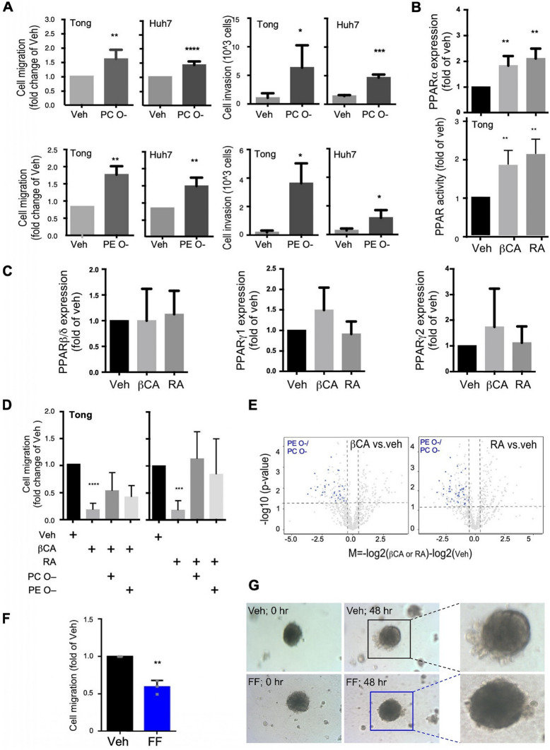 Figure 5
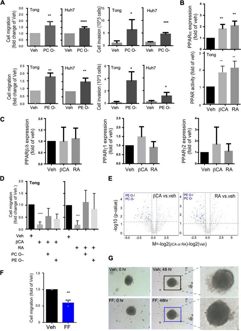 Figure 2
Peer Reviews
No public reviews on file for this paper yet. If you reviewed it on a platform where reviews are public (OpenReview, ICLR, NeurIPS, ICML), you can paste yours below so the community can read it here.
Videos
No videos yet. Explain this paper in a talk, walkthrough, or lecture? Add one.
Taxonomy
TopicsPeroxisome Proliferator-Activated Receptors · Cholesterol and Lipid Metabolism · Liver Disease Diagnosis and Treatment
**Correction: Journal of Biomedical Science (2025) 32:89 ** 10.1186/s12929-025-01178-y
After publication of this article [1], it was brought to our attention that figure 5 is incorrect.
The incorrect Fig. 5:Fig. 5PPARα mediated ether-lipids accumulation promote cell mobility and metastasis. A HCC cells, specifically Tong and Huh7 cells, were treated with ether-linked phospholipids (either PC O– or PE O–; 10 nM) to evaluate their migration and invasion activities. The presented migration/invasion data were averaged from three to four independent experiments. B The PPARα mRNA expression (upper panel) and the PPAR promoter activities (lower panel) upon treatments of βCA and RA. C The PPARβ (left panel), PPARγ1 (middle panel) and PPARγ2 (right panel) mRNA expression of HCC cells upon treatments of βCA and RA. D Effects of PPARα agonists (βCA, left side; RA, right side; 20 μM) on suppression of HCC cell migration. Counter effects of treatments with ether-lipids (PC O– and PE O–; 10 nM) and βCA-/RA-induced cell migration are displayed. E βCA/RA downregulated ether-lipid abundance in HCC cells. Volcano-plot showed differential expression of lipid species, where blue-dots represents PE O– and PC O–. F Migration of Tong cells, as determined through a wound-healing assay 48 h after fenofibrate (FF; a PPARα agonist) treatment. G 3D spheroid images of filopodia with or without FF treatment. All in vitro results were derived from a minimum of three consistent experiments; * for p < 0.05, ** for p < 0.01, *** for p < 0.001, and **** for p < 0.0001
The correct Fig. 5:Fig. 5PPARα mediated ether-lipids accumulation promote cell mobility and metastasis. A HCC cells, specifically Tong and Huh7 cells, were treated with ether-linked phospholipids (either PC O– or PE O–; 10 nM) to evaluate their migration and invasion activities. The presented migration/invasion data were averaged from three to four independent experiments. B The PPARα mRNA expression (upper panel) and the PPAR promoter activities (lower panel) upon treatments of βCA and RA. C The PPARβ (left panel), PPARγ1 (middle panel) and PPARγ2 (right panel) mRNA expression of HCC cells upon treatments of βCA and RA. D Effects of PPARα agonists (βCA, left side; RA, right side; 20 μM) on suppression of HCC cell migration. Counter effects of treatments with ether-lipids (PC O– and PE O–; 10 nM) and βCA-/RA-induced cell migration are displayed. E βCA/RA downregulated ether-lipid abundance in HCC cells. Volcano-plot showed differential expression of lipid species, where blue-dots represents PE O– and PC O–. F Migration of Tong cells, as determined through a wound-healing assay 48 h after fenofibrate (FF; a PPARα agonist) treatment. G 3D spheroid images of filopodia with or without FF treatment. All in vitro results were derived from a minimum of three consistent experiments; * for p < 0.05, ** for p < 0.01, *** for p < 0.001, and **** for p < 0.0001
The original article has been updated.