A case of cardiac sarcoidosis with a ventricular aneurysm visualized by four-dimensional left ventricular imaging using TrueVue glass
Yusuke Nakashima, Michio Yamada, Ayumi Omuro, Shinichi Okuda, Motoaki Sano

Abstract
Genes, proteins, chemicals, diseases, species, mutations and cell lines named across the full text — each resolved to its canonical identifier and authoritative record.
Click any figure to enlarge with its caption.
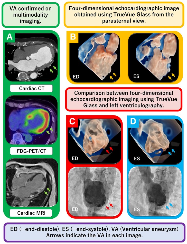 Figure 1
Peer Reviews
No public reviews on file for this paper yet. If you reviewed it on a platform where reviews are public (OpenReview, ICLR, NeurIPS, ICML), you can paste yours below so the community can read it here.
Videos
No videos yet. Explain this paper in a talk, walkthrough, or lecture? Add one.
Taxonomy
TopicsCardiac Structural Anomalies and Repair · Sarcoidosis and Beryllium Toxicity Research · Cardiac tumors and thrombi
A 65-year-old woman underwent percutaneous coronary intervention for a mid-left anterior descending artery lesion. Although no ventricular aneurysm (VA) was observed during the acute phase, follow-up echocardiography unexpectedly revealed a lateral wall VA. This location did not correspond to the expected ischaemic territory on CT (Panel A, top), indicating a diagnostic mismatch and prompting multimodality imaging. TrueVue Glass (Philips CVx system) was used to characterize this atypically-located VA. Compared with conventional 2D echocardiography, TrueVue Glass provided improved endocardial border delineation and enhanced spatial appreciation of aneurysmal morphology without contrast. Although not inherently diagnostic, it enabled flexible four-dimensional (4D) visualization resembling left ventricular angiography, allowing comprehensive structural assessment (Panel B, Supplementary data online, Video S1). FDG-PET/CT demonstrated focal left ventricular uptake (Panel A, middle), and cardiac MRI confirmed late gadolinium enhancement (LGE) involving the aneurysmal region (Panel A, bottom). Follow-up coronary angiography showed no restenosis or new lesions, and left ventriculography identified a true VA in LV segment 7 according to the AHA 17-segment model (see Supplementary data online, Video S2), consistent with TrueVue findings (Panels C and D, Supplementary data online, Video S3).
Based on the 2016 Japanese Circulation Society criteria—including the presence of a VA, reduced LV systolic function, localized FDG uptake, and LGE on MRI—the patient was diagnosed with cardiac sarcoidosis. TrueVue Glass, originally developed for foetal imaging, has recently been adapted for cardiac structural assessment. In this case, its non-invasive, real-time 4D visualization provided superior spatial understanding of the VA compared with 2D echocardiography, supporting the diagnosis of cardiac sarcoidosis.
Supplementary Material
qyag011_Supplementary_Data