Graphene oxide-modified PEEK composites: Properties and applications in orthopaedic repair — A review
Mingjing Zhang, Shuzhong Liu, Jinyi Xing, An Song, Liqi Ng, Nan Tao, Xin Su, Changning Sun, Chaozong Liu

TL;DR
Graphene oxide-modified PEEK composites show promise for orthopaedic repair by improving bone growth and antibacterial properties.
Contribution
This review highlights the novel integration of graphene oxide with PEEK to enhance osteogenic and antibacterial properties for orthopaedic applications.
Findings
GO-PEEK composites improve osteoblast adhesion, differentiation, and mineralized nodule formation.
In vivo studies confirm superior osseointegration and new bone formation with GO-PEEK.
Synergistic modifications with hydroxyapatite or antimicrobial peptides enhance both osteogenic and antibacterial outcomes.
Abstract
Critical-sized bone defect repair remains a major challenge in orthopaedics and tissue engineering. Polyetheretherketone (PEEK) has attracted wide attention due to its excellent mechanical compatibility and radiological transparency; however, its inherent bioinertness and insufficient antibacterial properties restrict its clinical utility. In recent years, the incorporation of graphene oxide (GO) has markedly improved the biological performance of PEEK. GO can increase surface hydrophilicity and roughness, enhance protein/ion adsorption, and promote osteoblast adhesion and differentiation, while simultaneously strengthening antibacterial and immunomodulatory effects without compromising, and in some cases even enhancing, mechanical performance. In vitro studies demonstrate that GO-PEEK stimulates osteogenic gene expression and mineralized nodule formation, while in vivo animal models…
Genes, proteins, chemicals, diseases, species, mutations and cell lines named across the full text — each resolved to its canonical identifier and authoritative record.
Click any figure to enlarge with its caption.
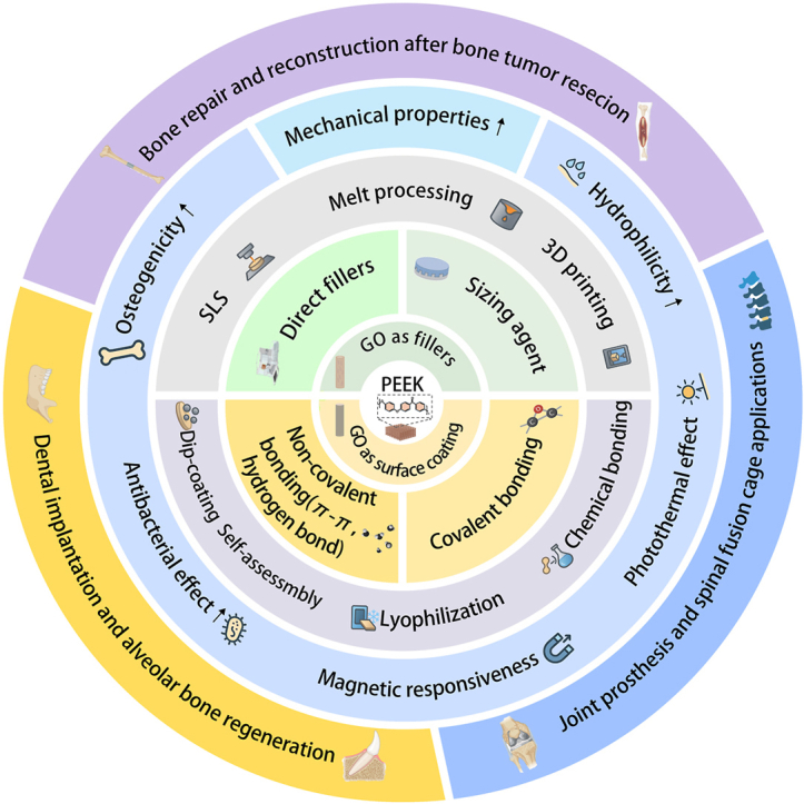 Figure 1
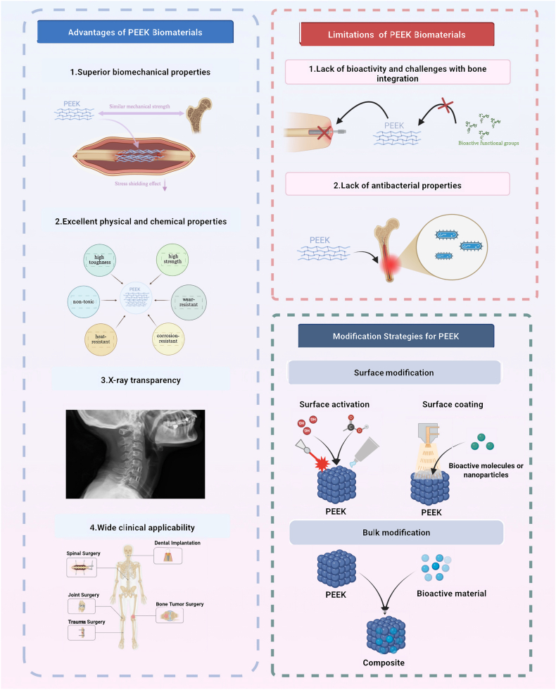 Figure 2
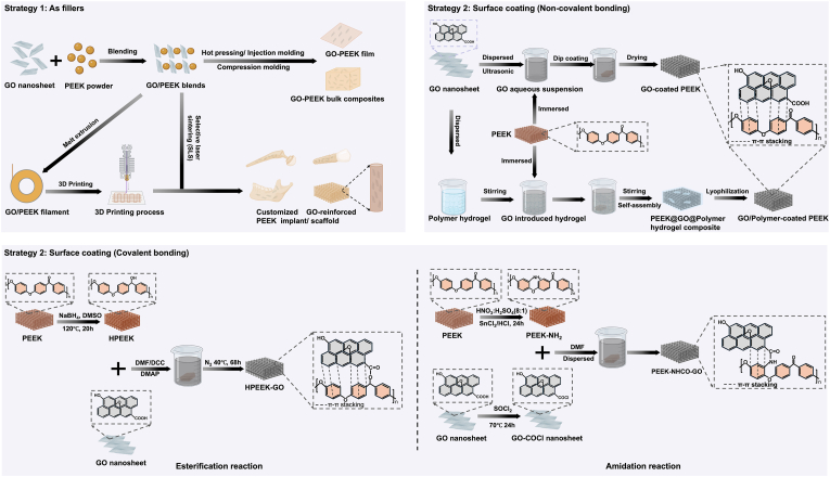 Figure 3
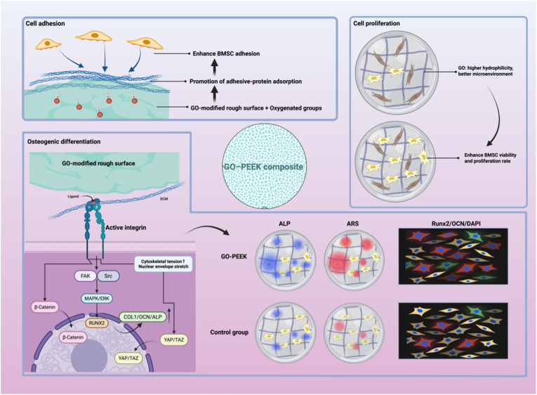 Figure 4
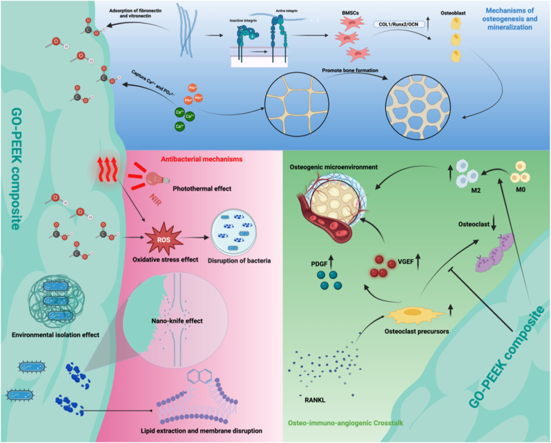 Figure 5
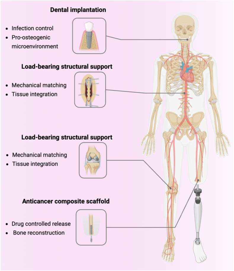 Figure 6
Peer Reviews
No public reviews on file for this paper yet. If you reviewed it on a platform where reviews are public (OpenReview, ICLR, NeurIPS, ICML), you can paste yours below so the community can read it here.
Videos
No videos yet. Explain this paper in a talk, walkthrough, or lecture? Add one.
Taxonomy
TopicsGraphene and Nanomaterials Applications · Bone Tissue Engineering Materials · Graphene research and applications
Introduction
1
Bone defects (BDs) refer to the loss or destruction of normal bone tissue, which may result from various etiologies, including trauma, osteoporosis, osteomyelitis, osteonecrosis, and bone tumors, with a wide range of severity [1,2]. When the extent of bone loss exceeds the body's inherent regenerative capacity, the defect is classified as a critical-size bone defect (CBD), which typically requires surgical intervention to facilitate effective bone regeneration [3]. Bone defects are not only common but also clinically significant, often resulting in long-term functional impairment and a marked decline in patients' quality of life [4,5]. This condition places substantial demands on clinical management and contributes to a growing socioeconomic burden. Despite considerable advances in bone repair strategies in recent years, the successful reconstruction of CBDs remains a major clinical and translational challenge [6,7]. BDs are not only highly prevalent but also clinically significant, often resulting in long-term functional impairment and a markedly reduced quality of life for affected patients [4,5].
Bone regeneration is a highly orchestrated and multifaceted pathophysiological process, encompassing a series of interdependent events such as osteogenesis, osteoclast-mediated bone resorption and remodeling, apoptosis, osteoblastic differentiation, biomineralization, angiogenesis, and immune regulation [8]. Autologous bone grafting remains the clinical gold standard for the treatment of bone defects. However, its application is constrained by limited donor availability and the risk of donor site morbidity, which can increase surgical complexity and impose additional physical and financial burdens on patients [9]. Allogeneic bone grafts can partly compensate for the scarcity of autologous sources, yet they carry inherent risks of immunogenic rejection and potential disease transmission. Additionally, the irregular morphology of many bone defects further complicates the use of conventional grafting approaches. In the absence of timely and effective intervention, patients are at increased risk of developing complications such as chronic pain, pathological fractures, infections, and non-union—conditions that severely compromise both physical function and quality of life [10]. Consequently, the development of safer, more effective artificial bone substitutes has become a key focus in regenerative medicine and orthopaedic research. Compared to autografts and allografts, synthetic bone graft materials offer numerous advantages, including scalable production, diverse raw material sources, cost-effectiveness, and avoidance of donor site morbidity [11]. More importantly, recent advancements in materials science and tissue engineering have substantially improved the mechanical properties, bioactivity, and customizability of synthetic bone scaffolds, opening new avenues for the repair and reconstruction of complex bone defects.
An ideal bone implant material should exhibit mechanical properties that are well-matched to those of native bone, along with favorable bioactivity to support and enhance bone regeneration [10,11]. Currently, synthetic bone graft materials commonly fall into three main categories: (A)Metallic materials (e.g., stainless steel, titanium and its alloy Ti-6Al-4V, cobalt-chromium alloys), which provide excellent mechanical strength and corrosion resistance, making them suitable for structural support at defect sites. However, their elastic moduli are substantially higher than that of natural bone, often resulting in stress shielding and subsequent secondary bone resorption [[10], [11], [12]]. (B)Ceramic materials (e.g., hydroxyapatite, tricalcium phosphate), which demonstrate high bioactivity and osteoconductivity but are limited by their intrinsic brittleness and poor fracture toughness. (C)Polymeric materials (e.g., polylactic acid [PLA], polycaprolactone [PCL]), which offer desirable features such as biodegradability, biocompatibility, and ease of processing. Nevertheless, their insufficient mechanical strength restricts their application in load-bearing bone defect repair [[13], [14], [15], [16], [17]]. Despite these advances, limitations such as inadequate immunocompatibility and suboptimal biological performance continue to impede the broader clinical translation of these materials. Consequently, the design and development of bone repair materials that combine appropriate mechanical strength, excellent biocompatibility, and tunable biological functionalities have emerged as a key priority in orthopaedic biomaterials research (Fig. 1).Fig. 1Advantages, limitations, and modification strategies of PEEK biomaterials. PEEK exhibits superior biomechanical compatibility, excellent physicochemical stability, X-ray transparency, and wide clinical applicability. However, its poor bioactivity and lack of antibacterial properties limit bone integration. Current modification strategies include surface activation, surface coating, and bulk modification with bioactive materials to enhance interfacial bonding and biological performance. Schematics created with BioRender.Fig. 1
Polyetheretherketone (PEEK) is a high-performance thermoplastic polymer that has garnered significant interest in the orthopaedic field due to its favorable physicochemical and mechanical properties, including excellent biocompatibility, high thermal and chemical stability, corrosion resistance, adjustable elastic modulus, intrinsic self-lubrication, ease of processing, and radiolucency [[18], [19], [20], [21]]. In contrast to conventional metallic implants, PEEK exhibits an elastic modulus more closely approximating that of native cortical bone, thereby reducing stress shielding and enhancing the mechanical compatibility at the bone–implant interface, which facilitates superior osseointegration [[22], [23], [24], [25]]. In addition, PEEK demonstrates outstanding imaging compatibility, producing minimal artifacts in both magnetic resonance imaging (MRI) and computed tomography (CT), thus enabling accurate postoperative assessment and monitoring [26,27]. Following its approval by the U.S. Food and Drug Administration (FDA) in 2013, PEEK-based implants have been widely adopted in various orthopaedic subspecialties, including spinal surgery, joint arthroplasty, trauma reconstruction, and orthopaedic oncology [[28], [29], [30], [31], [32], [33], [34]](Fig. 1).
Despite its numerous advantages, PEEK's inherent bioinertness and poor surface hydrophilicity significantly limit its widespread application in bone regeneration [35,36]. The lack of intrinsic functional groups that facilitate osteogenesis or antimicrobial activity results in suboptimal osseointegration and limited bioactivity [[37], [38], [39], [40], [41]]. To overcome these limitations, various modification strategies have been explored, including: (A) the fabrication of micro- and nano-scale surface structures via techniques such as sandblasting and sulfuric acid etching, which improve cell adhesion and enhance tissue anchorage([42], [43], [44], [45], [46]). (B) the incorporation of bioactive agents or functional polymers through surface coating, chemical grafting, or bulk blending methods. Common additives include titanium dioxide (TiO_2_) and hydroxyapatite (HAp), which have demonstrated promising osteoinductive potential [18,19,[47], [48], [49]]. Although these approaches have contributed to enhanced biological performance, challenges remain in achieving robust osteoinductivity, sustained antimicrobial activity, and integrated multifunctionality (Fig. 1).
In recent years, the integration of graphene and its derivatives has opened new avenues for the functionalization of orthopaedic implant materials. Among these, graphene oxide (GO) has attracted particular interest owing to its high density of oxygen-containing functional groups—such as carboxyl, hydroxyl, carbonyl, and epoxy—which impart excellent aqueous dispersibility and enable the modulation of cellular behavio [50,51]. GO-based materials have shown considerable promise in diverse biomedical applications, including drug delivery, antimicrobial therapy, cancer treatment, and tissue engineering, and are increasingly being explored as emerging candidates for bone repair strategies [13,16]. An ideal bone repair material must combine several key properties, including biocompatibility, osteoinductivity, antimicrobial efficacy, and mechanical compatibility. The incorporation of GO and other functional nanomaterials into PEEK scaffolds represents a compelling strategy to enhance the biological performance of the composite, while simultaneously conferring multifunctional capabilities such as antibacterial and anticancer activity [[52], [53], [54], [55], [56]].
In summary, the multifunctional characteristics of graphene oxide (GO) combined with the superior mechanical properties of polyetheretherketone (PEEK) offer a novel approach for the development of composite biomaterials exhibiting enhanced osseointegration, osteogenic activity, antibacterial efficacy, and anticancer functionality. Although significant challenges remain in further improving the biological performance of PEEK while preserving its inherent mechanical advantages, research on GO-PEEK composites has increasingly become a forefront and hotspot in the field of bone regeneration. This review systematically summarizes recent advances in GO-modified PEEK composites, focusing on their feasibility, underlying mechanisms, and future prospects in bone tissue repair.
Preparation methods of GO-PEEK composites
2
Preparation strategies
2.1
With the rapid progress of materials science, the integration of GO into PEEK has emerged as a promising approach for developing next-generation composites. Preparation strategies can be broadly divided into two categories. As a reinforcing phase, GO can be uniformly dispersed within the PEEK matrix to enhance mechanical strength and thermal stability [[57], [58], [59], [60], [61], [62], [63], [64], [65], [66], [67], [68], [69], [70], [71], [72], [73], [74], [75], [76], [77]]. Alternatively, it can serve as an interfacial enhancer by grafting onto carbon fibers (CF) or carbon nanotubes (CNTs) before incorporation, thereby strengthening interfacial bonding and load transfer. This approach markedly improves the mechanical and fatigue properties of the composites, making them particularly suitable for load-bearing orthopaedic implants ([66], [67], [68], [69], [70], [71], 77, 78). As a surface coating, GO improves the hydrophilicity and bioactivity of PEEK. The underlying interactions include non-covalent forces (π–π stacking, hydrogen bonding) ([79], [80], [81], [82], [83], [84], [85], [86], [87], [88], [89], [90], [91], [92], [93], [94], [95]). and covalent linkages via esterification or amidation reactions ([96], [97], [98], [99]). with covalent bonding providing superior adhesion and long-term stability (Fig. 2).Fig. 2Preparation strategies for GO/PEEK composites. Schematics created with BioRender.Strategy 1: GO nanosheets used as fillers are incorporated into the PEEK matrix via blending, hot pressing, injection molding, or 3D printing to form bulk composites or customized implants/scaffolds.Strategy 2: GO applied as surface coatings enhances interfacial properties and bioactivity. (a) Non-covalent bonding via π–π stacking and hydrogen bonding. (b) Covalent bonding through esterification or amidation reactions, yielding strong and stable interfacial adhesion.Fig. 2
Fabrication techniques and their advantages and limitations
2.2
For bone repair applications, the fabrication of GO-PEEK composites can be broadly divided into two categories: blending techniques and surface coating techniques, each with specific merits and constraints.
Blending techniques
2.2.1
These include hot pressing [[67], [68], [69], [70]], melt extrusion and injection molding [[71], [72], [73], [74]], and selective laser sintering (SLS)/3D printing [75,76,[100], [101], [102]], Compression molding [101,103,104]. Melt extrusion and injection molding provide excellent reproducibility and industrial scalability; however, the elevated processing temperatures may damage GO nanosheets, deplete functional groups, or induce aggregation, thereby weakening interfacial reinforcement [105]. SLS/3D printing allows the fabrication of patient-specific, porous structures highly suitable for bone scaffolds [106]. Nevertheless, high equipment cost, narrow processing parameters, and the stringent requirements for GO dispersion and thermal stability remain significant barriers to clinical translation [107](Fig. 2) (Table 1).Table 1GO combination strategies and their resulting enhancements in the physicochemical properties of PEEK composites.Table 1. Combination StrategyMatrixFillers/Coatings CompositionFabrication MethodInteractionsEffect of GO on Physicochemical PropertiesOptimum Composition/TrendRef.FillersSizing agentsPEEKGO: ∼1 wt%MWCNT: 0.5–1 wt%Hot pressingπ–π stacking↑Thermal conductivity(0.18 → 0.36 W/m·K); ↑Storage modulus (58.4 → 68.2 %)↑Crystallinity1 wt% GO + 0.5 wt% MWCNT; higher loadings → agglomeration and ↓ performance63Direct fillerPEEKGO: 0–1 wt %c-aminopropyl trimethoxysilane-modified graphene oxide (GO-Si)Hot pressingNot specified↑Wear resistance; ↓ Friction coefficient (lowest at 0.1 wt% GO-Si); ↑Thermal stability; ↑ Crystallinity; smoother worn surface observed0.1 wt% GO-Si, excessive GO → aggregation and ↑ friction64Sizing agentsPEEKGO: 0–0.75 wt%s-PSF coated CFHot pressingNot specified↑Flexural strength (to 847.29 MPa)↑Flexural modulus (to 63.77 GPa)↑Interlaminar shear strength(ILSS) (to 73.17 MPa); ↑ Tg (+4 °C); ↑ Density; ↓Water absorption0.5 wt% GO; higher GO → aggregation → decreased ILSS and flexural strength65Sizing agentsPEEKGO: ∼1 wt%CF: 30 wt%Melt extrusion and injection moldingNot specified↑hydrophilicity(↓ Contact angle (79.9° → 59.5°) vs CF/PEEK)GOCF/PEEK → highest wettability and bioactivity66Direct fillerPEEKGO: 0.1-1 wt%TDI–PEK-LMelt extrusion and injection molding/3D printingNot specified↑ Tensile (+6.8 %), ↑ modulus (+7.0 %), ↑ elongation (+31.6 %), ↑ flexural (+7.1 %), ↑ impact (+20.5 %); ↓ friction (−27.3 %) and ↓ wear rate (−18.3 %)0.1 wt% LFG: optimal mechanical strength and toughness;0.5 wt% LFG: best tribological (wear and friction) performance>0.5 wt% LFG: aggregation and ↓ ductility67Ssizing agentsPEEKGO: 0.5–2.0 wt%SiO_2:_ 30 wt%Melt blending and injection moldingπ-π∗ conjugation↑ Tensile strength (+12.6 %), ↑ modulus (+39.4 %), ↑ hydrophilicity; ↑ thermal stability1.5 wt% GO → best mechanical & surface performance, ≥2 wt% → GO restacking and reduced performance68Sizing agentsPEEKGO: ∼1 wt%CF: 30 wt%Melt extrusion and injection moldingNot specified↑Flexural strength (+51 %), ↑ Compressive strength (+46 %), ↑ Hardness (+30 %), ↑ Flexural modulus to 11.67 GPa, ↑ Compressive modulus to 6.12 GPa, ↓ ↑Hydrophilicitycontact angle (65.8° vs 73.6°)1 wt% AGO gave highest mechanical and surface properties tested; no trend beyond 1 wt% reported69Direct fillerPEEKGO: 0.1–5 wt%Melt extrusion and injection moldingπ-π∗ conjugation↑Compressive, flexural, and tensile properties, ↑hardness0.5 wt% GO; >0.5 wt% led to agglomeration and decreased mechanical properties70Direct fillerPEEKGO: 0–1.25 wt%HAp: 20 wt%Selectively laser sintering (SLS)π-π stacking↑ Compressive strength (+79.45 %); ↑ Modulus (+42.07 %); ↑ Thermal stability; ↓ Agglomeration below 1 wt% GO; Improved HAP dispersion1 wt% GO; >1 wt% → GO agglomeration ↓ mechanical strength71Direct fillerPEEK and PVA mixtureGO: 0–2 wt%Selective laser sintering (SLS)π-π stacking interactionHydrogen bonds (with PVA)↑ Compressive strength +97.16 %; ↑ Modulus +147.06 % (1 wt% GO); ↑ Thermal stability;↑ Hydrophilicity (contact angle ↓ from 85.52° → 78.16°); ↑ Water absorption and degradability; uniform PVA dispersion1 wt% GO optimum; >1.5 wt% → GO aggregation, defects, mechanical decline72Direct fillerSPEEKrGO: 0–1.5 wt%; HAp: 3.5–5 wt%Melt extrusion and 3D printingNot specified↑ Compressive strength with ↑ rGO↑ Thermal stability1.5 wt% rGO + 3.5 wt% HAp → highest compressive strength & thermal performance100Direct fillerPEEKGO: 0–1.5 wt%; HAp: 0–30 wt%Compression moldingπ-π stacking↓ Friction coefficient, ↓ Wear rate (1.60 × 10^−6^ mm^3^/N·m) ↑ Transfer-film stability and surface smoothness (Ra ≈1.36 μm)↑Dispersion and load transfer0.5 wt% GO + 10 wt% HAp → lowest wear rate & friction; higher reinforcement → agglomeration, COF↑, wear rate↑101Direct fillerPEEKGO: 0–0.75 wt%Mold casting and sinteringHydrogen bonding↑ GIC (+109.3 %) ↑ KIC (+35.6 %) ↑ Tensile strength (+14.7 %) ↑ Flexural strength (+5.0 %) ↓ Crystallinity ↑ Td_5_ → thermal stability maintainedGO-CTS = 0.25 wt% → best balance of toughness & strength; Pure GO peaks at 0.5 wt% but less effective102Direct fillerPEEKGO: 0–1.5 wt%HAp: 0–30 wt%Compression moldingπ-π stackingGO 0.5 wt% + HA 10 wt%, ↓Coefficient friction (COF), ↓Wear rate, ↑ Hardness0.5 wt% GO + 10 wt% HAp → lowest wear rate & friction; higher (>1 wt%) GO → agglomeration, COF↑, wear rate↑103Direct fillerPEEKGO: 0.5 wt%HAp: 10 wt%Compression moldingπ-π stacking↑ Tensile strength (+118 %) ↑ Elastic strain to 6.95–11.35 % ↑ Hardness (76.4 → 79.6 Hv)↑ T_m_ from 340 → 351 °C ↓ Surface roughness (Ra 1.62 → 1.36 μm) ↓ COF (0.025–0.141) ↓ Wear rate (3.39 × 10^−6^ → 1.69 × 10^−6^ mm^3^/N·m)0.5 wt% GO + 10 wt% HA → best mechanical & tribological behavior; higher GO not tested103Direct fillerPEEKGO: 5 wt%HAp: 0–40 wt%Mold casting and sinteringNot specified↑ Crystallinity up to 47.3 % (HAp 20 wt%); ↑ Thermal stability; ↑ Compressive strength (14.2 → 21.45 MPa at HAp 30 %); ↑ hydrophilicity5 wt% GO + 30 wt% HAp optimum → highest strength (21.45 MPa)>40 wt% HA → agglomeration and strength ↓ 13.95 Mpa104Direct fillerPEEKrGO: 1–3 wt%HAp: 0–30 wt%Melt extrusion and 3D printingNot specified↑ Compressive modulus (≈+60 %) and strength (≈+50 %) for PEEK-69/cHAp-30/rGO-1; PEEK-87/cHAp-10/rGO-3 → highest stress tolerance (25.32 GPa) and lowest deformation (9.29 mm)PEEK-87/HAp-10/rGO-3 wt% → best mechanical performance and lowest deformation; higher rGO >3 wt% not studied155Direct fillerPEEKGO: 1 wt%PDAZrO_2_: 10 wt%Melt extrusion and injection moldingπ-π stacking↑ Tensile strength (93.1 → 103.3 MPa) → ↑ Bending strength (↑17.2 %) → ↑ Compressive strength (↑9.8 %) → ↑ Hydrophilicity → Stable photothermal response (ΔT = 40.6 °C under 2 W, 808 nm) → Uniform nanoparticle dispersion1 wt% GO (PGPZ) → highest strength & photothermal stability167Surface coatingSurface coating (Non-convalent bonding)Sulfonated PEEK (SPEEK)GO: 0.5 wt%SPEEK: 1 wt%Dip-coating (X5 cycles)π–π stacking, hydrogen bonding↑Hydrophilicity(contact angle ↓ from 90° → 47.7°); Surface roughness↑; Wettability↑Elastic modulus ≈3–4 GPa (unchanged)0.5 wt% GO–SPEEK → best synergy of antibacterial and osteogenic properties18PEEKGO: 0.8 mg/mL pDA: 2 mg/mLBFP: 1 mMDip-coating (X6 cycles)π–π stacking↑Hydrophilicity (contact angle ↓ 76°→25°)Photothermal conversion ΔT ≈ +54 °C (808 nm, 0.5 W cm^−2^), stable for three cyclesGO/pDA/BFP hybrid exhibited highest photothermal efficiency and hydrophilicity without degradation.73SPEEKGO: 0.75 wt%Bioactive glassSpin-coatingNot specified↑ Hydrophilicity (contact angle ↓ 71° → 14.7°); strong adhesion (74.9 MPa); transparent homogeneous coating with ∼7 μm thickness0.75 wt% GO group exhibited highest hydrophilicity vs control group74SPEEKGO: 0.1 wt%SPEEKDip-coatingπ–π stacking and hydrogen bonding↑ Hydrophilicity (contact angle ↓ 82.7° → 10.9°)↑ Surface energy; ↑ Surface roughness (3D pores 200–500 nm);≈ Mechanical strength retained; ↑ Chemical activityGO–SPEEK → best hydrophilicity and bioactivity; GO > SPEEK > PEEK; stable coating without degradation75SPEEKZnO/GO mass ratio 3:1ZnO/GO mass ratio 4:1ZnO/GO mass ratio 5:1Dip-coatingπ–π stacking↑ Surface roughness (Ra↑), ↑ hydrophilicity (contact angle ↓ from 88.4° to 51.2°)ZnO:GO = 4:1 showed best antibacterial effect biocompatibility, and hydrophilicity. ZnO/GO mass ratio↑→ surface roughness ↑76PEEKrGO: 1–3 wt%Dip-coatingNot specified↑compressive strength, ↑surface roughness, ↑improved wettability3 wt% rGO → highest compressive strength, enhanced surface roughness, and improved wettability; ↑rGO → ↑grain density, ↑protein adsorption77PEEKGO: 0.75 wt% nHApDip-coatingπ-π stacking↓ Surface roughness (Ra = 21 nm vs 54 nm for PEEK);↑ Hydrophilicity (contact angle ↓ from 71.3° to 54.6°)GO 0.75 wt% yielded optimal balance of dispersion, adhesion, and biocompatibility80SPEEKGO: 1 wt% nisin: 0.04 mmol/mLDip-coatingNot specified↑ Hydrophilicity (CA ↓91°→24°) → ↑ O/N content (O 27.7 %, N 9.8 %) → stable GO coverage → porous anchoringGO + nisin synergy → highest hydrophilicity & stability81SPEEKGOMnFe_2_O_4_Hydrothermal synthesis (MFO/GO) → PDA-assisted coating on SPNot specified↑ NIR absorption (200–1000 nm) → ↑ photothermal efficiency (45.3 → 56.1 °C, 1 W cm^−2^) → GPx-mimetic GSH oxidation (54 → 67 % depletion under NIR) → ↑ ROS accumulationSP–P–MFO/GO shows best GSH consumption and photothermal stability82GO: 0.1 wt%Dip-coatingπ-π stacking↑hydrophilicity (contact angle ↓), ↑roughness, stable GO layer resistant to ultrasonicationCF concentration↑→Surface roughness↑; GO–40CF/PEEK exhibited highest surface roughness,and hydrophilicity8330 wt% CF-reinforced SPEEKrGO5 wt%HAp:10 wt%Spin coatingphysical embedding and interfacial bonding↓ Contact angle (112.5°→ 20°/54.5°/47°) → ↑ hydrophilicity; ↑ surface porosity → better coating anchorage; no structural change in PEEKHAp coating (no rGO) → highest hydrophilicity and bioactivity; rGO addition slightly reduced wettability87SPEEKGOPEEKPDASelf-assemblyπ–π stacking & H-bonding between PDA and GO, strong interfacial adhesion on PEEK↑ Hydrophilicity (Contact Angle 105.5°→38.5°) → ↑ protein adsorption; ↑ M2 macrophage polarization (17.6 %→47.4 %); ↓ pro-inflammatory cytokines (IL-1β, IL-18 ↓); ↑ osteogenic gene expression (RUNX2, COL-I ↑)PAG (PEEK–PDA–GO) showed optimal wettability and surface uniformity90SPEEKGO: 0.1 wt%Dip-coatingπ-π stacking↑ Hydrophilicity (contact angle 83.5° → 48.7°)GO-SPEEK (10 min sulfonation + GO 1 mg/mL) → optimal hydrophilicity91SPEEKGO: 0.1 wt%Ag^+^: 10 × 10 ^−3^MDip-coatingπ–π stacking and hydrogen bonding↑ Surface roughness & porosity (0.5–2 μm) → ↑ hydrophilicity (CA ↓ to ∼63°) → Ag^+^ slow release (47.9 % in 3 days) → durable structureGO layer (10–25 nm) + Ag ^+^ uniformly dispersed → best stability and antibacterial effect92CF-reinforced SPEEKGO: 0.1 wt%Dip-coatingπ-π stacking↑Surface roughness, ↑hydrophilicity; ↑Crystallinity; ↑Bending strength ↑ to ∼265 MPa; ↑Compressive modulus ∼1 GPa ↑ vs. CF/PEEKGO–SCF/PEEK exhibited best balance between bioactivity and mechanical integrity94PEEKGO: 0.4 wt%Spin-coating, dip-coating, plasma stabilizationπ-π stacking↑ Surface roughness → enhanced protein adsorption; ↑ O/C ratio → improved hydrophilicity; ↑ stability → better coating adhesionSpin-coated GO + CH_4_ + O_2_ plasma yielded uniform, stable, hydrophilic coating; dip-coated GO less stable in wet media95PEEKGO: 0.05 wt%PDASelf-assembly + Freexe dryingπ–π stacking & covalent bonding between PDA and GO; strong interfacial adhesion on PEEK↑Hydrophilicity(↓ Water contact angle (83.5° → 53°)); ↑ surface energyPEEK-PDA-GO exhibited best wettability and uniform surface morphology123CF-reinforced PEEKGO–HAP: 0, 25, 50, 100 ng/mLGelMA/PEGDAFreeze-dryingπ–π and electrostatic bonding↑ Surface porosity → ↑ NIR absorption → ↑ Photothermal conversion (33 °C→63 °C) → Stable cyclic heating → Enhanced hydrophilicity and cell adhesionGH100 (100 ng/mL GO–HAP) → best photothermal & stability performance205Convalent bondingEpoxy resinGO: 0.1 wt%-0.5 wt%HPEEK-gMechanical stirring + ultrasonication + curingCovalent ester linkage (–COO–) between GO and HPEEK → improved interfacial adhesion and dispersion↑ Storage modulus (+42 %) → ↑ hardness (+65 %) → ↑ fracture toughness (+31 %) → ↑ tensile strength (+7 %) → ↑ Tg (173–215 °C)0.5 wt% HPEEK-g-GO → optimum mechanical and thermal enhancement112Aminated PEEKGO: 5–20 wt%hot-press sinteringCovalent amide bonding (–NHCO–) between GO and PEEK → enhanced interfacial coupling and filler orientation↑ Thermal conductivity (0.25 → 0.97 W/m·K, ↑380 %) → ↑ tensile stress (128 → 183 MPa) → ↑ Td (525.8 °C) → ↓ Tm/Tc (crystallinity suppression)20 wt% GO → highest TC and Td; excessive GO reduces mechanical strength113
Surface coating techniques
2.2.2
Typical methods include dip-coating ([79], [80], [81], [82], [83], [84], [85], [86], [87], [88], [89], [90], [91], [92], [93]), vacuum-assisted filtration [108,109], self-assembly [[110], [111], [112]], and freeze-drying [54,113,114]. Non-covalent coatings are facile and minimally invasive to the substrate, enabling the introduction of antibacterial and osteogenic functions. Their limitations lie in weak interfacial adhesion and poor long-term stability, with delamination risks under physiological conditions. Covalent coatings form stable chemical bonds with the PEEK substrate, offering superior adhesion and long-term osseointegration potential. However, chemical modifications may compromise GO functional groups or trigger side reactions, necessitating strict control of processing parameters and biosafety. Recent studies on GO/HA/PEEK composite scaffolds have demonstrated in vitro mechanical and osteogenic properties comparable to cortical bone, underscoring their short-term feasibility [115] (Fig. 2) (Table 1).
In previous studies on GO-PEEK composites, the control of synthesis parameters has been identified as a critical determinant of their overall performance. Among these, GO concentration is the most frequently regulated factor. Most reports indicate that maintaining the GO content within 0.1–5 wt% markedly improves surface hydrophilicity, cellular adhesion, and osteogenic activity, whereas higher concentrations (>5 wt%) often result in agglomeration, interfacial defects, and excessive reactive oxygen species (ROS) generation, thereby compromising mechanical properties and increasing cytotoxicity [116]. The oxidation degree of GO is equally significant. A higher oxidation degree introduces abundant carboxyl, hydroxyl, and epoxy groups that facilitate protein adsorption and calcium deposition, but may simultaneously reduce conductivity and structural stability. In contrast, moderately oxidised GO has been shown to offer a more favorable balance between bioactivity and stability [117]. Surface exposure, largely determined by dispersion methods and fabrication processes, also plays a decisive role: uniformly dispersed and appropriately exposed GO nanosheets strongly promote cellular responses, whereas GO encapsulated within the PEEK matrix contributes little to biological performance [118]. Moreover, lateral size and layer thickness exert additional influences: small-flake or monolayer GO provides high surface area and uniform coating, while large-flake or multilayer GO is more effective for enhancing mechanical reinforcement [119]. Collectively, the interplay of GO concentration, oxidation degree, and surface exposure defines a “parameter window effect” that governs osteogenesis, antibacterial performance, and mechanical reinforcement. Optimising these parameters is therefore critical for advancing both fundamental research and clinical translation of GO-PEEK composites [120](Table 1).
In summary, melt extrusion or 3D printing combined with GO blending holds promise for load-bearing and structurally complex implants, whereas surface coating-particularly covalent modification-remains essential for enhancing bioactivity and osseointegration. Future development may favor hybrid approaches, such as integrating interfacial reinforcement with surface functionalization, to achieve synergistic optimization of mechanical robustness and biological performance.
Properties of GO-PEEK composites
3
Microstructure and surface characteristics
3.1
The incorporation of graphene oxide (GO) substantially modifies the surface morphology and interfacial properties of PEEK. GO nanosheets increase surface roughness and micro/nanostructures, which enhance protein adsorption, cell adhesion, and osteogenic differentiation [93,121]. Polar functional groups (–COOH, –OH) further promote hydrophilicity and Ca^2+^ deposition, thereby activating osteogenic signaling pathways. Within the bulk matrix, well-dispersed GO establishes strong interfacial bonding through π–π stacking, hydrogen bonding, or covalent grafting, facilitating stress transfer and improving composite stability. Conversely, GO aggregation may act as a stress concentrator and impair performance [57,58]. Emerging evidence indicates that GO can also modulate immune responses and promote angiogenesis, synergistically supporting osseointegration and long-term implant stability [122,123]. Thus, GO not only optimizes the physicochemical characteristics of PEEK but also endows it with biofunctional surfaces relevant for bone tissue engineering.
Mechanical properties
3.2
PEEK is widely used in orthopaedics due to its elastic modulus comparable to cortical bone; however, its bioinert surface remains a major barrier to osseointegration [35,36]. The introduction of GO as a filler enhances mechanical performance by improving interfacial adhesion and stress transfer via mechanical interlocking, π–π stacking, and hydrogen bonding [[57], [58], [59], [60], [61], [62],[65], [66], [67], [68], [69], [70], [71], [72], [73], [74],76,77,81]. Functionalized GO further amplifies these effects: amidated GO (AGO) improves hydrogen bonding with sulfonated PEEK (SPEEK), while phosphorylated GO (PGO), sulfonated GO (SGO), and epoxy-modified GO all demonstrate superior dispersion and interfacial stability [60,62,64,65]. Alternative strategies, such as polydopamine coating, in situ growth of metal–organic frameworks, and toluene-2,4-diisocyanate (TDI) functionalization, have achieved strong reinforcement and are promising for additive-manufactured PEEK components [58,61,72]. In addition, GO serves as an effective sizing agent for carbon nanotubes (CNTs) and carbon fibers (CFs), markedly improving stress transfer, toughness, and fatigue resistance [[66], [67], [68], [69], [70], [71],77]. While GO coatings exert minimal effects on bulk mechanical strength, they preserve robustness and significantly improve surface bioactivity [79,81,[85], [86], [87], [88], [89], [90],93,111,124]. Collectively, GO modification enables PEEK to maintain mechanical integrity while overcoming its intrinsic surface bioinertness(Table 1).
Photothermal effect and magnetic targeting properties
3.3
Beyond structural and mechanical advantages, GO imparts unique functional properties to PEEK composites. Its strong near-infrared (NIR) absorption and high photothermal conversion efficiency enable localized hyperthermia, with functionalized GO shown to inhibit melanoma growth under NIR irradiation [[125], [126], [127], [128], [129], [130], [131], [132], [133], [134]]. Integrating this capability into PEEK implants offers the potential for dual roles of structural support and local tumor ablation. Magnetic graphene oxide (MGO) further combines GO's physicochemical advantages with magnetic targeting. MGO has been demonstrated to promote osteogenic differentiation of bone marrow mesenchymal stem cells (BMSCs) under external magnetic fields while enabling controlled drug delivery [[134], [135], [136], [137], [138]]. MGO-based systems have shown synergistic chemo–photothermal efficacy in breast cancer and osteosarcoma models, and they hold promise as biosensors for detecting circulating tumor cells [[139], [140], [141], [142], [143], [144], [145], [146], [147], [148], [149], [150], [151]]. These multifunctional capabilities highlight GO-PEEK composites as next-generation implant materials that integrate mechanical compatibility with therapeutic and diagnostic functionalities, offering considerable translational potential in orthopaedic oncology and regenerative medicine.
Biological performance of GO-PEEK composites
4
Biocompatibility and cellular response
4.1
Biocompatibility is a fundamental prerequisite for bone repair materials, as it dictates the interactions between implants and host tissues and determines their physicochemical stability in vivo. An ideal orthopedic implant should minimize adverse host responses, particularly inflammation. [100,103,[152], [153], [154]]. Current evidence demonstrates that GO coatings exhibit negligible cytotoxicity toward osteoblasts and fibroblasts in vitro and only induce mild inflammatory reactions in vivo, underscoring their biosafety and clinical potential [79,80,104,155].
Through π–π stacking and hydrogen bonding, GO can firmly adhere to PEEK surfaces, significantly improving surface hydrophilicity and energy, thereby enhancing protein adsorption and promoting osteoblast/fibroblast adhesion and proliferation [88]. Multifunctional coatings that integrate GO with polydopamine, bioactive peptides, or inorganic components on sulfonated PEEK (SPEEK) have been reported to upregulate alkaline phosphatase (ALP) activity, calcium matrix deposition, and osteogenic gene expression [92]. Reduced GO (rGO) incorporated into 3D-printed PEEK scaffolds also shows promise for biomimetic bone repair [80], while GO combined with bioactive glass improves coating adhesion without compromising compatibility with osteoblasts and gingival fibroblasts [70]. In carbon fiber-reinforced PEEK (CFR-PEEK), GO modification enhances surface bioactivity and accelerates the early osteogenic response of bone marrow mesenchymal stem cells (BMSCs) [80]. Furthermore, GO-SPEEK has demonstrated the ability to induce hydroxyapatite (HAp) deposition and promote extracellular matrix mineralization in simulated body fluid [91].
Mechanistically, the biological activity of GO is largely attributed to its ability to improve hydrophilicity and provide functional groups (–COOH, –OH) that facilitate Ca^2+^ binding, mineral nucleation, and subsequent cell adhesion and osteogenic differentiation [122,123]. Recent findings further suggest that GO coatings may also enhance osseointegration via immunomodulation and pro-angiogenic effects, offering new strategies for complex bone defect repair [122].
Osteogenic activity and mineralization
4.2
In recent years, GO–PEEK composites have demonstrated unique potential in promoting osteogenic differentiation and mineral deposition. Their biological actions can be systematically interpreted from four interrelated perspectives: mechanotransduction, surface chemistry, osteoimmunomodulation, and angiogenesis, which collectively contribute to the “osteogenesis–mineralization” cascade (Fig. 3).Fig. 3Schematic illustration of the cellular responses to GO–PEEK composites.GO-modified PEEK enhances BMSC adhesion through increased surface roughness and oxygen-containing functional groups, promoting adhesive protein adsorption. Improved hydrophilicity provides a favorable microenvironment for cell proliferation. The GO–PEEK surface further activates integrin-mediated signaling pathways (FAK/Src and MAPK/ERK), leading to upregulated expression of osteogenic markers (COL1, OCN, ALP, RUNX2, and YAP/TAZ) and enhanced osteogenic differentiation. Schematics created with BioRender.Fig. 3
Direct mechanisms (mechanotransduction)
4.2.1
The wrinkled morphology and nanoscale roughness of GO nanosheets provide multiple anchorage points and local tension gradients for cells, facilitating focal adhesion maturation (vinculin/FAK complex assembly) and activating the integrin–FAK–Src–MAPK cascade. This subsequently upregulates key osteogenic genes such as Runx2, COL1, and OCN, driving the “mechanosensing–signal transduction–osteogenic differentiation” process of MSCs [156,157](Fig. 3).
In parallel, the nanoscale topography of the substrate induces cytoskeletal reorganization and nuclear membrane stretching, enhancing β-catenin nuclear translocation and activating the Wnt/β-catenin pathway, which synergistically promotes osteogenic lineage commitment through multi-pathway crosstalk [158,159].
Moreover, graphene coatings significantly upregulate cell-surface integrins (e.g., α5β1), thereby activating the integrin/FAK axis and facilitating YAP/TAZ nuclear localization [156]. Previous studies in neuronal regeneration have also shown that GO stiffness and nanoscale roughness can co-enhance YAP/TAZ activation, which in osteogenesis promotes MSC proliferation and differentiation, cooperatively regulating Runx2 and ALP expression and subsequent mineral matrix deposition [160,161](Fig. 3).
Indirect mechanisms (chemo–topography)
4.2.2
GO is inherently rich in polar functional groups such as –COOH and –OH, which markedly increase surface energy and hydrophilicity. These functional moieties preferentially adsorb fibronectin (FN) and vitronectin (VN), exposing RGD/LDV adhesion motifs and enhancing α5β1/αvβ3–integrin-mediated cell attachment and spreading, which in turn promotes proliferation and ALP expression [159,162,163].
In addition, the negatively charged GO surface can capture Ca^2+^ and PO_4_^3−^ ions, lowering the nucleation energy barrier for hydroxyapatite (HAp) formation and establishing a sequential process of ion enrichment → heterogeneous nucleation → mineral propagation. This cascade is accompanied by the amplification of BMP/Smad and integrin–FAK signaling, leading to ordered mineral deposition and matrix maturation [164,165](Fig. 3).
Time- and concentration-dependent synergy
4.2.3
The osteogenic process of GO–PEEK composites follows a “initiation–accumulation–maturation” biological rhythm: At the early stage (1–7 days), oxygen-containing groups (–OH, –COOH) on GO enhance fibronectin adsorption by approximately 2.1-fold, facilitating MSC adhesion and proliferation while activating Runx2 and ALP. Consequently, ALP activity increases by ∼45 % compared with pure PEEK. During the intermediate phase (7–14 days), mineralization accelerates—Alizarin Red staining reveals a 60 % increase in mineral nodule formation compared with neat PEEK, with calcium and phosphate deposition reaching ∼1.2 mmol/L, comparable to native bone matrix levels. In the late phase (≥21 days), osseointegration progresses steadily; in vivo studies up to 12 weeks report a 35 % increase in bone–implant contact (BIC) ratio relative to unmodified PEEK, consistent with the long-term bone bridging behavior of PEEK fusion cages [71,75].
Concentration dependence exhibits a strict dose–window effect: The optimal GO loading lies between 0.05 and 0.1 wt%, at which the mineralized nodule area after 14 days increases by ∼25 % compared with 0.02 wt% without apparent cytotoxicity. Below 0.05 wt%, osteogenic signaling remains insufficient, while concentrations above 0.5 wt% generate excessive reactive oxygen species (ROS), impairing mineralization and reducing calcium deposition by ∼30 % after 21 days.
This concentration-dependent behavior parallels the trends observed in other polymeric GO composites, as discussed in the fabrication section(166, 167) (Table 1).
Osteo-immune and angiogenic crosstalk
4.2.4
GO also exerts its effects through immunomodulation and angiogenesis. It suppresses RANKL-induced osteoclast maturation and bone resorption, thereby restoring the osteoblast–osteoclast balance [168]. Simultaneously, GO promotes the release of platelet-derived growth factors and VEGF, enhancing angiogenic signaling, improving oxygen/nutrient supply, and facilitating recruitment of osteoprogenitor cells during early bone repair [122,169]. Moreover, GO surface chemistry and nano-topography promote macrophage polarization toward an M2 reparative phenotype, mitigating inflammation and favoring MSC osteogenic differentiation. These findings are consistent with reports that GO-SPEEK scaffolds enhance new bone formation in osteoporotic models [170](Fig. 4).Fig. 4**Schematic illustration of the multifunctional biological mechanisms of GO–PEEK composites.**GO–PEEK composites promote osteogenesis by enhancing protein adsorption, activating integrin-mediated signaling, and stimulating osteogenic differentiation of BMSCs. The material also exhibits antibacterial effects through photothermal conversion, oxidative stress, and membrane disruption. Additionally, GO–PEEK regulates the osteo–immuno–angiogenic microenvironment by modulating macrophage polarization (M0→M2), osteoclast activity, and the release of VEGF, PDGF, and RANKL, thereby facilitating bone regeneration. Schematics created with BioRender.Fig. 4
Synergistic framework and design implications
4.2.5
Taken together, the osteogenic activity of GO-PEEK arises from a four-axis synergy: mechanotransduction, surface chemistry, osteo-immune regulation, and angiogenesis. Nano-topography initiates mechanosignaling, polar functional groups enhance protein/ion enrichment and nucleation, while immune regulation and angiogenesis reshape the local microenvironment. This integrated mechanism results in enhanced osteogenic differentiation, accelerated mineral deposition, and superior osseointegration [168].
Importantly, these effects follow a “window effect” determined by GO sheet size, surface density, and oxidation state. Suboptimal levels fail to establish stable protein coronas and ion-rich niches, whereas excessive levels may trigger ROS overproduction and cellular stress. Therefore, future designs should optimize GO within a safe–effective range, balancing osteogenic potency with biocompatibility. Particularly in challenging contexts such as osteoporosis and large-segment defects, establishing a “mineralization–immunity–angiogenesis” tri-axis equilibrium may represent a key strategy for clinical translation of GO-PEEK composites [166].
Antibacterial properties
4.3
PEEK and its derivatives (e.g., SPEEK) have emerged as promising candidates for orthopedic implants owing to their excellent mechanical strength, favorable biocompatibility, and mechanical similarity to native bone. However, their inherent lack of antibacterial activity remains a major clinical limitation, as implant-associated infections represent a leading cause of implant failure [171]. The incorporation of graphene oxide (GO) offers a potential strategy to overcome this challenge. GO-PEEK composites not only preserve the mechanical and biological advantages of PEEK but also confer multiple antibacterial mechanisms, exhibiting broad-spectrum inhibitory activity against both Gram-positive and Gram-negative bacteria [124].
Primary antibacterial mechanisms
4.3.1
The antibacterial effects of GO can be summarized into five major categories.
- (A)Physical disruption/Nano-knife effect: The sharp edges of GO nanosheets can penetrate bacterial membranes, leading to leakage of intracellular contents, irreversible damage, and eventual cell death [172].
- (B)Oxidative stress: Oxygen-containing functional groups (–OH, –COOH, –O–) on GO promote the generation of reactive oxygen species (ROS). Excessive ROS induces oxidative damage to bacterial proteins, lipids, and DNA, resulting in metabolic dysfunction and death [173].
- (C)Nutrient isolation: GO sheets can envelop bacterial membranes, disrupting nutrient transport and metabolic exchange, thereby suppressing bacterial viability [174].
- (D)Lipid extraction and membrane disruption: The hydrophobic sp^2^ carbon framework of GO interacts with bacterial membranes, extracting phospholipids and compromising structural integrity [175].
- (E)Photothermal/photodynamic effects: The π–π conjugated structure of GO enables broadband light absorption. Under near-infrared (NIR) irradiation, GO can simultaneously induce localized heating and ROS overproduction, achieving synergistic bactericidal effects [167,176].
Mechanistic features in GO-PEEK composites
4.3.2
While these mechanisms remain largely applicable to GO-modified PEEK, their effectiveness depends on the degree of GO surface exposure and binding stability. Physical disruption and nutrient isolation effects may be attenuated due to immobilization of GO sheets within the matrix, yet strong interfacial adhesion minimizes the risk of nanosheet detachment and potential cytotoxicity. Consequently, antibacterial activity in GO-PEEK is predominantly maintained by direct contact-mediated killing and ROS-related pathways, which effectively inhibit bacterial adhesion and delay biofilm formation [75,91].
Synergistic modifications and snhancement strategies
4.3.3
To further enhance antibacterial efficacy while maintaining biocompatibility, GO has been combined with various antibacterial agents:
Metallic/inorganic nanoparticles: GO/ZnO coatings display superior synergistic antibacterial activity compared with single components [177]; GO/nHA coatings improve both osseointegration and antibacterial effects [178]; GO/Ag-modified SPEEK surfaces achieve broad-spectrum antibacterial activity with lower cytotoxicity to osteogenic cells than Ag alone [179].
Bioactive molecules: SPEEK-GO-nisin coatings demonstrate potent antibiofilm formation and antibacterial properties [81]; GO combined with polydopamine (PDA) enhances antibacterial activity beyond that of either material alone [180].
Design considerations and optimization
4.3.4
Although GO functionalization significantly augments the antibacterial performance of PEEK, balancing antibacterial efficacy with host cell compatibility remains a critical challenge. At high concentrations, GO may induce mild cytotoxicity; however, mammalian cells generally exhibit greater resilience and self-repair capacity, mitigating overall risks [181]. Future optimization should focus on: (A) precisely controlling GO concentration, lateral size, and oxidation state to avoid ROS-induced cytotoxicity; (B) refining deposition techniques to ensure stable adhesion and controlled exposure; and (C) developing multi-component hybrid systems to achieve dual antibacterial and osteogenic functionality(Fig. 4).
Summary,The antibacterial capability of GO-PEEK arises from the synergistic contributions of physical disruption, oxidative stress, nutrient isolation, membrane destabilization, and photothermal/photodynamic effects. Rational control of GO exposure, binding stability, and integration with complementary agents can suppress bacterial adhesion and biofilm formation while maintaining host cell safety, thereby advancing the clinical translation of GO-PEEK for orthopedic implants.
Applications of GO-PEEK composites in bone repair
5
In vitro studies
5.1
In vitro experiments are fundamental for evaluating the biological performance of GO-PEEK composites [182]. Common endpoints include cell adhesion, spreading, proliferation, and osteogenic differentiation. Studies have shown that GO-modified SPEEK significantly enhances adhesion and proliferation of osteoblasts and mesenchymal stem cells, while promoting calcium matrix deposition and upregulation of osteogenic genes [79]. When combined with dopamine, peptide molecules, or inorganic bioactive components, GO-modified SPEEK exhibits further improvements in ALP activity and mineralization [183]. In addition, reduced GO (rGO) integrated with 3D-printed PEEK scaffolds has demonstrated potential for biomimetic bone repair [72], while GO/bioactive glass composite coatings provide both strong interfacial bonding and excellent cytocompatibility [184]. Relevant studies have further confirmed that GO-SPEEK coatings significantly improve antibacterial activity and bioactivity while maintaining the intrinsic mechanical properties of the substrate [79]. GO-modified microporous SPEEK has also been shown to enhance osseointegration [185]. Nevertheless, several limitations remain. Precise control of GO concentration and surface exposure is challenging, and excessive levels may induce ROS-related cytotoxicity [186]. In addition, the long-term stability and degradation behavior of these coatings under complex physiological conditions are not yet fully understood [187]. Balancing antibacterial efficacy with host cell compatibility therefore represents a critical direction for further optimization [188] (Table 2).Table 2. Summary of in vitro and in vivo bone-repair performance of GO–PEEK composites.Table 2. Sample typeSample sizeIn vitro modelIn vivo modelMain findingsKey resultsPotential applicationRef.GO-coated SPEEKØ 8.5 × 2 mm disksCell: MG63Bacteria: E. coli and S. aureusNot PerformedIn vitro: GO–SPEEK exhibited improved hydrophilicity, enhanced MG-63 cell adhesion, proliferation, and osteogenic differentiation (↑ALP, RUNX2, COL1A1, OCN). Moderate antibacterial activity against E. coli (53.9 % at 0.5 wt%, 77.3 % at 1 wt%) compared with SPEEK.0.5 wt% GO coating optimally enhances biocompatibility and antibacterial activity via improved surface wettability.Orthopedic implants18GO-grafted CF reinforced PEEKØ 5 × 1 mm discsCells: Bone marrow mesenchymal stem cells (BMSCs) isolated from male Sprague Dawley(SD) ratsSD rats, bilateral calvarial defect (4–8 weeks)In vitro: GO-CF/PEEK improved BMSC adhesion (4 × CF/PEEK) and proliferation.In vivo: No systemic toxicity; BV/TV, Tb.N ↑; Tb.Sp ↓; mineral deposition rate ↑2.2 × vs CF/PEEK.GO–CF/PEEK exhibited excellent cytocompatibility promoted osteogenesis and new bone formation via enhanced surface bioactivity and BMSCs mechanotransductionOrthopaedic/dental implants66GO-grafted CF reinforced PEEKØ 5 × 1 mm discsCells: BMSCs isolated from male SD ratsSD rats, bilateral calvarial defect (4–8 weeks)In vitro: No cytotoxicity, enhanced BMSCs adhesion, proliferatin and osteogenic differentiation (↑Runx2, ALP, COl1a1, OCN)In vivo: No systemic toxicity, ↑BV/TV and ↑Tb.N, ↓Tb.Sp, and 2.2 × higher mineral deposition rate compared to CF/PEEKAGO–CF/PEEK demonstrated excellent biocompatibility and promoted osteogenesis and new bone formation through enhanced surface bioactivity and BMSC responsiveness.Orthopaedic/dental implants69GO-reinforced PEEKØ 8 × 1 mm discsCells: BMSCs isolated from SD ratsNot PerformedIn vitro: BMSCs on 0.1–1.0 wt% GO/PEEK spread well with elongated pseudopodia; 0.5 wt% GO → best adhesion and proliferation; no cytotoxicity (CCK-8 ↑ over 1–7 days)GO enhances BMSC adhesion and proliferation via improved hydrophilicity; 0.5 wt% GO = optimal for biocompatibilityOrthopaedic/dental implants70GO/HAp-reinforced PEEK10 × 10 × 5 mm scaffoldsCells: MG63Male New Zealand white rabbits, radial diaphysis segmental bone defect (60 days)In vitro: GO improved MG-63 cell adhesion, proliferation, and differentiation (↑ALP), ↑apatite formationIn vivo: New bone bridged defect after 60 days; no systemic toxicity or inflammation.GO acts as an interface phase to enhance interfacial bonding and bioactivity; promotes osteogenesis and bone regeneration via improved surface bioactivityBone tissue engineering scaffolds71GO-reinforced PVA/PEEKΦ12 × 13 mm scaffoldsCells: MG63Male New Zealand rabbits, bone defect in middle of the radius diaphysis (8 weeks)In vitro: Enhanced adhesion, proliferation, and ALP activity ↑ with time; ↑hydrophilicity and ↑degradation rate.In vivo: Radiographs and histology show complete defect bridging at 8 weeks, abundant new bone with ossein formation; no inflammation.GO improves mechanical and biological performance; promotes osteogenesis and bone regenerationBone tissue engineering scaffolds72GO/BFP-SPEEKØ 8.5 × 2.5 mm (in vitro);Ø 4 × 10 mm (in vivo)Cells: MC3T3-E1Bacteria: E. coli and S. aureusRabbit, femoral defect (4–8 weeks)In vitro: Enhanced MC3T3-E1 proliferation and osteogenic differentiation (↑ALP, RUNX2, OCN); >90 % antibacterial efficiency under NIR.In vivo: Accelerated osseointegration (↑BV/TV, ↑push-out strength), no inflammationGO/pDA/BFP 2D coating synergistically promotes osteogenesis and photothermal antibacterial effectOrthopedic implants73GO/BG-coated SPEEKNot specifiedCells: MG63 and HGF-1 gingival fibroblastsBacteria: E. coli and S. aureusNot performedIn vitro: GO addition (0.75 wt%) enhanced antibacterial effect (larger inhibition zones, esp. vs S. aureus); no cytotoxicity to MG-63 or HGF-1, and induced apatite formation vs no GO groupBG/GO composite coating significantly improved hydrophilicity, adhesion, cytocompatibility, and antibacterial activity, suggesting promise for dental implant applicationsDental implants74GO/pDA-SPEEKØ 10 × 2 mm discsCells: MC3T3-E1Bacteria: E. coli and S. aureusNot performedIn vitro: GO–SPEEK exhibited highest antibacterial rate (E. coli ↓ 86 %, S. aureus ↓ 94 %), superior MC3T3-E1 adhesion and spreading with elongated filopodia, significantly increased cell viability, and upregulated osteogenic genes (↑RUNX2, OCN, COL-I)GO coating synergizes with sulfonation to markedly enhance surface hydrophilicity and bioactivity, enabling strong osteogenic differentiation and antibacterial activity without affecting mechanical stabilityOrthopaedic/dental implants75GO/ZnO-coated SPEEK10 × 10 × 2 mm discsCells: L929;Bacteria: S. sanguinis, F. nucleatum and P. gingivalisNot performedIn vitro: Cell response: No cytotoxicity observed, L929 cells adhered and spread well.Antibacterial activity: inhibition rates of S. sanguinis ≈ 97 %, P. gingivalis ≈ 89 %, F. nucleatum ≈ 39 %. Significantly reduced CFU counts and suppressed biofilm formation;ZnO/GO-SPEEK coating provided effective antibacterial activity against peri-implant pathogens while maintaining excellent cytocompatibility; potential for dental implant abutment applicationsOrthopaedic implants76rGO-coated PEEK20 × 10 × 5 mm porous scaffoldsNot specificedNot performedIn vitro: rGO–PEEK exhibited enhanced cell adhesion, proliferation, and viability over 14 daysrGO–PEEK scaffolds showed improved surface morphology, cytocompatibility, and mechanical strength; 3 wt% rGO identified as optimal for bone implant applicationsBone implants77GO/nHA-coated PEEKØ 16 mm discs: MG63 and GF-1 fibroblastsBacteria: E. coli and S. aureusNot performedIn vitro: Cell response: HGF viability (75.1 %) > MG-63 (68.2 %)Antibacterial: S. aureus↓ ≈ 99 % and E. coli (>2 log reduction); slight antibacterial effect of nHA alonenHA/GO coating significantly improved antibacterial performance and maintained acceptable cytocompatibility, supporting potential as a bioactive, antibacterial surface for PEEK-based dental implants.Dental implants80GO/nisin-PEEK/SPEEKNot specifiedS. aureusNot performedIn vitro: PEEK/SPEEK → no inhibition → dense biofilm → GO → inhibition zone 10 mm → nisin → 19 mm → GO–nisin → 27 mm → no bacterial adhesion or biofilm → severe membrane rupture under SEMGO + nisin = synergistic antibacterial & anti-adhesion → dual mechanism: ROS (GO) + membrane disruption (nisin)Orthopedic/dental implants81GO/MFO-PEEK/SPEEKNot specifiedBacteria: E. coli and S. aureusNot performedIn vitro: Without NIR → partial killing via Fe^3+^/Mn^3+^ release and GSH oxidation → with NIR (808 nm, 10 min) → 99.99 % bactericidal rate → visible bacterial rupture (SEM)Combined GPx-mimetic & photothermal effects cause ROS imbalance and hyperthermia → synergistic antibacterial mechanismBone implants82GO-coated CF-PEEK10 × 10 × 1 mm (in vitro), Ø5 mm × 1 mm (in vivo)Cells: BMSCs and L929SD rats, calvarial defect, (1–3 months)In vitro: GO increased cell attachment, proliferation ( × 3 vs CF/PEEK), ↑ALP activity (4.19 ± 0.08 U/mg protein vs 1.70 ± 0.07), ↑mineralizationIn vivo: ↑bone ingrowth; no cytotoxicity (RGR >75 %), no systemic toxicity in heart/liver/kidneyGO wrapping promoted osteogenic differentiation, mineralization, and osseointegration while maintaining biosafety and mechanical stabilityOrthopedic/dental implants83GO/HAp-coated CFR–SPEEK10 × 10 × 2 mm discsCells: MC3T3–E1Not performedIn vitro: ↑ Cell viability on all coatings (p < 0.0001); HAp-coated CFR–SPEEK → highest proliferation at 120 h (vs 10 %RGO); ↑ Filopodia extensions and adhesion on all coated surfacesSulfonation + HAp/rGO coating enhanced surface wettability and biocompatibility; HAp-only coating showed best osteogenic cell responseBone-contact implant surfaces/orthopedic fixation87GO/FDA-PEEK/SPEEKNot specifiedCells: RAW264.7 macrophages and BMSCs(co-culture)OVX rat, femoral defectIn vitro: ↓ STAT3–NLRP3/caspase-1/IL-1β signaling → ↓ inflammation; ↑ IL-4, IL-10 → anti-inflammatory; ↓ M1 (CCR7^+^ 51.6 %→18.0 %) ↑ M2 (CD206^+^ 17.6 %→47.4 %); ↑ALP, OCN, RUNX2 expression; ↓ osteoclast TRAP^+^ cells → less bone resorption;In vivo: ↑ BV/TV, Tb.Th, BMD → enhanced osseointegrationSPEEK@PDA–GO inhibited osteoclastogenesis, promoted M2 macrophage polarization and osteogenesis via STAT3/NLRP3 axis suppressionOsteoporotic implants90GO-SPEEK10 × 10 × 1 mm discsCells: MC3T3-E1;Bacteria: P. gingivalis and S. mutansNot performedIn vitro:Cell response: ↑ adhesion and proliferation, and osteogenesis: ↑ALP, ↑ARS mineralisation ↑Antibacterial: P. gingivalis ↓ 80.8 %, S. mutans ↓ 66.4 % vs PEEKGO coating on 3D SPEEK enhances hydrophilicity, antibacterial activity and osteogenic differentiation without affecting mechanical properties; promising for orthopaedic/dental implants.Orthopaedic/dental implants91GO/Ag–SPEEKNot specifiedCells: MC3T3-E1;Bacteria: E. coli, S. aureus, P. aeruginosa, K. pneumoniae and C. albicansNot performedIn vitro: Antibacterial: GO–SPEEK → ↓ CFU (10 %) → limited effect; Ag/GO–SPEEK → ↓ CFU (E. coli ↓25 %, S. aureus ↓22 %) → clear inhibition zones; ↓ biofilm formation (OD_575_ ↓38.6 %) → disrupted bacterial membranes → Ag^+^ sustained release (0.36 μg/mL @ 3 days)Cell response: biocompatible with osteoblastsAg/GO double decoration → dual antibacterial mechanism (Ag^+^ release + GO anti-adhesion) → suppressed biofilm formation → low cytotoxicityBone implants92GO-coated CF-SPEEK10 × 10 × 3 mm (in vitro)/Ø 5 mm discs (in vivo)Cells: L929 and BMSCsSD rats, skull defect, (4–12 weeks)In vitro: Cell reponse: Cell viability >75 %;↑Osteogenic markers (↑ALP, Runx-2, Col-I, OCN ↑ > 2-fold vs PEEK);In vivo: Micro-CT showed abundant new bone and improved osseointegration; no visceral toxicitySulfonation + GO coating synergistically improved hydrophilicity, apatite formation, osteogenic gene expression and osseointegration, while maintaining mechanical strength and biosafetyOrthopedic/dental implants94GO-coated PEEK1 × 1 cm^2^ discsCells: C2C12 murine cell linesNot performedIn vitro: ↓ Cytotoxicity (<6 % LDH release) → high cell viability; ↑ Cell adhesion & spreading → enhanced myogenic differentiation; plasma-GO > dip-GO in stabilityGO coatings showed excellent cytocompatibility and enhanced adhesion; plasma treatment improved stability and cell responseMuscle or bone interface materials95GO/PDA-coated PEEKNot specifiedCells: human gingival fibroblasts (HGFs)Bacteria: P. gingivalis, F. nucleatum, and S. mutans.Not performedIn vitro: ↓ CFU counts (PAG < PA < P); ↑ ROS-mediated bacterial membrane rupture; ↓ virulence gene expression (Fim, Gtf, FadA ↓); ↑ fibroblast adhesion & viability (>95 %)PDA–GO (PAG) surface achieved strong antibacterial activity via oxidative stress and membrane disruption while maintaining cytocompatibilityDental implants123GO/HAp-reincorced PEEK12.4 mm × 12.4 mm × 6 mm scaffoldsCells: NIH3T3 mouse embryonic fibroblastNot PerformedIn vitro: GO/HAp-PEEK (5 wt% GO, 30 wt% HAp) scaffolds exhibited highest cell viability compared to GO-PEEK scaffold, 30 wt% HAp promote cells adhesion and proliferation, while 40 wt% slightly inhibits5 wt% GO significantly improves PEEK-HAp interfacial bonding and homogeneity; 30 % HA provides an optimal balance between mechanical and cellular activityBone tissue engineering scaffolds104GO/HAp reinforced PEEK10 mm × 10 mm × 1 mm scaffoldsNot specificedNot PerformedIn vitro: Biological activity and osteogenesis are significantly improvedAddition of rGO + cHAp improved PEEK's bioactivity and osteogenesis; rGO/cHAp well-dispersed in PEEK matrix, enhancing cytocompatibilityOrthopaedic implants155GO/PDA@ZrO-PEEKNot specifiedCells: L929 and BMSCsBacteria: E. coli and S. aureusRabbit, cranial defect model (4–12 weeks)In vitro: BMSC adhesion & proliferation → ↑ ALP & ARS under NIR → antibacterial rate: 99.4 % (E. coli), 95.8 % (S. aureus) → no hemolysisIn vivo: new bone fills lock grooves → Mortise-and-tenon integration after 8 wkPGPZ (+NIR) exhibited superior photothermal-enhanced osteogenesis and osseointegration without toxicity or inflammationBone implants167GO/HAp-PEEKNot specifiedCells: BMSCs and Human Umbilical Vein Endothelial Cells(HUVECs)Bacteria: E. coli and S. aureusNot performedIn vitro: Without NIR → partial antibacterial effect (via GO–bacteria contact) → With NIR → T↑ to 63 °C → bacterial membrane damage & protein denaturation → antibacterial rate 97.79 % (S. aureus), 96.09 % (E. coli) → excellent BMSC & HUVEC viability (no cytotoxicity)GO–HAP cryogel modification provided synergistic photothermal and biocompatibility enhancementBone implant206
Future in vitro assessments should emphasize [1]: direct comparisons with reference materials such as pristine PEEK and titanium [2]; short-term (hours to 3 days) and long-term (7–21 days or more) dynamic observations [3]; multi-endpoint evaluations, including protein adsorption, osteogenic gene/protein expression, and mineralization assays; and [4] the influence of GO content on bioactivity and potential cytotoxicity. Overall, in vitro studies not only verify the advantages of GO-PEEK but also provide mechanistic insights and guidance for compositional optimization.
In vivo studies
5.2
Building upon in vitro evidence, GO-PEEK composites have been tested in various animal bone defect models, including calvarial, femoral, and tibial defects, to evaluate osteogenesis and osseointegration [189]. Key parameters include bone–implant contact (BIC), bone volume fraction (BV/TV), bone mineral density (BMD), micro-CT analysis, push-out/pull-out mechanical testing, and histological assessment. Results consistently indicate that GO-modified SPEEK or CFR-PEEK exhibits superior bone integration and new bone formation compared with controls [190,191]. Notably, GO-PEEK composites have also been associated with reduced inflammatory responses and enhanced angiogenesis in vivo, suggesting potential utility for complex bone defect repair [72] (Table 2).
Critical considerations from animal studies include [1]: strong and stable binding of GO to the PEEK surface to avoid delamination [2]; precise control of GO content and distribution for long-term stability; and [3] balancing immunomodulatory and osteogenic effects. These findings provide essential preclinical evidence supporting the clinical translation of GO-PEEK.
Clinical translation potential
5.3
At present, no GO-PEEK implants have entered human clinical trials [192]; however, their translational prospects have attracted increasing attention. Based on current preclinical evidence, the clinical advantages of GO-PEEK are mainly reflected in three aspects.
- (i)an elastic modulus more closely matched to bone tissue, which may reduce stress shielding and improve long-term stability [193,194];
- (ii)significant enhancement of antibacterial and osteogenic performance by GO modification in vitro and in vivo, potentially reducing infection risk and promoting osseointegration [195,196];
- (iii)multifunctional properties—including antibacterial, osteoinductive, and immunomodulatory effects—that provide integrated therapeutic strategies for complex defects and high-infection-risk scenarios [90].
Recent studies have also demonstrated that pristine PEEK implants have been successfully applied in maxillofacial and neurosurgical defect repair with favorable preliminary outcomes, thereby offering a material and engineering foundation for GO-based composites [197]. In addition, systematic reviews suggest that GO-based biomaterials possess unique advantages and versatility in bone repair [198].
Post-tumor (drug-loading + osteogenesis) reconstruction
5.3.1
Segmental defects following bone tumor resection require both structural reconstruction and prevention of local recurrence and infection [199]. GO nanosheets, rich in carboxyl and hydroxyl groups, can act as carriers for chemotherapeutics, antibacterial agents, and osteogenic factors, thus enabling a dual function of “localized release + bone regeneration” [200]. Compared with systemic delivery, local administration achieves higher drug concentrations at the lesion site with reduced systemic toxicity, while concurrently suppressing bacterial adhesion and biofilm formation [201]. Moreover, the photothermal/photodynamic (PTT/PDT) properties of GO under NIR irradiation enable tumor cell ablation and synergize with drug or antibacterial release, constituting a “therapy–repair integration” strategy that combines oncological control with bone regeneration [202].
Potential anti-tumor applications of GO-PEEK include.
- (a)Localized chemotherapy delivery (LC-Tx): GO can load small-molecule chemotherapeutics (e.g., anthracyclines, taxanes) or targeted inhibitors, offering tunable release kinetics and reduced systemic toxicity. Co-delivery of antibacterial agents may further minimize infection risk [203].
- (b)Photothermal/photodynamic synergy (PTT/PDT): GO's broadband absorption and ROS generation capacity allow NIR-triggered local ablation of residual tumor cells, with potential synergism when combined with chemotherapy or antibacterial release [167,204,205].
- (c)Immune microenvironment modulation: GO's surface chemistry and topography can influence macrophage polarization and inflammatory cascades. Coupled with immunoregulatory molecules, this approach may optimize “inflammation control–bone regeneration,” improving postoperative outcomes [206].
- (d)Osteogenesis and structural repair: GO-PEEK has demonstrated improved osteogenic signaling and bone–implant integration in vitro and animal models, facilitating defect healing and biomechanical restoration [132].
- (e)Imaging and follow-up: Leveraging the radiolucency and MRI compatibility of PEEK, GO-PEEK implants reduce artifacts and enhance accuracy in postoperative imaging, thereby supporting long-term monitoring and recurrence surveillance [207](Fig. 5).Fig. 5Representative biomedical applications of GO–PEEK composites. GO–PEEK composites are applied in dental implantation for infection control and promotion of a pro-osteogenic microenvironment; in load-bearing implants such as spinal, joint, and trauma supports for mechanical matching and tissue integration; and in anticancer scaffold for controlled drug release and bone reconstruction. Schematics created with BioRender.Fig. 5
Dental implants and alveolar bone repair
5.3.2
In dentistry, infection remains one of the primary causes of implant failure. Both GO-SPEEK coatings and GO-modified microporous SPEEK exhibit strong antibacterial activity and osteogenic potential in vitro, effectively reducing bacterial adhesion and biofilm formation while promoting osteoblast adhesion, proliferation, and differentiation [208]. These features render GO-PEEK particularly suitable for low-load sites such as alveolar bone repair, where it can reduce postoperative infection risk and accelerate osseointegration. Considering the ease of follow-up and replacement of dental implants, the alveolar bone represents a promising early entry point for the clinical translation of GO-PEEK(Fig. 5).
Joint implants and spinal fusion devices
5.3.3
PEEK has been extensively utilized in spinal fusion devices and various internal fixation implants across orthopaedic subspecialties, including joint and trauma surgery [33]. Its outstanding radiolucency under X-ray and MRI, favorable mechanical compatibility with bone tissue, and excellent machinability have made it one of the preferred materials for orthopaedic load-bearing applications [209]. However, its bioinertness and susceptibility to infection remain limitations. GO modification has the potential to mitigate bacterial adhesion, reduce infection risk, and enhance long-term fusion stability [210]. Nevertheless, given the complex biomechanical environment, further studies are required to assess the long-term stability of GO coatings, interfacial bonding strength, and safety with respect to wear debris. A rational translational pathway would involve initial application in small-scale fusion devices or lower-risk cases, followed by stepwise expansion to large-scale, load-bearing applications(Fig. 5).
Challenges and future directions
6
Material-level challenges
6.1
The long-term stability and degradation behavior of graphene oxide (GO) remain key barriers to clinical translation. In physiological and immune environments, GO is readily degraded by reactive oxygen species secreted by neutrophils and macrophages, leading to compromised coating integrity and particle-related risks [211,212]. Furthermore, inappropriate GO concentration or oxidation state may provoke excessive ROS production, resulting in cytotoxicity or chronic inflammation [213]. Future work should focus on establishing a structure–property–safety paradigm that systematically correlates GO sheet size, oxidation degree, and functional group exposure with stability and biocompatibility. In parallel, the integration of GO with biodegradable polymers or ceramics may enable controlled degradation of functional layers, maintaining mechanical support while mitigating long-term risks [214,215].
Functional-level challenges
6.2
The multifunctionality of GO raises significant concerns regarding the balance of biological effects. Antibacterial activity and osteogenic stimulation can be antagonistic: potent antibacterial actions may suppress osteoblast viability, whereas osteogenic promotion may attenuate antibacterial efficacy [216,217]. Moreover, the photothermal and photodynamic (PTT/PDT) effects of GO rely on near-infrared (NIR) irradiation, yet the limited penetration depth and substantial attenuation in deep bone or peri-metallic regions restrict their clinical feasibility [218]. Addressing these issues requires temporally staged multifunctional strategies, in which antibacterial efficacy is prioritized in the immediate postoperative phase, followed by the sequential release of osteogenic and immunomodulatory cues to achieve dynamic synergy [[219], [220], [221]]. Additional refinements may be achieved through surface functionalization and bioactive ligand conjugation, enabling selective modulation of bacterial, osteogenic, and immune cellular responses [222].
Translational-level challenges
6.3
The industrialization and clinical translation of GO-PEEK remain hindered by systemic challenges. Batch-to-batch consistency is difficult to ensure, as variations in GO sheet size, oxidation state, and surface exposure lead to unpredictable biological outcomes [216]. Process standardization is insufficient, with a paucity of GMP-compliant protocols and traceable quality systems capable of meeting regulatory requirements [217,223]. Sterilization compatibility remains unresolved: conventional methods (γ-irradiation, ethylene oxide, steam sterilization) may alter GO chemistry, undermining antibacterial and osteogenic performance [224]. In addition, the increased manufacturing complexity and cost associated with GO functionalization raise questions of economic viability, which must be justified by demonstrable clinical benefits such as reduced infection, accelerated healing, and fewer revision surgeries. Such benefits require validation in prospective clinical trials and real-world studies [169].
Future translational strategies should prioritize: (i) defining a design space that links GO concentration, exposure, and clinical outcomes within quality-by-design frameworks [120]; (ii) developing 3D printing–compatible, patient-specific processes that ensure coating uniformity and long-term stability in complex geometries [225]; and (iii) implementing a stepwise clinical pathway, beginning with low-load, high-monitorability indications (e.g., dental implants, cranial defects) and progressively expanding to load-bearing long bone prostheses and spinal/joint/tumor implantss, supported by cost-effectiveness analyses [226].
Conclusions and outlook
7
GO-PEEK has emerged as a next-generation orthopaedic biomaterial that retains the mechanical and imaging advantages of PEEK while imparting enhanced antibacterial, osteogenic, and immunomodulatory properties [91]. GO-SPEEK coatings and GO-modified microporous SPEEK have demonstrated promising outcomes in dental implantation, post-tumor reconstruction, and spinal/joint implants, highlighting their translational potential [79].
Nonetheless, three critical hurdles remain: (i) material-level concerns regarding long-term stability and particle-associated risks [227,228]; (ii) functional-level challenges in reconciling antibacterial, osteogenic, and immune-regulatory effects, along with the clinical feasibility of NIR-based stimulation [229]; and (iii) translational-level barriers including batch consistency, process standardization, sterilization compatibility, and cost-effectiveness validation [230,231].
Looking ahead, progress should be driven along three interconnected axes—materials, functionality, and translation. Key priorities include.
- (A)the establishment of structure–property–safety models and rigorous multiscale in vivo validation [232];
- (B)the implementation of temporally staged, multifunctional release strategies to harmonize antibacterial, immunomodulatory, and osteogenic effects [233];
- (C)the development of GMP-compliant manufacturing workflows and regulatory-ready evidence chains supported by real-world data [234]; and
- (D)the integration of GO-PEEK with 3D printing, digital modeling, and intelligent drug-delivery systems to enable personalized, smart implants [235].
In conclusion, GO-PEEK presents a compelling clinical value proposition characterized by strong material advantages, synergistic functional enhancements, and well-defined application scenarios. With robust advances in safety profiling, process standardization, and evidence-based translation, GO-PEEK is well positioned to advance from laboratory validation to clinical reality, offering a multifunctional solution for infection-prone and complex bone repair.
Declaration of Generative AI in scientific writing
During the preparation of this work the author(s) used ChatGPT-4 in order to improve readability and refine the language. After using this tool/service, the author(s) reviewed and edited the content as needed and take(s) full responsibility for the content of the publication.
Authors’ contributions
MJZ and SZL conceived the review topic and wrote the manuscript. JYX, MJZ, and NT prepared and drew the figures. AS, LQNg, CNS and XS contributed to the literature collection, data organization, and interpretation. SZL and CZL provided critical revision for important intellectual content and technical accuracy. All authors have read and approved the final manuscript
Funding
The work was funded by the first batch of Young Scholars Support Programme of Peking Union Medical College (project no: 2022002) & EU via HORIZON - MSCA-2024-DN- RENOVATE project (project no: 101227121). The funders have no role in the study design, data collection and analysis, decision to publish or preparation of the manuscript.
Declaration of competing interest
The authors declare that they have no known competing financial interests or personal relationships that could have appeared to influence the work reported in this paper.
The reference list from the paper itself. Each links out to its DOI / PubMed record.
- 1Shi W.P.Jiang Y.P.Wu T.Y.Zhang Y.Z.Li T.Advancements in drug-loaded hydrogel systems for bone defect repair Regen Ther 2520241741853823030810.1016/j.reth.2023.12.010PMC 10789937 · doi ↗ · pubmed ↗
- 2Zhang B.Yin X.H.Zhang F.Hong Y.R.Qiu Y.S.Yang X.Y.Customized bioceramic scaffolds and metal meshes for challenging large-size mandibular bone defect regeneration and repair Regen Biomater 10202310.1093/rb/rbad 057PMC 1028791237359729 · doi ↗ · pubmed ↗
- 3Han Y.F.Wu Y.Wang F.X.Li G.F.Wang J.Wu X.Heterogeneous DNA hydrogel loaded with Apt 02 modified tetrahedral framework nucleic acid accelerated critical-size bone defect repair Bioact Mater 3520241163829845110.1016/j.bioactmat.2024.01.009PMC 10828543 · doi ↗ · pubmed ↗
- 4Habibovic P.(∗) strategic directions in osteoinduction and biomimetics Tissue Eng Part A 2323–242017129512962903274510.1089/ten.TEA.2017.0430 · doi ↗ · pubmed ↗
- 5Bharadwaz A.Jayasuriya A.C.Recent trends in the application of widely used natural and synthetic polymer nanocomposites in bone tissue regeneration Mater Sci Eng C Mater Biol Appl 110202011069810.1016/j.msec.2020.110698 PMC 743390432204012 · doi ↗ · pubmed ↗
- 6He P.Zhao Y.B.Wang B.Liu G.Y.Zhang L.Li M.A biodegradable magnesium phosphate cement incorporating chitosan and rh BMP-2 designed for bone defect repair J Orthop Transl 49202416718010.1016/j.jot.2024.08.004PMC 1152512539483125 · doi ↗ · pubmed ↗
- 7Liao S.N.Li S.H.Liu Z.Q.Lu W.T.He Y.T.Xia K.A bioswitchable si RNA delivery system: RN Ai therapy based on tetrahedral framework nucleic acids for bone defect repair Nanoscale 1646202421531215443948048510.1039/d 4nr 04105 d · doi ↗ · pubmed ↗
- 8Yu D.Tang Z.Bao S.Guo S.Chen C.Wu Q.Immunoregulatory neuro-vascularized osseointegration driven by different nano-morphological Ca Ti O(3) bioactive coatings on porous titanium alloy scaffolds Adv Healthc Mater 1492025 e 240464710.1002/adhm.20240464739989094 · doi ↗ · pubmed ↗