Real-World Outcomes and Choroidal Vascular Structural Changes After Switching to Faricimab in Neovascular Age-Related Macular Degeneration
Lidia Remolí-Sargues, Clara Monferrer-Adsuara, Verónica Castro-Navarro, Belén López-Salvador, Ester Francés-Muñoz, Emma Marín-Payá, Juan Marín-Montiel, Enrique López-Sánchez

TL;DR
This study examines the effects of switching to faricimab in patients with neovascular age-related macular degeneration, finding improved vision and longer treatment intervals without changes in choroidal structure.
Contribution
The study provides new insights into the anatomical and visual outcomes of switching to faricimab in nAMD patients.
Findings
Best-corrected visual acuity improved significantly after 6 months of faricimab treatment.
Central macular thickness decreased significantly during the 12-month follow-up.
Treatment intervals increased from 7.53 to 12.47 weeks after switching to faricimab.
Abstract
Objectives: The objective of this study was to investigate choroidal structural alterations and evaluate the outcomes of switching to faricimab in patients with neovascular age-related macular degeneration (nAMD) previously treated with other anti-vascular endothelial growth factor (anti-VEGF) therapies after 12 months of follow-up. Methods: We performed a retrospective study of 30 eyes from 30 patients with nAMD who were switched to faricimab. The choroidal vascularity index (CVI), best-corrected visual acuity (BCVA), central macular thickness (CMT), subfoveal choroidal thickness (CST), and the presence of subretinal fluid, intraretinal fluid, and wet macula were assessed at baseline and after 6 and 12 months. Results: CVI remained stable during follow-up (p > 0.05). BCVA improved significantly after 6 months (p = 0.041), but not at 12 months (p = 0.075). A significant reduction in CMT…
Genes, proteins, chemicals, diseases, species, mutations and cell lines named across the full text — each resolved to its canonical identifier and authoritative record.
Click any figure to enlarge with its caption.
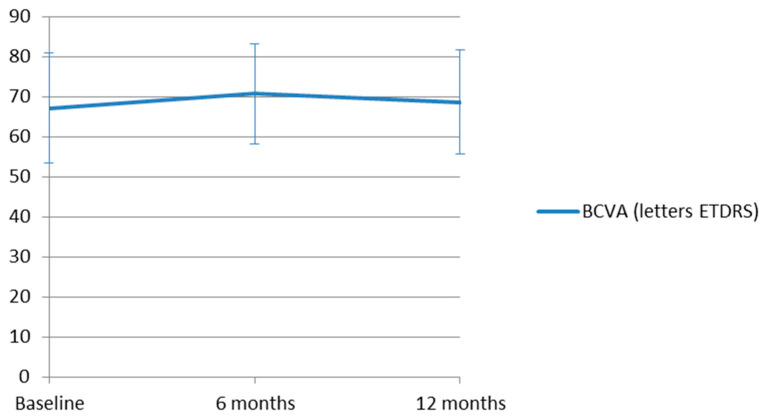 Figure 1
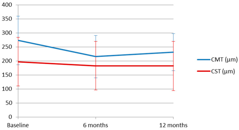 Figure 2
Peer Reviews
No public reviews on file for this paper yet. If you reviewed it on a platform where reviews are public (OpenReview, ICLR, NeurIPS, ICML), you can paste yours below so the community can read it here.
Videos
No videos yet. Explain this paper in a talk, walkthrough, or lecture? Add one.
Taxonomy
TopicsRetinal Diseases and Treatments · Ophthalmology and Visual Impairment Studies · Retinal Imaging and Analysis