MRI Diagnosis of Gymnast’s Wrist (Distal Radial Physeal Stress Injury)
Amal El Madani, Lokmane Taihi

TL;DR
MRI can help diagnose wrist injuries in young gymnasts caused by repetitive stress, enabling timely treatment.
Contribution
The paper presents a case highlighting MRI's role in diagnosing distal radial physeal stress injuries in gymnasts.
Findings
MRI showed focal widening of the distal radial physis and bone marrow edema in a 16-year-old gymnast.
Conservative treatment led to resolution of symptoms in the reported case.
Abstract
Gymnast’s wrist is an uncommon stress‑related injury of the distal radial physis caused by repetitive axial loading in skeletally immature athletes. We report the case of a 16‑year‑old elite gymnast presenting with progressive wrist pain without acute trauma. Magnetic resonance imaging demonstrated focal widening of the volar distal radial physis with adjacent metaphyseal and epiphyseal bone marrow edema, consistent with a stress‑related physeal injury. Conservative management with activity modification and physical therapy resulted in symptom resolution. Teaching point: In young athletes with wrist pain, stress‑related physeal injury should be considered, and MRI allows early diagnosis and appropriate management.
Genes, proteins, chemicals, diseases, species, mutations and cell lines named across the full text — each resolved to its canonical identifier and authoritative record.
Click any figure to enlarge with its caption.
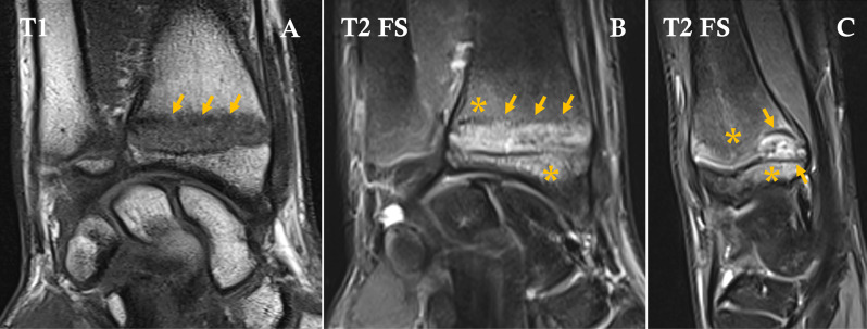 Figure 1
Peer Reviews
No public reviews on file for this paper yet. If you reviewed it on a platform where reviews are public (OpenReview, ICLR, NeurIPS, ICML), you can paste yours below so the community can read it here.
Videos
No videos yet. Explain this paper in a talk, walkthrough, or lecture? Add one.
Taxonomy
TopicsSports injuries and prevention · Muscle and Compartmental Disorders · Orthopedic Surgery and Rehabilitation