Surface-Collision Analysis of Microscale-Confined 129Xe in Pyrex Vapor Cells Based on Stem-Transport and Gradient Diffusion Dynamics
Shangtao Jiang, Tengyue Wang, Xuyang Qiu, Heng Yuan

TL;DR
This paper studies how surface collisions in Pyrex vapor cells affect the spin coherence of 129Xe atoms and proposes a method to optimize cell geometry for better performance in NMR gyroscopes.
Contribution
A new structural coupling factor and geometry correction model are introduced to predict and reduce transverse relaxation rates in Pyrex vapor cells.
Findings
The model predicts transverse relaxation rates with high accuracy (R2=0.982) across eight configurations.
Geometry optimization reduces the relaxation rate by 41.8%, from 0.225 to 0.131s−1.
The framework enables in-situ comparison of surface depolarization effects across fabrication protocols.
Abstract
Surface collisions at Pyrex walls limit the spin coherence in nuclear magnetic resonance gyroscopes (NMRG) vapor cells, while the cavity–stem junction introduces geometry dependent exchange that perturbs the transverse spin relaxation time T2 of 129Xe atoms. We combine T2 measurements with Monte Carlo simulations of confined diffusion and surface collisions to decompose the relaxation of Xe atoms and derive a cavity–stem geometry correction for wall relaxation. A structural coupling factor (SCF) is introduced to compress stem length and aperture diameter into a dimensionless metric for diffusion-limited mixing, enabling prediction of the transverse relaxation rate versus geometry. Across eight simulated configurations, the model yields R2=0.982 and agrees with experiments within 7–9%, comparable to the measurement uncertainty (±0.015s−1). Using the validated framework, geometry…
Genes, proteins, chemicals, diseases, species, mutations and cell lines named across the full text — each resolved to its canonical identifier and authoritative record.
Click any figure to enlarge with its caption.
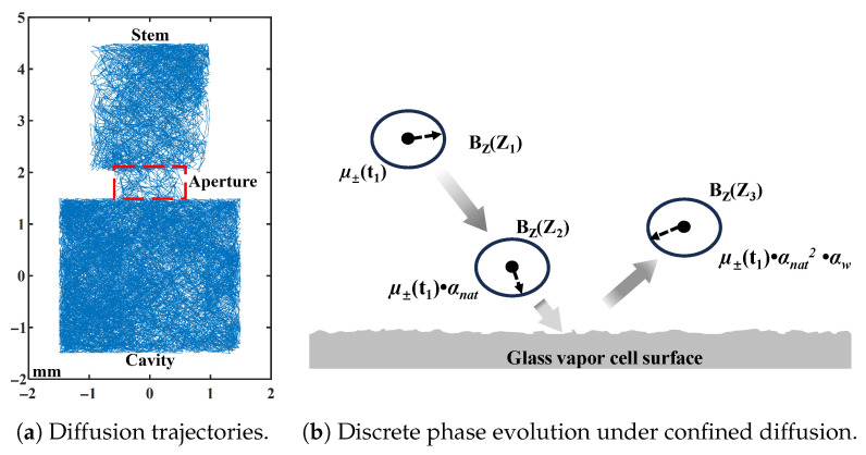 Figure 1
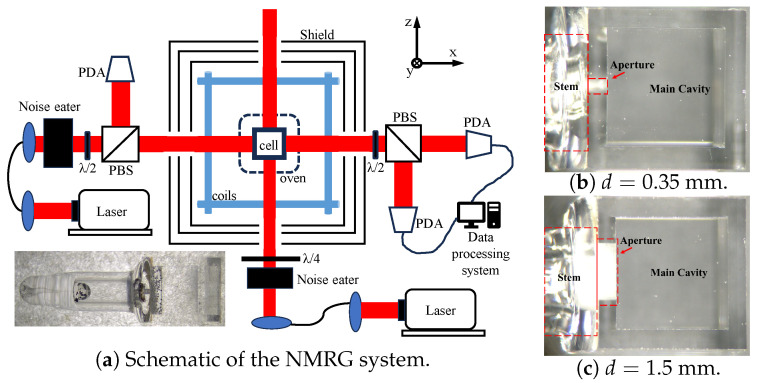 Figure 2
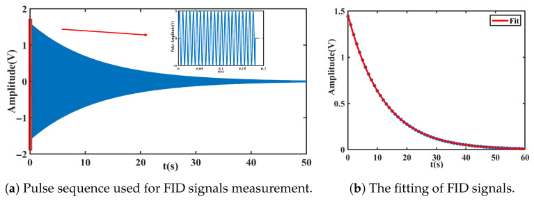 Figure 3
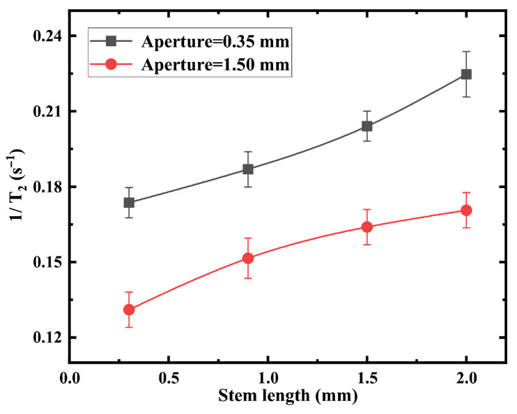 Figure 4
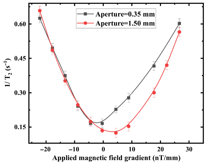 Figure 5
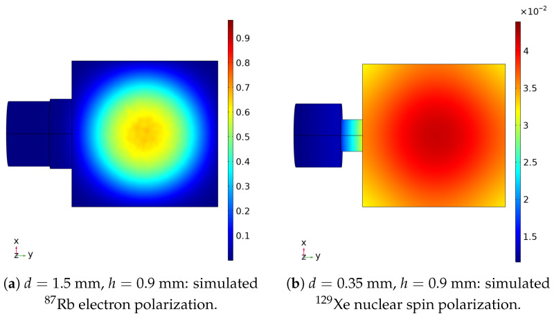 Figure 6
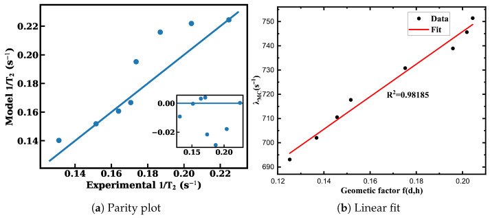 Figure 7
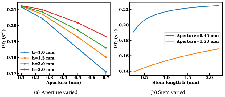 Figure 8
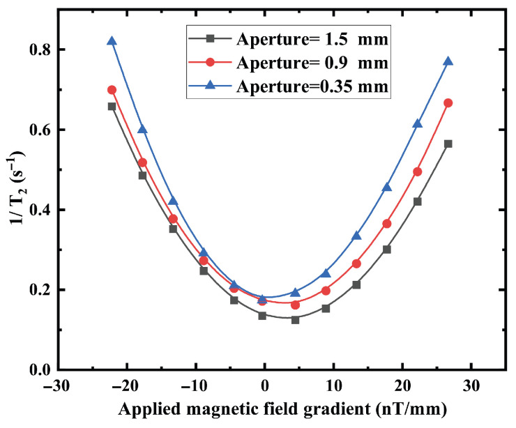 Figure 9
- —Innovation Program for Quantum Science and Technology
Peer Reviews
No public reviews on file for this paper yet. If you reviewed it on a platform where reviews are public (OpenReview, ICLR, NeurIPS, ICML), you can paste yours below so the community can read it here.
Videos
No videos yet. Explain this paper in a talk, walkthrough, or lecture? Add one.
Taxonomy
TopicsAtomic and Subatomic Physics Research · Advanced NMR Techniques and Applications · Advanced MRI Techniques and Applications
1. Introduction
Nuclear magnetic resonance gyroscopes (NMRGs) and alkali-metal–noble-gas comagnetometers require long-lived noble-gas coherence to achieve high stability and compact size in inertial sensing applications [1,2]. In these systems, the transverse relaxation time of ^129^Xe determines the linewidth of free-induction-decay (FID) measurement and coherent integration time, thereby imposing a central performance constraint in miniaturized vapor cells [3]. Diffusion-driven transverse relaxation in inhomogeneous magnetic fields is well described within the Bloch-Torrey and restricted-diffusion framework [4,5,6], where relaxation arises from the interplay of diffusion, magnetic-field gradients, and boundary interactions. As the cell dimensions shrink and the surface-to-volume ratio increases, wall-induced depolarization becomes an increasingly dominant limitation to coherence performance [7,8].
Microfabrication has enabled vapor cells with coupled cavity–stem geometries, where aperture diameter and stem length strongly influence diffusion pathways and wall-collision statistics [9]. Donley et al. demonstrated microfabricated noble-gas cells with preserved nuclear coherence [10], and Kitching highlighted geometry constraints in chip-scale atomic devices [11]. Wang et al. experimentally investigated stem-length effects on polarization-induced gradient relaxation in Xe comagnetometers [12]. Subsequent studies focused on performance improvement through gradient compensation, buffer-gas optimization, surface treatments, and signal-processing strategies [13,14,15,16]. Wu et al. analyzed surface-coating effects on Xe relaxation [17], and Xu et al. examined relaxation mechanisms in confined vapor cells [18]. Parallel theoretical efforts developed statistical and transport-based descriptions of confined spin dynamics and boundary-related relaxation [16,19,20]. Despite these advances, geometry-induced diffusion redistribution and intrinsic surface depolarization are often treated implicitly or remain coupled within geometry-specific simulations, limiting predictive comparison across stem–aperture configurations and hindering geometry-normalized assessment of inner-surface effects.
In contrast to prior studies that primarily focus on performance optimization or treat geometry-dependent effects implicitly within simulations, this work establishes an experimentally constrained and geometry-resolved description of wall-induced transverse relaxation. By explicitly linking cavity–stem structure to relaxation behavior through a compact structural descriptor, the proposed framework enables geometry-normalized comparison and provides a physically transparent basis for predictive design of miniature ^129^Xe vapor-cell systems.
2. Principle and Method
2.1. Structure-Weighted Diffusion and Boundary-Induced Relaxation
Transverse relaxation of diffusing ^129^Xe atoms in static field inhomogeneity is treated within the standard Bloch-Torrey/restricted-diffusion framework [4,5,21,22], with low-field gas scaling discussed by Cates et al. [6]. The gradient-induced contribution is written as
where is the gyromagnetic ratio, is the magnetic field along the z-axis. For the linear magnetic field gradients, [23], then
where and are set by restricted diffusion and the geometry of vapor cells. In cavity–aperture–stem cells, the aperture diameter d and stem length h redistribute diffusion trajectories through the cavity–stem exchange. A reduced two-region form is expressed by,
The equation highlights that geometry enters through the stem occupancy , defined as the probability for atoms to reside in the stem region, which represents a structure-weighted sampling of magnetic-field inhomogeneity and internal boundaries. In later sections, this exchange effect is condensed into a structural coupling factor (SCF) for design-oriented comparison.
2.2. Polarization-Field Mapping and Gradient Metrics from COMSOL Simulations
The field inhomogeneity sampled by Xe atoms is generated by polarized Rb electrons. Following standard spin-exchange optical pumping (SEOP) descriptions [24,25,26], the coupled Bloch-Torrey equations for Rb and ^129^Xe polarization are solved in COMSOL Multiphysics 6.4 software to obtain steady-state and [27]:
where and are the spin polarization of Rb and Xe atoms, B is the magnetic field, D is the diffusion coefficient of atoms, is the optical pumping rate, and R is the relaxation rate. The Rb polarization is converted to the effective field experienced by Xe via the Fermi-contact interaction,
which provides and its gradients as the input for evaluating .
The nuclear polarization field is retained to support controlled model reduction for stemmed cells. Specifically, COMSOL-derived polarization/field metrics are used later to justify the separation (Equation (10)) only in the experimentally relevant range where the effective gradient sampling shows weak dependence on h.
2.3. Monte Carlo Modeling of FID Signals and Effective Surface Parameterization
Relaxation in the full cavity–aperture–stem geometry is evaluated by a 3D Monte Carlo random walk [4,21]. The diffusive step is and the phase evolves as . Representative trajectories illustrate geometry-dependent exchange and boundary sampling, and the discrete phase accumulation used to compute is shown in Figure 1. The total decay rate is extracted by exponential fitting of .
We decompose the total relaxation as
where is the wall collisions of atoms, represents the effective spin-preservation probability per wall collision and denotes Monte Carlo extracted wall-collision frequency.
3. Experimental Setup and Procedure
3.1. Experimental Setup
Figure 2a shows the experimental configuration. Two DBR lasers were used as pump and probe sources, tuned to the Rb D_1_ and D_2_ transitions, respectively. The pump beam was circularly polarized and expanded to a 2.5 mm Gaussian profile with stabilized power of 4 mW. Optical pumping polarized ^87^Rb atoms, which transferred polarization to Xe nuclei via spin exchange [28]. The probe beam was linearly polarized and slightly detuned to detect transverse spin precession through Faraday rotation [29]. A lock-in amplifier (Zurich Instruments, Zürich, Switzerland, HF2LI) performed phase-sensitive detection. Gradient coils provided magnetic-field-gradient generation and compensation. A four-layer magnetic shield reduced the environmental magnetic interference [30].
The cubic cavity has an inner side length of 3 mm, corresponding to a volume of approximately 27 mm^3^, while the stem contributes less than 5% additional volume in the vapor cell. The cell contains ^87^Rb, 2 Torr ^129^Xe, 8 Torr ^131^Xe, and 300 Torr N_2_, and operates at the temperature of 393 K. Cells with aperture diameters of 0.35 mm and 1.5 mm were fabricated, and the initial 2 mm stem was progressively shortened to tune the structural coupling factor (Equation (13)). Figure 2b,c shows the vapor cells fabricated for experimental validation. The geometry was systematically tuned by progressively shortening the stem while preserving the integrity of the cubic sensing cavity. Although laser-based trimming may introduce localized thermal stress or surface roughness, the processed region lies outside the active sensing volume and does not participate in optical pumping or detection. No systematic change in the baseline transverse relaxation rate beyond measurement repeatability was observed after processing.
3.2. Measurement of Free Induction Decay Signals
As illustrated in the inset of Figure 3a, a pulse tips the Xe nuclear magnetization into the transverse plane. After the pulse is turned off, the transverse magnetization undergoes free precession at the Larmor frequency while decaying exponentially in time. The underlying principles follow standard spin-echo theory [31,32] as described in classical magnetic resonance literature [33]. The decay envelope is fitted to extract the transverse relaxation time , and repeated measurements are performed to evaluate the associated statistical uncertainty. A representative single-shot free-induction-decay signal is shown in Figure 3b.
4. Results and Discussion
Figure 4 shows the compensated transverse relaxation rate as a function of the length of the vapor cell stem. Shortening the stem consistently reduces the relaxation rate for both aperture diameters. The larger aperture configuration achieves systematically lower relaxation, confirming the simulation trend. Reducing h and enlarging d can suppress the transverse relaxation rate from to , corresponding to a reduction of . For the stem-length dependence (Figure 5), comparison between the repeated measured relaxation rates gives mean absolute percentage errors of 4–5%.
Figure 5 confirms the quadratic dependence of the transverse relaxation rate on magnetic-field gradient magnitude, consistent with diffusion theory in confined geometries. The curvature of the quadratic dependence decreases for geometries with larger apertures or shorter stems, indicating reduced sensitivity to gradient-induced dephasing. For the gradient-dependent measurements (Figure 4), quadratic fitting of versus applied gradient yields for mm and for mm, with maximum relative deviations below 5%. The repeated experimental uncertainties (error bars) of are predominantly within , with a few data points showing or . Considering that mainly ranges from to , the corresponding relative uncertainties are approximately 3– (typically around ), indicating influence of the reproducibility and data dispersion on the overall trend.
These results demonstrate that geometry modifies the effective sampling of magnetic-field gradients by diffusing atoms, rather than merely shifting the baseline relaxation rate. The observed trend cannot be explained solely by cavity volume change, indicating a geometry-mediated diffusion effect. To distinguish gradient-induced relaxation from wall-collision contributions, we extracted effective polarization-gradient metrics from COMSOL point clouds [34].
By solving Equation (11), the distribution of Rb electron polarization can be simulated. The parameters can be referred to our former work [35], as is shown in Figure 6. The COMSOL steady-state electron polarization is used to derive an effective field
Gradients are computed from exported COMSOL point clouds using local linear reconstruction on irregular nodes, as is shown in Table 1. For geometry comparison, we use the cubic scaling normalization
Within the investigated range, gradient metrics vary mainly with d and only weakly with h (Appendix A). We therefore write
and approximate
with
To capture the stem-length dependence, we introduce (Appendix B)
As is shown in Figure 7a, simultaneous least-squares fitting to eight experimental data points yields
The model gives with maximum relative deviation below 6.5%. The SCF form follows from diffusion conductance through a circular aperture ( with ) together with a two-compartment exchange model in which independent decorrelation channels add in rate; see Appendix B for the full derivation and definitions. The coefficient 1.62 is fixed by Monte Carlo exchange statistics under the present geometry and operating conditions.
To validate the proposed geometric wall-collision factor
As is shown in Figure 7b, a linear regression between and yields
Under the condition of dominated wall-collisions relaxation,
The values in Table 2 are averaged over all experimental geometries yields
After extracting , the Monte Carlo simulation of the FID process was carried out under the same diffusion and geometry conditions. Substituting the linear relation of gives a fully analytic relaxation model.
The simulated FID decay is then directly compared with the experiment.
To quantitatively evaluate the agreement between experiment, Monte Carlo simulation combining analytical model, the relaxation trends were compared under two structural variations: (i) stem length h and (ii) applied magnetic-field gradient.The relative deviation was calculated as
First, the relaxation rate exhibits a consistent monotonic dependence on stem length across both experimental (Figure 4) and theoretical results (Figure 8b). Increasing h leads to an increase in , reflecting enhanced wall-collision probability and extended diffusion residence in the stem region. This trend is reproduced by the theoretical model and Monte Carlo simulations, indicating that the geometry-weighted wall contribution is captured correctly at the leading order.The relative deviation between experiment and theory for the eight data points remains within approximately 6∼9%.
Second, under applied magnetic-field gradients, the relaxation rate follows a quadratic dependence, consistent with diffusion-mediated dephasing theory. To quantitatively evaluate the agreement between theoretical trend (Figure 9) and experimental data (Figure 5). Across the full magnetic field range, the relative deviations are predominantly below 5%, with an average relative deviation of approximately 3–4%, and a maximum deviation below 10% occurring at low-field conditions.
The semi-empirical linear magnetic field gradient assumptions and simplified wall-collision boundary conditions introduce theoretical uncertainties in the Monte Carlo model. In addition, laser-based stem shortening, surface roughness and stress, or contamination may contribute to experimental uncertainty. Together, these factors account for the small but measurable discrepancies between simulation and experiment. A baseline offset exists between model prediction and experiment, and small differences in quadratic curvature (concavity) appear under gradient scans. Quantitatively, for the mm aperture, the root-mean-square error (RMSE) is approximately , indicating excellent agreement. In contrast, for the mm aperture, the RMSE increases to approximately .
This suggests that under smaller aperture conditions, either the wall-relaxation contribution or the effective gradient correction term may be slightly underestimated. Alternatively, additional surface-related depolarization mechanisms may contribute in the experiment but are not fully captured in the present model. The overall deviation magnitude is approximately while the measured relaxation range is – , corresponding to a relative error of approximately 6– . Considering that the experimental uncertainty of is on the order of 3–10%, the observed deviations fall within or close to the experimental error bounds. This confirms quantitative consistency within experimental uncertainty.
The model assumes linear magnetic-field gradients and idealized wall-collision boundary conditions in the Monte Carlo simulations. Each FID configuration was measured five times; the error bars reflect the standard deviation from exponential fitting. Within these uncertainties, the trends are reproducible, and validation across 20 geometries (Table 1) provides an internal consistency check on the extracted wall parameter and gradient contribution, respectively.
5. Conclusions
This work combines systematic measurements with Monte Carlo simulations of surface-collision statistics and introduces SCF to describe diffusive exchange between the sensing cavity and the stem. Based on this framework, we establish a relaxation model that relates the measured transverse relaxation rate to stem length and aperture size. Fitting is performed on eight experimentally tested configurations with a coefficient of determination , and the framework is cross-checked against 20 simulated geometries. Repeated FID measurements yield uncertainties (error bars) within . The model-predicted relaxation rates differ from the experimental values by approximately 6– , remaining within or near the experimental uncertainty range. Guided by the model, geometry optimization reduces the relaxation rate from to , corresponding to a improvement in coherence performance. From a materials perspective, this Pyrex surface-collisional analysis supports in-situ, -based comparison of effective surface depolarization across fabrication routes and surface treatments, while explicitly accounting for the cavity–stem junction that can bias the apparent relaxation. This offers a practical tool for microscale vapor-cell design and evaluation: once cavity–stem exchange and gradient–diffusion effects are accounted for, can be used to compare effective surface depolarization across fabrication, cleaning, and coating protocols. Future work will refine surface-loss models, correlate the effective surface term with independent surface metrology, and extend the approach to other cell architectures and materials.
The reference list from the paper itself. Each links out to its DOI / PubMed record.
- 1Kornack T.W. Ghosh R.K. Romalis M.V. Nuclear Spin Gyroscope Based on an Atomic Comagnetometer Phys. Rev. Lett.20059523080110.1103/Phys Rev Lett.95.23080116384290 · doi ↗ · pubmed ↗
- 2Donley E.A. Nuclear Magnetic Resonance Gyroscopes IEEE Sens. J.20101118431858
- 3Wei K. Zhao T. Fang X. Xu Z. Liu C. Cao Q. Wickenbrock A. Hu Y. Ji W. Fang J. Ultrasensitive Atomic Comagnetometer with Enhanced Nuclear Spin Coherence Phys. Rev. Lett.202313006320110.1103/Phys Rev Lett.130.06320136827554 · doi ↗ · pubmed ↗
- 4Torrey H.C. Bloch Equations with Diffusion Terms Phys. Rev.195610456356510.1103/Phys Rev.104.563 · doi ↗
- 5Mc Gregor D.D. Transverse Relaxation of Spin-Polarized Gas Due to a Magnetic Field Gradient J. Magn. Reson.19909051352510.1103/physreva.41.26319903396 · doi ↗ · pubmed ↗
- 6Cates G.D. Schaefer S.R. Happer W. Relaxation of Spins Due to Field Inhomogeneities in Gaseous Samples at Low Magnetic Fields and Low Pressures Phys. Rev. A 1988372877288510.1103/Phys Rev A.37.28779900016 · doi ↗ · pubmed ↗
- 7Sun Q. Wang L. Liu M. Surface Engineering Effects on Noble-Gas Spin Relaxation in Vapor Cells Materials 2023163312
- 8Zhou P. Zhao R. Lin J. Advances in Atomic Magnetometry for Inertial Applications Materials 2024171125