DHX9 sustains hematopoietic stem cell function in cooperation with H3 acetylation
Minhui Shi, Mengqing Gao, Huixin Luo, Chong Wang, Xueyang Hu, Yacen Xiong, Yan Chen, Xingxing Ren, Shu Zhu, Huaiping Zhu

TL;DR
This study shows that DHX9 helps maintain hematopoietic stem cells by regulating histone acetylation, and its absence leads to blood cell production issues.
Contribution
DHX9 is identified as a novel regulator of hematopoietic stem cell function through its role in maintaining H3 acetylation.
Findings
Dhx9 deletion causes bone marrow failure and impaired hematopoietic reconstitution due to loss of HSCs.
DHX9 maintains H3 acetylation at hematopoietic gene promoters to promote transcription activation.
Restoring H3K27ac levels partially rescues hematopoietic defects in DHX9-deficient cells.
Abstract
Hematopoietic stem cells (HSCs) self-renew to sustain stem cell pools and differentiate into all types of blood cells, whose properties are tightly regulated by epigenetic and transcriptional networks. Here, we identified DHX9 as a critical regulator of HSC maintenance. Dhx9 deletion caused bone marrow failure and impaired hematopoietic reconstitution in murine primary and secondary transplantation recipients due to loss of HSCs and defective self-renewal capacity. Further investigations revealed that Dhx9 deficiency led to aberrant cell cycle entry, increased apoptosis, and elevated ROS, which compromise HSC function. Mechanistically, DHX9 interacts with CBP/p300 acetyltransferase and maintains H3 acetylation at hematopoietic gene promoters to facilitate transcription activation. Inhibition of CBP/p300 disrupted their expression, whereas the enhancement of H3K27ac levels partially…
Genes, proteins, chemicals, diseases, species, mutations and cell lines named across the full text — each resolved to its canonical identifier and authoritative record.
Click any figure to enlarge with its caption.
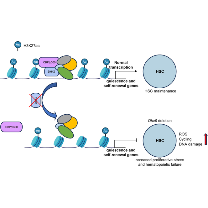 Figure 1
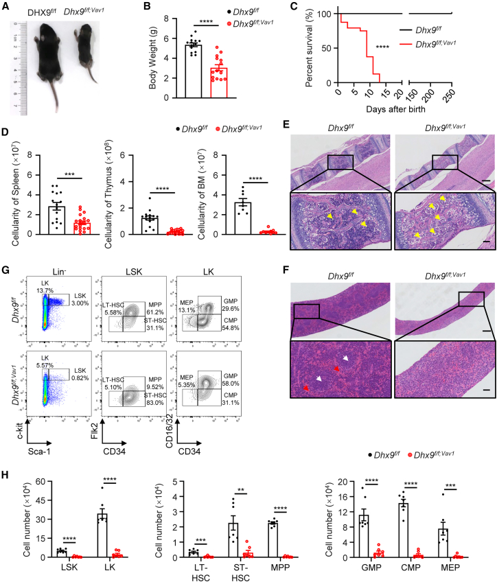 Figure 2
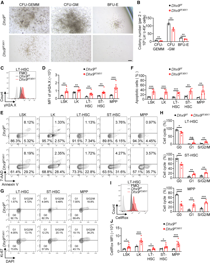 Figure 3
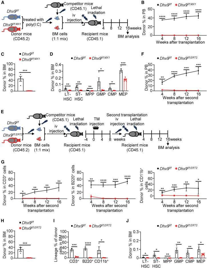 Figure 4
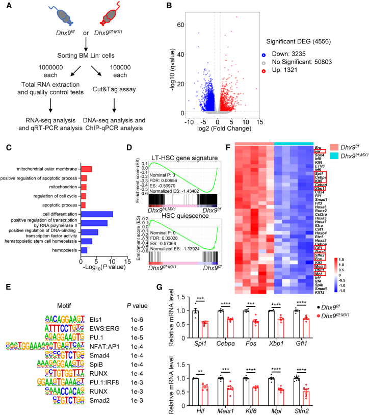 Figure 5
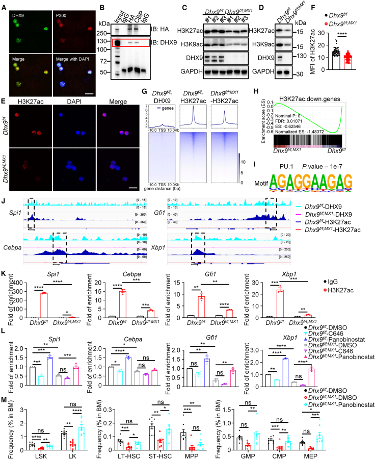 Figure 6
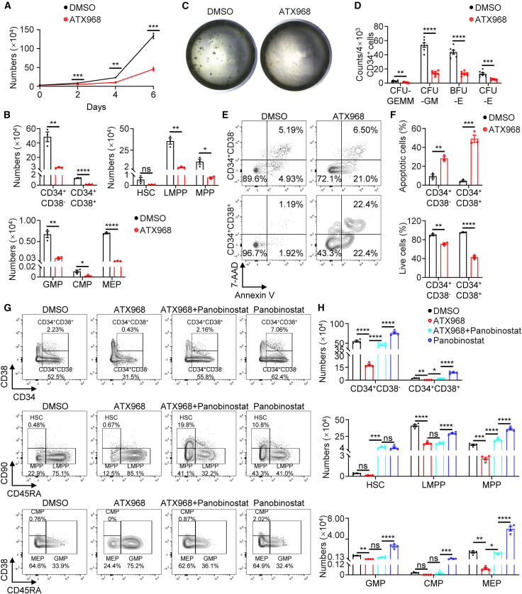 Figure 7
Peer Reviews
No public reviews on file for this paper yet. If you reviewed it on a platform where reviews are public (OpenReview, ICLR, NeurIPS, ICML), you can paste yours below so the community can read it here.
Videos
No videos yet. Explain this paper in a talk, walkthrough, or lecture? Add one.
Taxonomy
TopicsEpigenetics and DNA Methylation · Hematopoietic Stem Cell Transplantation · Genomics and Chromatin Dynamics
Introduction
Hematopoietic stem cells (HSCs) can renew themselves and differentiate into all lineages of blood cells throughout their lifetime (Boulais and Frenette, 2015). HSC function is tightly regulated by intrinsic mechanisms and extrinsic cues, which result in an intricate molecular state to balance self-renewal and differentiation (Mendelson and Frenette, 2014; Morrison and Scadden, 2014). Intrinsic factors involved in cellular metabolism, and epigenetic, or transcriptional regulation, have been reported to regulate HSC functions (Dzierzak and Bigas, 2018; Meng et al., 2023; Suda et al., 2011). Epigenetic mechanisms that dictate chromatin accessibility for transcription factors are crucial for cell fate decisions (Butler and Dent, 2013). Previous studies have shown that DNA methylation and histone modification dynamics, such as H3K27ac, H4K16ac, and H3K79me2, are required for HSC self-renewal and differentiation (Meng and Nerlov, 2025).
DEAH-box helicase 9 (DHX9), also known as RNA helicase A (RHA), a member of the DExD/H-box superfamily II of helicases, plays a central role in many cellular processes, including DNA replication, transcription, translation, RNA processing and transport, and maintenance of genomic stability (Ren et al., 2024). Global knockout of Dhx9 is embryonic lethal in mice because of a differentiation defect in the embryonic ectoderm during gastrulation (Lee et al., 1998), and embryonic stem cells depleted of Dhx9 are unable to differentiate (Leone et al., 2017), indicating that DHX9 is critical in early embryonic development. The RHA-1/L1TD1/LIN28 complex promotes the translation of the stem cell factor Oct4 by recruiting RHA-1 into the translation complex in human embryonic stem cells (hESCs) (Närvä et al., 2012), indicating a role for DHX9 in hESC renewal. These data indicate that DHX9 is essential during development. However, the role of DHX9 in HSC biology is unknown.
In this study, a critical role of DHX9 in HSC function was reported in mice with the conditional deletion of Dhx9. Deletion of Dhx9 in hematopoietic cells led to hematopoietic failure of multilineage blood cells. Serial transplantation revealed that Dhx9 deficiency resulted in impaired HSC reconstitution. We demonstrated that DHX9 promotes histone H3 acetylation at lysines 9 and 27 (K9, K27) at hematopoietic gene loci, possibly through interactions with CBP/p300. Genome-wide analysis via CUT&Tag revealed that H3K27ac signals at promoter regions of hematopoietic genes and regulators were decreased in Dhx9 knockout cells, which is consistent with their decreased expression according to transcriptomic profiling. Notably, these genes also overlapped with the binding peaks of DHX9. Our data suggest that DHX9-mediated gene transcription occurs through H3 acetylation, serving as a mechanism for controlling HSC self-renewal.
Results
Genetic deletion of DEAH-box helicase 9 in hematopoietic cells leads to defective hematopoiesis
We first analyzed public datasets ([GSE111085](GSE111085) and [GSE16334](GSE16334)) and found that Dhx9 expression was markedly reduced in patients with myelodysplastic syndrome (MDS) and Fanconi anemia (FA). Consistently, clinical samples collected from patients with aplastic anemia (AA) also showed decreased DHX9 expression compared to those from healthy donors (Figures S1A–S1C), suggesting that DHX9 might be involved in the regulation of hematopoiesis. To explore the role of DHX9 in hematopoiesis, we generated mice with the specific deletion of Dhx9 in hematopoietic cells (Dhx9^f/f;Vav1^) by crossing Dhx9^f/f^ (WT) (Ren et al., 2023) mice with Vav1-Cre mice (Figure S1D). We confirmed the effective deletion of DHX9 in splenocytes, thymocytes, and bone marrow (BM) cells in Dhx9^f/f;Vav1^ mice (Figures S1E and S1F).
The Dhx9^f/f;Vav1^ mice were undersized, accompanied by reduced thymus size and pale organ, and had a significantly shortened lifespan of approximately 2 weeks after birth (Figures 1A–1C and S1G). Complete blood count analysis showed significantly reduced white blood cells (WBCs), red blood cells (RBCs), hematocrit, and hemoglobin with an increased mean corpuscular volume (MCV). While there is no change in the levels of platelet (PLT) (Figure S1H). Reduced cellularity was observed in the spleen, thymus, and BM of Dhx9^f/f;Vav1^ mice (Figure 1D). Histological analysis further showed that Dhx9^f/f;Vav1^ mice had decreased BM cells, increased adipose infiltration in vertebrae (Figure 1E), and a disorganized structure of white and red pulp in the spleen (Figure 1F), suggesting defective hematopoiesis. We then examined the number of primitive hematopoietic stem and progenitor cells (HSPCs) in the BM of Dhx9^f/f^ mice and Dhx9^f/f;Vav1^ mice. Loss of Dhx9 significantly reduced the absolute number of Lin^−^Sca-1^+^c-Kit^+^ (LSK) cells, Lin^−^Sca-1^−^c-Kit^+^ (LK) cells, long-term repopulating HSCs (LT-HSCs: Flk2^−^CD34^−^LSK), short-term repopulating HSCs (ST-HSCs: Flk2^−^CD34^+^LSK), multipotent progenitors (MPP: Flk2^+^CD34^+^LSK), and fate-committed progenitors, including granulocyte-macrophage progenitors (GMPs: CD16/32^+^CD34^+^LK), common myeloid progenitors (CMPs: CD16/32^+^CD34^−^LK), and megakaryocyte-erythroid progenitors (MEPs: CD16/32^−^CD34^−^LK) (Figures 1G and 1H). Together, these data suggest that DHX9 is required for multilineage development in murine hematopoiesis.Figure 1Dhx9 deletion results in the lethality and depletion of hematopoietic stem cells(A) A representative image of 12-day-old Dhx9^f/f^ and Dhx9^f/f;Vav1^ mice.(B) Body weights of Dhx9^f/f^ and Dhx9^f/f;Vav1^ mice (n = 14).(C) Kaplan‒Meier survival curves of Dhx9^f/f^ and Dhx9^f/f;Vav1^ mice (n = 28).(D) Total nucleated cell (TNC) numbers in the spleen (left), thymus (middle), and in the femurs and two tibias (n ≥ 8).(E and F) Representative hematoxylin and eosin (HE)-stained vertebrae (E) and spleens (F) from Dhx9^f/f^ and Dhx9^f/f;Vav1^ mice. Scale bars, 250 μm (top) and 50 μm (bottom). Yellow arrows, adipocytes; red arrows, red pulp; white arrows, white pulp.(G) Representative fluorescence-activated cell sorting (FACS) analyses of LK cells, LSK cells, HSPCs, and lineage-restricted progenitors from Dhx9^f/f^ and Dhx9^f/f;Vav1^ mice.(H) Absolute numbers of different HSPC populations from Dhx9^f/f^ and Dhx9^f/f;Vav1^ mice (n = 7).All mice used in the experiments were 7–14 days old. The data are presented as the mean ± SEM. ^∗∗^p < 0.01; ^∗∗∗^p < 0.001; ^∗∗∗∗^p < 0.0001; statistical significance was determined using the log rank test for (C) and unpaired two-tailed Student’s t test for (B), (D) and (H).See also Figure S1.
Knockout of DEAH-box helicase 9 impairs hematopoietic stem cell survival
To determine the role of DHX9 in adult hematopoiesis, we generated Dhx9^f/f;MX1^ (cKO) mice, in which Cre expression was induced by injecting polyinosine-polycytidylic acid [poly(I:C)], leading to the reduced expression of DHX9 in BM (Figures S2A and S2B). After poly(I:C) injection, Dhx9^f/f;MX1^ mice ultimately resulted in lethality and recapitulated the phenotype of Dhx9^f/f;Vav1^ mice, with reduced volume of thymus, pale kidneys and blood, and decreased BM cellularity (Figures S2C–S2E). Hematologic analysis further revealed similar alterations in both models, except that cKO mice showed a transient increase in PLT counts. A consecutive five-day assessment confirmed that PLT levels initially rose but subsequently returned to baseline, ultimately showing no significant difference from controls, consistent with the phenotype observed in Dhx9^f/f;Vav1^ mice (Figures S2F and S2G).
Flow cytometric analysis was conducted to further assess the impact of Dhx9 deletion on HSPC homeostasis (Figure S2H). Consistent with previous findings, a marked reduction in multiple HSPC subsets was observed following poly(I:C) administration in Dhx9^f/f;MX1^ mice (Figure S2I). This decrease was further corroborated using an alternative gating strategy, which similarly revealed a significant decline in HSPC populations, including the HSC^SLAM^ population (CD150^+^CD48^−^LSK) and MPP^SLAM^ population (CD150^−^CD48^−^LSK) (Figure S2J), indicating that Dhx9 deletion leads to HSC exhaustion. Interestingly, the proportion of CD41^+^CD150^+^ cells within the LK population remained comparable (Figure S2K).
To characterize the role of DHX9 in HSPCs, we sorted Lin^-^c-Kit^+^ cells and performed a colony-forming unit (CFU) assay in vitro. Dhx9-deficient cells showed decreased number and size of multipotential colony-forming units (CFU-GEMM), granulocyte-macrophage colony-forming units (CFU-GM), and erythroid burst-forming units (BFU-E) (Figures 2A and 2B). We next assessed DNA damage, apoptosis, and cell cycle in WT and Dhx9-deficient HSPCs. γH2A.X levels, a marker of DNA damage, were strongly increased in cKO HSPCs (Figures 2C and 2D). In addition, a significant increase in the frequency of apoptotic cells in the total BM, suggested that Dhx9 deletion resulted in elevated apoptosis in HSPCs (Figures 2E and 2F). Cell cycle analysis revealed an increased proportion of the S/G2/M phase in LT-HSCs, ST-HSCs and MPPs after Dhx9 deletion (Figures 2G and 2H), indicating cell cycle entry of HSPCs, which could lead to the loss of quiescence and exhaustion of the HSPC pool. Given the pivotal role of low intracellular reactive oxygen species (ROS) levels in maintaining HSC quiescence (Hu et al., 2018; Singh et al., 2018), we assessed and found significantly increased ROS production in cKO mice (Figures 2I and 2J). These findings indicate that DHX9 is required for HSC survival and maintenance.Figure 2Dhx9-deficient HSCs have increased cycling and apoptosis(A) Representative images of CFU assays of Lin^-^c-Kit^+^ cells from WT and cKO mice. Scale bars, 100 μm.(B) Hematopoietic colonies were enumerated from methylcellulose cultures plated with 2 × 10^3^ Lin^-^c-Kit^+^ cells (n = 4).(C and D) Representative FACS analyses (C) and quantitation of γH2A.X mean fluorescence intensity (MFI) (D) in WT (n = 6) and cKO (n = 7) hematopoietic cells.(E–H) Representative FACS plots show apoptosis (E) and cell cycle (G) in HSPCs. The percentages of apoptotic HSPCs (annexin V^+^) (F) and HSPCs in the G0, G1, and S/G2/M phases (H) were quantified (n = 5).(I and J) ROS levels in WT (n = 6) and cKO (n = 8) HSPCs were determined via CellRox Green.All mice used in these experiments were analyzed seven days after the first poly(I:C) injection. The data are presented as the mean ± SEM. ^∗^p < 0.05; ^∗∗^p < 0.01; ^∗∗∗^p < 0.001; ^∗∗∗∗^p < 0.0001; ns, not statistically significant; unpaired two-tailed Student’s t test.See also Figure S2.
DEAH-box helicase 9 is required for hematopoietic stem cell reconstitution and self-renewal in vivo
We next determined the role of DHX9 in HSC reconstitution in vivo. Since homozygous knockout of Dhx9 (Dhx9^f/f;Vav1^ or Dhx9^f/f;MX1^) causes rapid lethality due to hematopoietic failure, we performed a competitive BM transplantation (BMT) assay (Figure 3A), which can ensure the survival of recipient mice with severe defects caused by Dhx9 deletion. Donor chimerism, represented by the total CD45.2^+^ frequency in peripheral blood (PB), was assessed every 4 weeks after transplantation. Notably, the frequency of total chimerism from cKO mice was considerably lower at 4 weeks and did not recover 16 weeks after transplantation (Figure 3B). At the end of the experiment, BM chimerism was markedly reduced in Dhx9^f/f;MX1^ recipients (Figure 3C). Donor-derived HSPC populations from Dhx9^f/f;MX1^ mice also decreased robustly (Figure 3D), suggesting impaired HSC reconstitution.Figure 3DHX9 is required for HSC reconstitution and self-renewal(A) Schedule of competitive BMT. BM cells from Dhx9^f/f^ or Dhx9^f/f;MX1^ mice (CD45.2) were collected one week after the first dose of poly(I:C) injection and transplanted with WT competitor cells (CD45.1) into lethally irradiated WT recipients (CD45.1); iv, intravenous injection.(B) CD45.2^+^ donor chimerism in the PB of recipient mice was measured every 4 weeks, and the results are graphed. WT (n = 4) and cKO (n = 5).(C) Percentages of CD45.2^+^ cells in the BM of BMT recipient mice 16 weeks after transplantation. WT (n = 4) and cKO (n = 5).(D) Percentages of CD45.2^+^ HSPC subpopulations in the BM of primary transplant recipient mice at 16 weeks. WT (n = 4) and cKO (n = 5).(E) Schematic of competitive reconstitution assays using the UBC-CreERT2 system. BM cells from 8-week-old Dhx9^f/f^ and Dhx9^f/f;ERT2^ mice (CD45.2) were transplanted into lethally irradiated WT recipient mice (CD45.1) with the same number of competitor BM cells (CD45.1) (5×10^5^ cells). Four weeks after transplantation, the recipient mice were injected with tamoxifen 5 times every second day. PB analyses were performed every 4 weeks, and BM analysis was performed 8 weeks after tamoxifen injection, while secondary BMT was performed by injecting 1×10^6^ total BM cells from mice receiving primary transplants into secondary recipients. TM, tamoxifen.(F–J) The contribution of CD45.2^+^ cells in total CD45^+^, T, B, and myeloid cells from PB (F, G) or BM (H, I), and HSCs and other hematopoietic populations in the BM (J) was analyzed in secondary BMT recipients. Dhx9^f/f^ (n = 3) and Dhx9^f/f;ERT2^ (n = 5).The data are presented as the mean ± SEM. ^∗^p < 0.05; ^∗∗^p < 0.01; ^∗∗∗^p < 0.001; and ^∗∗∗∗^p < 0.0001; unpaired two-tailed Student’s t test.See also Figure S3.
To determine whether the Dhx9 depletion-mediated HSC repopulation defects are cell-intrinsic and independent of deletion timing and HSC-homing ability, we introduced a Dhx9^f/f;ERT2^ mouse model for BMT in which Dhx9 deletion was induced by injecting tamoxifen following BMT (Figure 3E). Before tamoxifen injection, Dhx9^f/f;ERT2^ mice had body weights and blood cell counts similar to controls (Figures S3A and S3B). However, tamoxifen administration resulted in reduced body weight and severe anemia, phenocopying Dhx9^f/f;MX1^ mice (Figures S3C–S3E). Furthermore, BM cells isolated from Dhx9^f/f^ and Dhx9^f/f;ERT2^ mice were subsequently transplanted into lethally irradiated recipient mice, followed by tamoxifen treatment 4 weeks later (Figure 3E). Prior to tamoxifen administration, the percentages of donor-derived cells from Dhx9^f/f;ERT2^ mice were comparable to those from Dhx9^f/f^ controls (Figure S3F). However, after Dhx9 depletion, a significant reduction in reconstitution was observed in PB of recipient mice (Figure S3G). At the end of 8 weeks, the contribution of CD45.2^+^ donor-derived cells in total CD45^+^, T (CD3^+^), B (B220^+^), and myeloid (CD11b^+^) cells, and LSK, LK, and HSCs in the BM was drastically reduced in Dhx9^f/f;ERT2^ transplant recipients (Figures S3H–S3K), confirming the defects associated with long-term HSC reconstitution. Collectively, these results suggest that the role of DHX9 in HSC maintenance in vivo is cell-intrinsic and is not due to defects in homing.
To further examine the effect of Dhx9 loss on HSC self-renewal, we performed secondary BMT (Figure 3E). Notably, defective reconstitution of long-term hematopoiesis was demonstrated by decreased chimerism in PB for 16 weeks after secondary transplantation in Dhx9^f/f;ERT2^ mice (Figures 3F and 3G). Similarly, reduced percentages of donor-derived total and multilineage cells, as well as HSPC populations, were observed in the BM of Dhx9^f/f;ERT2^ mice at the endpoint of transplantation (Figures 3H–3J). These data indicate that Dhx9-deleted mice have inferior BM-repopulating LT-HSCs with reduced self-renewal. In summary, these data suggest that the loss of Dhx9 impaired the long-term self-renewal capacity of HSCs.
DEAH-box helicase 9 sustains the hematopoietic gene expression program
To gain mechanistic insights into how DHX9 maintains HSC function, we characterized the transcriptome profiles of Lin^−^ cells in WT and cKO mice (Figure 4A). We identified 1321 significantly upregulated and 3235 downregulated genes after Dhx9 knockout (fold change >2, Padj < 0.05) (Figure 4B). Gene Ontology (GO) analysis of the upregulated genes showed enrichment in pathways associated with mitochondrion, cell cycle, and apoptosis in cKO cells (Figure 4C), which was consistent with increased ROS, cell cycle, and DNA damage observed in these cells (Figures 2E–2J). In addition, genes associated with hematopoiesis, HSC homeostasis, and cell differentiation were downregulated in cKO mice (Figure 4C). Consistent with the observed hematopoietic defects, gene set enrichment analysis (GSEA) revealed that the dramatically downregulated gene sets included those related to HSC quiescence (Forsberg et al., 2010) and LT-HSC signature (Figure 4D) (Ficara et al., 2008). Single-cell RNA-seq analysis of Lin^-^c-Kit^+^ cells further demonstrated that gene signatures associated with LT-HSC signature were markedly reduced in the HSC cluster of Dhx9-deficient mice, accompanied by a pronounced decrease in the proportion of HSCs and an increase in cycling HSC compared with controls (Figures S4A–S4C). Furthermore, motif analysis predicted the enrichment of PU.1 (SPI1), RUNX, and ERG DNA binding motifs in the promoters of these downregulated genes (Figure 4E), which was consistent with BM failure. Among the significantly downregulated genes following Dhx9 depletion, several key hematopoietic transcription factors that maintain HSC quiescence and self-renewal, including Spi1, Cebpa, Gfi1, Xbp1, Fos, Hlf, Meis1, and Klf6, as well as other critical stem cell genes such as Mpl and Slfn2 were found (Figure 4F), which were further validated by qRT-PCR analysis (Figure 4G). In conclusion, these results indicate that DHX9 is required for the expression of hematopoiesis-maintenance genes.Figure 4DHX9 sustains the hematopoietic gene expression program(A) Schematic illustration of the experimental design.(B) Volcano plot shows the genes differentially expressed between WT and cKO Lin^−^ hematopoietic cells. Genes with an adjusted p value (Padj) < 0.05 are highlighted (blue: genes downregulated in cKO Lin^−^ cells; red: genes upregulated in cKO Lin^−^ cells).(C) Pathway analysis of genes associated with RNA-seq downregulated (blue) or upregulated (red) regions in cKO Lin^−^ cells.(D) GSEA of cKO Lin^−^ hematopoietic cells compared with WT Lin^−^ hematopoietic cells. ES, enrichment score; FDR, false discovery rate.(E) Transcription factor motifs enriched in TSS (−1 kb to +1 bp) of cKO Lin^−^ cell-downregulated genes.(F) Heatmap shows hematopoiesis-related genes, as revealed by transcriptomic studies. The genes in red boxes represent critical hematopoietic genes for maintaining HSC quiescence and self-renewal.(G) mRNA expression of the significantly changed hematopoiesis-related genes in cKO Lin^−^ hematopoietic cells (n ≥ 4).The data are presented as the mean ± SEM. ^∗∗^p < 0.01; ^∗∗∗^p < 0.001; ^∗∗∗∗^p < 0.0001; unpaired two-tailed Student’s t test.
DEAH-box helicase 9 interacts with CBP/p300 and maintains histone acetylation for transcriptional activation
Previous studies have shown that DHX9 binds to the transcriptional coactivators CBP/p300 and RNA Pol II at the promoter regions to regulate gene transcription (Nakajima et al., 1997; Ren et al., 2023). Based on this, we hypothesized that DHX9 cooperates with CBP/p300 in regulating HSC quiescence and self-renewal genes. We observed the colocalization of DHX9 and p300 in murine Lin^−^ cells via immunofluorescence (Figure 5A). This interaction was further confirmed through co-immunoprecipitation in 293T cells, where DHX9 was eluted from CBP immunoprecipitates (Figure 5B). Since CBP/p300 are well-known acetyltransferases (Weinert et al., 2018), we assessed histone acetylation and found that Dhx9 depletion led to a global reduction in histone H3 acetylation, including the downregulation of histone H3 acetylation at lysines 9 and 27 (H3K9ac, H3K27ac) (Figures 5C–5F and S4D). We therefore hypothesized that DHX9 could sustain hematopoietic gene transcription in cooperation with histone acetylation by interacting with CBP/p300 at gene promoters.Figure 5. Cooperation between DHX9 and CBP/p300 maintains hematopoietic homeostasis via H3K27 acetylation(A) Representative confocal microscopy images of Dhx9^f/f^ Lin^−^ cells staining by DHX9 and p300 are shown as indicated. Scale bars, 20 μm.(B) 293T cells were co-transfected with pLVX-DHX9 and pEnCMV-*CREBBP-*HA-SV40-Neo. After 48 h, cell lysates were collected, then immunoprecipitated, and blotted as indicated.(C and D) Western blot analyses of H3K9ac and H3K27ac in TNCs (C) or freshly isolated Lin^−^ cells (D) from the BM of WT and cKO mice.(E and F) Confocal microscopy images (E) and quantification of H3K27ac MFI (F) in WT and cKO Lin^−^ cells. Each dot represents a single cell from 3 mice per group. Scale bars, 20 μm.(G) Heatmaps show CUT&Tag signals of DHX9 or H3K27ac centered on the TSS region in Lin^−^ cells sorted from WT and cKO mice.(H) Leading-edge plots show the enrichment of genes associated with H3K27ac-downregulated regions based on GSEA of WT and cKO Lin^−^ cells. ES, enrichment score; FDR, false discovery rate.(I) Transcription factor motifs enriched in H3K27ac genomic regions downregulated in cKO Lin^−^ cells.(J) IGV genome browser tracks show CUT&Tag sequencing data of DHX9 (WT Lin^−^ cells)–, DHX9 (cKO Lin^−^ cells)–, H3K27ac (WT Lin^−^ cells)–, and H3K27ac (cKO Lin^−^ cells)–bound promoter regions of hematopoiesis-related genes.(K) ChIP-qPCR analysis of H3K27ac-bound sites in hematopoiesis-related gene (Spi1, Cebpa, Gfi1, Xbp1) promoters with or without Dhx9 (n = 3).(L) qRT-PCR analysis of hematopoiesis-related genes in WT and cKO hematopoietic cells after treatment with 40 μM C646 or 20 nM panobinostat (n = 3).(M) Ratios of different HSPC populations in the BM of mice treated with DMSO or 5 mg/kg panobinostat via intraperitoneal injection every two days for a total of five doses, starting seven days after the first poly(I:C) injection (n = 8).The data are presented as the mean ± SEM. ^∗^p < 0.05; ^∗∗^p < 0.01; ^∗∗∗^p < 0.001; ^∗∗∗∗^p < 0.0001; ns, not statistically significant; unpaired two-tailed Student’s t test (F and K), one-way ANOVA with Tukey’s test (L and M).See also Figures S4 and S5.
To confirm our hypothesis, we additionally performed CUT&Tag analysis to characterize the genomic distribution of H3K27ac and DHX9 in murine Lin^−^ cells and compared the results with transcriptomic data to address their effects on hematopoietic gene transcription (Figure 4A). Peak analysis of DHX9-occupied regions revealed that DHX9 was localized mostly to distal intergenic (34.37%), other intron (23.72%), promoter (21.28%), and first intron (10.23%) regions (Figure S4E). Compared with random genomic distributions (Mould et al., 2015), these data suggest an enrichment of promoter localization of DHX9-binding regions. Furthermore, we found that genes whose promoter regions were bound by DHX9 were significantly downregulated in cKO Lin^−^ cells (Figure S4F), indicating a regulatory role of DHX9 in gene transcription. Motif analysis of DHX9-binding peaks revealed the enrichment of most hematopoietic transcription factors, such as PU.1, IRF8, and ERG DNA binding motifs (Figure S4G), which was consistent with the downregulated motifs in the transcriptomic results (Figure 4E).
Density map analysis of H3K27ac CUT&Tag signal revealed that H3K27ac in all gene transcription start sites (TSS) ± 10 kb regions was moderately decreased in cKO cells (Figure 5G). Specifically, we found that loss of H3K27ac occupancy in TSS regions was strongly associated with transcriptional downregulation upon DHX9 depletion (Figure 5H). In addition, hematopoietic cell lineage signatures and related TF motifs, such as PU.1, which was among the decreased H3K27ac motifs, as well as the DHX9-binding motif (Figures 5I, S4G, and S4H), were consistent with the transcriptional profiles following Dhx9 deletion (Figure 4).
To further investigate the role of H3K27ac in DHX9-mediated transcriptional regulation of HSC function, we assessed H3K27ac enrichment at the promoter regions of key hematopoietic genes that are significantly downregulated upon Dhx9 deletion. Analysis using the IGV genome browser revealed a reduction in H3K27ac peaks at these hematopoietic genes in cKO cells compared to controls. Notably, these genes also coincided with DHX9-binding sites (Figures 5J and S4I). The decreased H3K27ac levels and DHX9 occupancy across these loci were validated by chromatin immunoprecipitation (ChIP)-qPCR (Figures 5K, S4J, and S4K). Electrophoretic mobility shift assays (EMSA) were performed using biotin-labeled DNA probes derived from the XBP1 promoter region identified by CUT&Tag. Incubation with nuclear extracts from DHX9-overexpressing 293T cells led to a supershift upon the addition of a DHX9 antibody (Figure S4L), suggesting that DHX9 functionally associates with chromatin and may promote CBP/p300-mediated histone acetylation. To further confirm the involvement of H3K27ac in DHX9-driven gene expression, we treated Dhx9^f/f^ -hematopoietic cells with the CBP/p300 inhibitor C646, resulting in a downregulation of key hematopoietic genes. Conversely, exposure to the broad-spectrum histone deacetylase inhibitor (HDACi) panobinostat increased the expression of these genes in WT cells. Similar expression trends were observed in cKO cells, although the magnitude of these changes was generally less pronounced (Figures 5L, S5A, and S5B).
To further explore the role of H3K27ac in hematopoiesis in vivo, cKO mice were treated with panobinostat. Panobinostat treatment markedly increased the ratios of multiple HSPC populations, with a trend toward higher total BM cellularity (Figures 5M, S5C, and S5D). Collectively, these data suggest that DHX9 cooperates with CBP/p300 to preserve H3K27 acetylation, thereby maintaining hematopoietic gene expression.
Restoring H3K27ac levels partially rescues hematopoietic defects caused by DEAH-box helicase 9 inhibition in umbilical cord blood hematopoietic stem cells
We next investigated whether the functional cooperation of DHX9 and H3K27ac could be recapitulated in human HSPCs. CD34^+^ cells were sorted from human umbilical cord blood (UCB) cells and were treated with ATX968, a DHX9 inhibitor (Castro et al., 2025) for 6 consecutive days. We observed that ATX968 significantly reduced total cell numbers in CD34^+^ cells compared to controls (Figure 6A). Flow cytometric analysis revealed that DHX9 inhibition led to marked reductions in CD34^+^CD38^−^, CD34^+^CD38^+^, LMPP (CD34^+^CD38^−^CD45RA^+^CD90^−^), MPP (CD34^+^CD38^−^CD45RA^−^CD90^−^), GMP (CD34^+^CD38^+^CD45RA^+^), CMP (CD34^+^CD38^high^CD45RA^−^), and MEP (CD34^+^CD38^low^CD45RA^−^) (Figures 6B and S6A–S6C). CFU assays further showed that ATX968 treatment significantly reduced the colony numbers of CFU-GEMM, CFU-GM, BFU-E, and erythroid colony-forming units (CFU-E) (Figures 6C and 6D). This data suggests that DHX9 inhibition impaired hematopoiesis was functionally relevant but not simply a change in cell surface marker expression. In line with in vivo data in Dhx9-deficient mice (Figures 2E and 2F), ATX968 treatment significantly increased apoptosis compared to controls in CD34^+^ cells (Figures 6E and 6F), supporting a role of DHX9 in maintaining HSPC survival.Figure 6. Restoring H3K27ac levels partially rescues the damage caused by DHX9 inhibition in umbilical cord blood HSCs(A) Growth curves of CD34^+^ cells following treatment with DMSO or 40 μM ATX968 (n = 3).(B) Absolute numbers of different HSPC populations analyzed by flow cytometry following 6-day treatment with DMSO or ATX968 treatment (n = 3).(C) Representative images of the colony-forming unit assay.(D) Hematopoietic colonies were enumerated (n = 7).(E and F) Representative gating strategies used in FACS analyses for apoptotic cells (E) and quantification of apoptosis ratios (F) (n = 3).(G) Representative gating strategies used in FACS analyses for the frequencies of CD34^+^CD38^−^, CD34^+^CD38^+^ cells, and indicated lineage progenitors.(H) Absolute numbers of different HSPC populations analyzed by flow cytometry following 6-day treatment with DMSO, ATX968, and/or 1 nM panobinostat treatment (n = 4).The data are presented as the mean ± SEM. ^∗^p < 0.05; ^∗∗^p < 0.01; ^∗∗∗^p < 0.001; ^∗∗∗∗^p < 0.0001; ns, not statistically significant; unpaired two-tailed Student’s t test (A, B, D, and F), one-way ANOVA with Tukey’s test (H).See also Figure S6.
To test whether restoring histone acetylation could rescue the impaired hematopoietic phenotype, we co-treated CD34^+^ cells with ATX968 and panobinostat. Strikingly, panobinostat co-treatment significantly restored HSPC numbers and reduced apoptosis (Figures 6G, 6H, and S6D–S6F). These findings suggest that DHX9 is essential for the maintenance of human hematopoietic homeostasis, at least in part through its regulation of H3K27 acetylation. Notably, the capacity of HDACi to reverse ATX968-induced defects underscores its therapeutic potential for mitigating hematopoietic dysfunction caused by DHX9 impairment.
Discussion
The mechanisms underlying self-renewal and multipotent capabilities of HSCs remain incompletely understood (Wilkinson et al., 2020). We found that DHX9 is required for multilineage hematopoiesis under steady-state conditions. Deletion of Dhx9 results in decreased cellularity in the spleen, thymus, and BM, as well as decreased WBCs and RBCs in the PB. Given that global Dhx9 knockout is embryonic lethal (Lee et al., 1998), its effects on hematopoiesis could potentially reflect broad impacts on cell survival rather than selective hematopoietic defects. To address this, we performed competitive BMT, which revealed that Dhx9-deficient cells failed to reconstitute hematopoiesis in primary and secondary recipients (Figure 3), suggesting a critical role of DHX9 in HSC self-renewal. Additionally, apoptosis and cellular stress assessment further demonstrated that DHX9 maintains HSC mainly by sustaining its survival and self-renewal capacity beyond general effects on cell viability (Figures 2, 4, and 5). It is worth noting that our transplantation experiments were performed using total BM cells rather than highly purified HSCs, and genotype-dependent variation in HSPC abundance may have partially contributed to the observed reconstitution outcomes. We also cannot completely rule out the possibility that rare HSC clones escaping DHX9 deletion disproportionately contributed to donor chimerism. Therefore, these data should be interpreted with caution. Future purified HSC transplantation studies are needed to clarify the role of DHX9 in sustaining HSCs, and analyses of donor-derived cells could further validate this conclusion. A similar consideration applies to the RNA-seq and CUT&Tag experiments performed on Lin^−^ cells, where differences in HSPC composition between genotypes could have partially influenced the resulting transcriptional and chromatin landscapes.
Interestingly, in Dhx9^f/f;Vav1^ mice, although HSC and multiple progenitor cell types were markedly reduced (Figure 1H), PLT counts remained unchanged (Figure S1H). This likely reflects the strong buffering capacity of PLT homeostasis, which is regulated not only by the progenitor pool but also by megakaryocyte maturation, ploidy, shear stress-mediated PLT release, and microenvironmental cues (Furniss et al., 2024). In addition, DHX9 loss may reshape megakaryopoiesis. The remaining Dhx9-deficient MEPs and megakaryocytes could become functionally hyperactive, exhibiting higher ploidy and enhanced PLT formation. Alternatively, Dhx9 deficiency may induce a chronic low-grade stress state that subtly biases HSCs (Haas et al., 2015; Roch et al., 2015) or primes MEPs toward increased megakaryocytic differentiation, resembling sustained “emergency” PLT production. Consistent with these possibilities, our flow cytometric (Figure S2K) and single-cell RNA-seq analyses (Figure S4B) suggest that DHX9 loss preferentially impairs the erythroid differentiation potential of MEPs while largely preserving megakaryocytic differentiation.
DHX9 is a multifunctional helicase that acts on both RNA and DNA substrates, including complex nucleic acid structures such as R-loops, G-quadruplexes, and H-DNA (Liu et al., 2024b). DHX9 participates in diverse roles involving RNA processing, gene transcription, and translation. Previous studies have reported that DHX9 can bind to transcription factors and activate gene transcription in various cell contexts (Huo et al., 2010; xRen et al., 2023). Here, we found transcriptional profiling of Dhx9-deficient cells showed a significant downregulation of hematopoietic genes, including Spi1, Cebpa, Gfi1, and Xbp1, which are essential for maintaining HSC quiescence and self-renewal. Mechanistically, we found that DHX9 could colocalize with CBP/p300 and promote H3 acetylation. Consistent with prior reports highlighting the role of H3K27ac in sustaining HSC self-renewal and lineage priming (Chagraoui et al., 2021; Liu et al., 2024a), we observed that H3K27ac binding peaks were decreased in Dhx9^f/f;MX1^ cells on TSS regions of the whole genome, especially on promoters of key hematopoietic regulators. These findings suggest that DHX9 functions as a coactivator or chromatin scaffold that facilitates the recruitment or stabilization of CBP/p300 at promoter regions of hematopoietic regulatory genes to maintain high levels of H3K27 acetylation and robust transcriptional activity. In line with this, pharmacological inhibition of CBP/p300 by C646 mimicked the transcriptional defects observed in Dhx9-deficient cells, whereas the restoration of histone acetylation with the broad-spectrum HDACi panobinostat partially rescued these impairments. Although these results imply that DHX9 contributes to transcriptional activation by cooperating with CBP/p300, whether DHX9 binds DNA directly or acts indirectly through associated protein complexes remains to be determined. As a full-length purified DHX9 protein is currently unavailable, future experiments using purified DHX9 and in vitro DNA binding assays will be required to resolve this question.
To further test this relationship, we treated Dhx9-deficient mice or ATX968-treated human UCB cells with panobinostat. Interestingly, panobinostat restored the frequencies of LT-HSCs, ST-HSCs, CMPs, GMPs, and MEPs in Dhx9-deficient mice, but failed to rescue MPPs. In contrast, panobinostat effectively rescued MPP defects caused by DHX9 inhibition in human UCB HSCs. This discrepancy may reflect distinct regulatory dependencies between murine and human hematopoiesis. Indeed, recent single-cell analyses have revealed that while the overall hematopoietic hierarchy is conserved between mice and humans, subtle differences exist in specific hematopoietic populations and regulatory programs (Buenrostro et al., 2018; Gao et al., 2021; Kucinski et al., 2024). Additionally, complete loss of DHX9 in mice may induce irreversible transcriptional or epigenetic alterations in MPPs that cannot be fully compensated by HDACi, whereas transient DHX9 inhibition in human HSCs allows panobinostat to restore both MPP phenotype and function.
It should be noted that panobinostat is a non-selective HDACi and not specific to H3K27ac. Our analysis focused on H3K27ac as a representative active histone mark given its established role in HSC regulation, but panobinostat likely induces broad chromatin changes, including increases in other active acetylation marks (H3K9ac, H4ac) and decreases in repressive marks (H3K9me3) (Legoff et al., 2021; Moshref et al., 2021). Therefore, while our data indicate that elevated H3K27ac correlates with rescue of the DHX9-deficient phenotype, we cannot fully exclude potential indirect effects arising from these broader epigenetic alterations.
Dhx9 deficiency caused a decrease in H3K27ac and downregulated the expression of hematopoietic regulators. Since the deregulation of histone marks in HSCs could predetermine stem cell aging and the development of myeloid malignancies (Adelman et al., 2019; Verovskaya et al., 2019), we speculate that DHX9 could play a role in myeloid malignancies. Indeed, a recent study has suggested an oncogenic role for DHX9 in acute myeloid leukemia (AML) (Xiong et al., 2025). Notably, the therapeutic implications of targeting DHX9 may differ fundamentally depending on disease context. In hematologic malignancies such as AML, where DHX9 may be aberrantly activated or co-opted by oncogenic programs, inhibiting its helicase activity could suppress leukemogenic chromatin remodeling and gene expression. In contrast, in BM failure syndromes characterized by reduced DHX9 expression or function, our findings raise the possibility that restoring downstream chromatin acetylation—such as through HDACi—may help rescue impaired gene expression and hematopoietic output. These divergent roles underscore the need for disease-specific strategies when targeting the DHX9–CBP/p300–H3K27ac regulatory axis and highlight the broader principle that chromatin-modifying factors may act as context-dependent therapeutic vulnerabilities in hematopoietic disorders.
In summary, we identify DHX9 as a critical regulator of hematopoietic gene transcription and HSC function. Our findings underscore the importance of a permissive chromatin landscape in maintaining HSC quiescence and self-renewal, offering mechanistic insights and potential therapeutic avenues for diseases associated with the epigenetic dysregulation of hematopoiesis.
Methods
Animals
For tissue-specific or inducible deletion of Dhx9, Dhx9^f/f^ mice were crossed with Vav1-Cre, MX1-Cre, or UBC-CreERT2 (The Jackson Laboratory; JAX:007179) mice. MX1-Cre mice were injected intraperitoneally with 250 μg poly (I:C) every other day for 3 doses. Mice were analyzed 7 days after the first poly(I:C) administration unless otherwise indicated. For the induction of UBC-CreERT2 activity, the mice (aged 6–8 weeks) were injected intraperitoneally with five doses of tamoxifen dissolved in corn oil, 120 mg/kg body weight per dose, doses separated by 48 h. Mouse sex was randomized. The mice were maintained in a specific pathogen-free facility under strict 12:12 h light‒dark cycles, with lights on from 8 a.m. to 8 p.m. All animal procedures were approved by the Ethics Committee of USTC (USTCACUC26120123058).
Competitive repopulation assay
For competitive BMT, 5 × 10^5^ total BM cells from Dhx9^f/f^ or Dhx9^f/f;MX1^ (CD45.2) or Dhx9^f/f;ERT2^ (CD45.2) mice were mixed with the same number of competitor total BM cells (CD45.1), and transplanted into lethally irradiated (a split dose of 10.5 Gy) recipient mice (CD45.1) via intravenous injection. Reconstituted donor HSPCs from the BM were analyzed after 16 weeks of BMT, and 1×10^6^ BM cells obtained from primary recipient mice were transplanted into lethally irradiated (10.5 Gy) secondary WT (CD45.1) recipient mice for the second transplantation.
Flow cytometry
For mouse HSPCs staining, BM cells were harvested, treated with ACK lysis buffer, and incubated with Alexa Fluor 700 anti-mouse CD34 at 4°C for 1 h, further incubated with a cocktail of PE-conjugated primary antibodies containing mouse specific lineage markers: anti-CD2, anti-Ter119, anti-CD3ε, anti-Gr-1, anti-B220 and anti-CD41, Pacific Blue anti-mouse Sca-1, APC anti-mouse c-Kit, Brilliant Violet 510 anti-mouse CD45, PE-CF594 Rat anti-mouse Flk2, and FITC anti-mouse CD16/32, or FITC anti-mouse CD48, PE/Cy7 anti-mouse CD150 at 4°C for 0.5 h. For the analysis of megakaryocyte progenitors (MkPs: CD45^+^Lin^−^Sca-1^−^c-Kit^+^CD41^+^CD150^+^), BM cells were stained with antibodies against CD45, c-Kit, Sca-1, CD150, CD48, CD41 and Lineage Cocktail. For the lineage cell analyses, cells from BM and PB were stained with antibodies against CD45.1, CD45.2, B220, CD3, and CD11b. For the reconstituted HSPCs, BM cells were stained with the lineage cocktail and antibodies against Sca-1, c-Kit, CD16/CD32, CD34, Flk2, CD45.1, and CD45.2.
For human HSPCs staining, cultured cells were collected on the sixth day after treatment, washed once with PBS, and incubated with antibodies against lineage cocktail, CD34, CD90, CD38, and CD45RA at 4°C for 30 min. Flow cytometry was performed on a BD LSR FORTESSA, and data were analyzed using FlowJo V10.
RNA sequencing and analysis
Lin^−^ cells were sorted from the BM of Dhx9^f/f^ and Dhx9^f/f;MX1^ mice. Total RNA was isolated via the use of TRIzol reagent. Libraries were prepared and sequenced on the Illumina NovaSeq 6000 platform with a 2 × 150 paired-end configuration. The raw data were processed by filtering out reads containing adapters, PCR primers, or fragments thereof, and reads whose quality was lower than 20, to obtain clean read data, which were used for all subsequent analyses. The clean data were initially aligned to the mouse genome (GRCm39) via HISAT2 software (v2.0.1). Differential gene expression analyses were conducted using the DESeq2 Bioconductor package. Genes with an adjusted p value <0.05 and an absolute fold change (FC) ≥ 2 were considered significantly differentially expressed genes.
Quantification and statistical analysis
All statistical analyses were performed by GraphPad Prism 8. The error bars represent the standard error of the mean (SEM). p values were calculated by two-tailed unpaired t tests for comparisons between two groups, by one-way ANOVA with Dunnett’s or Tukey’s test for comparisons among more than two groups. p < 0.05 (confidence interval of 95%) was considered statistically significant. In the Figures, ^∗^, ^∗∗^, ^∗∗∗^, and ^∗∗∗∗^ were used to indicate p < 0.05, 0.01, 0.001, and 0.0001, respectively. Statistical parameters were indicated in the figure legends. In our study, n refers to the number of biological replicates from individual mice or UCB samples analyzed per group, unless otherwise stated. For all other experiments, n ≥ 3, and the experiments were performed in three or more fully independent replicates.
Resource availability
Lead contact
Further information and requests for resources and reagents should be directed to and will be fulfilled by the lead contact, Huaiping Zhu ([email protected]).
Materials availability
This study did not generate new unique reagents.
Data and code availability
- •RNA-seq and CUT&Tag sequencing data have been deposited at Gene Expression Omnibus (GEO: GSE278362 and GSE278361), and scRNA-seq data have been deposited at Sequence Read Archive (SRA: PRJNA1356327; https://www.ncbi.nlm.nih.gov/sra/PRJNA1356327). All datasets are publicly available as of the date of publication.
- •Original Western blot images have been deposited at Mendeley Data at [Reserved https://doi.org/10.17632/wvpdmpx7v5.1] and are publicly available as of the date of publication. Microscopy data reported in this article will be shared by the lead contact upon request.
- •This article does not report original code.
- •Any additional information required to reanalyze the data reported in this article is available from the lead contact upon request.
Acknowledgments
We thank Zhongjun Dong for gifting the Vav1-Cre mice and Yongzhong Liu for gifting the MX1-Cre mice. We thank Lijun Xia for insightful discussions. This work was supported by grants from the 10.13039/501100001809National Natural Science Foundation of China [(31870897) (H.Z.) and (82200197) (M.G)].
Author contributions
M.S. and M.G. designed and performed the experiments, analyzed the data, and wrote the article; H.L., C.W., X.H., Y.X., and Y.C. helped to conduct the animal experiments; S.Z. and X.R. provided scientific counseling and materials; H.Z. conceived the project, designed the study, and revised the article; all authors discussed the article.
Declaration of interests
The authors declare no competing interests.
Declaration of generative AI and AI-assisted technologies in the writing process
The authors declare that no generative AI or AI-assisted technologies were used in the preparation of this work.
The reference list from the paper itself. Each links out to its DOI / PubMed record.
- 1Adelman E.R.Huang H.T.Roisman A.Olsson A.Colaprico A.Qin T.Lindsley R.C.Bejar R.Salomonis N.Grimes H.L.Figueroa M.E.Aging Human Hematopoietic Stem Cells Manifest Profound Epigenetic Reprogramming of Enhancers That May Predispose to Leukemia Cancer Discov.920191080110110.1158/2159-8290.Cd-18-147431085557 PMC 7080409 · doi ↗ · pubmed ↗
- 2Boulais P.E.Frenette P.S.Making sense of hematopoietic stem cell niches Blood 12520152621262910.1182/blood-2014-09-57019225762174 PMC 4408288 · doi ↗ · pubmed ↗
- 3Buenrostro J.D.Corces M.R.Lareau C.A.Wu B.Schep A.N.Aryee M.J.Majeti R.Chang H.Y.Greenleaf W.J.Integrated Single-Cell Analysis Maps the Continuous Regulatory Landscape of Human Hematopoietic Differentiation Cell 173201815351548.e 1610.1016/j.cell.2018.03.07429706549 PMC 5989727 · doi ↗ · pubmed ↗
- 4Butler J.S.Dent S.Y.R.The role of chromatin modifiers in normal and malignant hematopoiesis Blood 12120133076308410.1182/blood-2012-10-45123723287864 PMC 3952453 · doi ↗ · pubmed ↗
- 5Castro J.Daniels M.H.Brennan D.Johnston B.Gotur D.Lee Y.T.Knockenhauer K.E.Lu C.Wu J.Nayak S.A Potent, Selective, Small-Molecule Inhibitor of DHX 9 Abrogates Proliferation of Microsatellite Instable Cancers with Deficient Mismatch Repair Cancer Res.85202575877610.1158/0008-5472.Can-24-039739589774 PMC 11831107 · doi ↗ · pubmed ↗
- 6Chagraoui J.Girard S.Spinella J.F.Simon L.Bonneil E.Mayotte N.Mac Rae T.Coulombe-Huntington J.Bertomeu T.Moison C.UM 171 Preserves Epigenetic Marks that Are Reduced in Ex Vivo Culture of Human HS Cs via Potentiation of the CLR 3-KBTBD 4 Complex Cell Stem Cell 2820214862.e 610.1016/j.stem.2020.12.00233417871 · doi ↗ · pubmed ↗
- 7Dzierzak E.Bigas A.Blood Development: Hematopoietic Stem Cell Dependence and Independence Cell Stem Cell 22201863965110.1016/j.stem.2018.04.01529727679 · doi ↗ · pubmed ↗
- 8Ficara F.Murphy M.J.Lin M.Cleary M.L.Pbx 1 regulates self-renewal of long-term hematopoietic stem cells by maintaining their quiescence Cell Stem Cell 2200848449610.1016/j.stem.2008.03.00418462698 PMC 2416441 · doi ↗ · pubmed ↗