Extensive hyperpigmented psoriatic plaques on the bilateral lower extremities
Pawan Banduji Itankar, Gaurav Rajendra Sawarkar

Abstract
Genes, proteins, chemicals, diseases, species, mutations and cell lines named across the full text — each resolved to its canonical identifier and authoritative record.
Click any figure to enlarge with its caption.
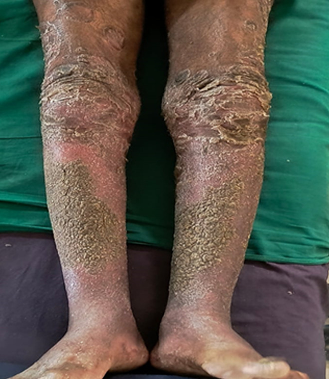 Figure 1
Peer Reviews
No public reviews on file for this paper yet. If you reviewed it on a platform where reviews are public (OpenReview, ICLR, NeurIPS, ICML), you can paste yours below so the community can read it here.
Videos
No videos yet. Explain this paper in a talk, walkthrough, or lecture? Add one.
Taxonomy
TopicsInfectious Diseases and Tuberculosis · Spondyloarthritis Studies and Treatments · Skin Diseases and Diabetes
Image in medicine
A 52-year-old male manual labourer from a humid, tropical region presented with persistent reddish-black, scaly lesions on both lower limbs, extending from the knees to the ankles, for the past four months. The lesions began as mild dryness and scaling but progressively worsened, especially with scratching. He reported frequent itching, particularly at night, occasional pain, and a foul odour from the lesions, suggesting possible secondary bacterial infection. There was no history of systemic illness, trauma, or new medications. Over-the-counter emollients and antihistamines provided only temporary relief. On examination, well-demarcated maculopapular plaques with scaling, raised edges, and hyperpigmentation were noted on both lower limbs. Multiple joint pain, swelling, and stiffness were observed. Based on the clinical findings, a diagnosis of psoriatic arthritis (PA) was considered, as psoriasis commonly precedes joint involvement. Psoriasis is a chronic inflammatory skin condition characterized by scaly, erythematous plaques, and psoriatic arthritis is a frequent complication that affects the joints. The foul odour and rough skin texture suggest secondary infection from chronic scratching. Treatment includes topical corticosteroids (e.g., clobetasol) to reduce inflammation, vitamin D analogues (e.g., calcipotriene) to control scaling, phototherapy if needed, systemic agents (methotrexate or biologics) if joint symptoms arise, and antibiotics for secondary infection. Prognosis involves a chronic, relapsing course requiring long-term management and regular follow-up to prevent joint damage.
maculopapular plaques with scaling, raised edges, and hyperpigmentation on both lower limbs from the knee to the ankle