Lack of Oxygen and/or Glucose Differentially Potentiates Aβ40e22q- and Aβ42-Induced Cerebral Endothelial Cell Death, Barrier Dysfunction and Angiogenesis Impairment
Ashley Carey, Tetyana Buzhdygan, Silvia Fossati

TL;DR
This study shows how low oxygen and glucose levels worsen the harmful effects of specific amyloid-beta proteins on brain blood vessels in Alzheimer's disease.
Contribution
The study identifies distinct molecular mechanisms by which hypoxia and glucose deprivation potentiate Aβ-induced cerebral endothelial dysfunction.
Findings
Hypoperfusion exacerbates AβQ22-induced apoptosis and inflammatory activation in cerebral endothelial cells.
Hypoperfusion strongly enhances Aβ42-mediated necrosis and increases specific protein markers like MMP2 and IFNγ.
Glucose deprivation and hypoxia differentially affect cell death and barrier dysfunction pathways in cECs.
Abstract
Cerebrovascular damage/dysfunction promote cerebral hypoperfusion early within Alzheimer’s Disease (AD). Cerebral hypoperfusion is also a common consequence of cardiovascular risk factors/diseases, typically manifesting in midlife when AD pathology initiates, and contributing to AD onset/progression. We demonstrated that AβQ22 (vasculotropic Dutch mutant) and Aβ42 promote cerebral endothelial cell (cEC) apoptosis, barrier permeability, and angiogenic impairments. Prior research indicates hypoperfusion promotes analogous EC dysfunction. Aβ accumulates within a hypoperfused environment in AD, but whether Aβ exposure of cECs under hypoperfusion potentiates dysfunction through activation of shared molecular mechanisms remains unknown. We treated cECs with Aβ40-Q22/Aβ42, glucose deprivation (GD), or both under normoxia or hypoxia. Cell death, barrier dysfunction/permeability, proinflammatory…
Genes, proteins, chemicals, diseases, species, mutations and cell lines named across the full text — each resolved to its canonical identifier and authoritative record.
Click any figure to enlarge with its caption.
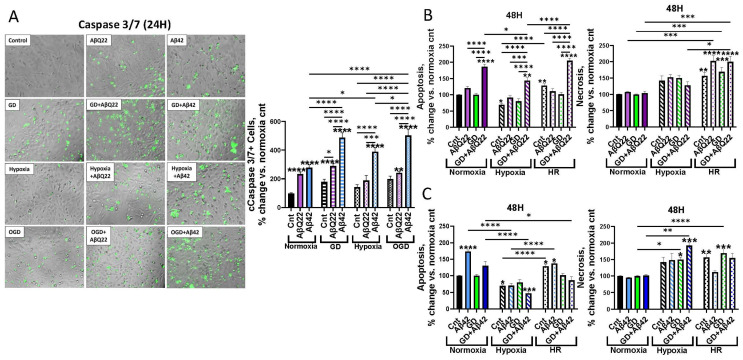 Figure 1
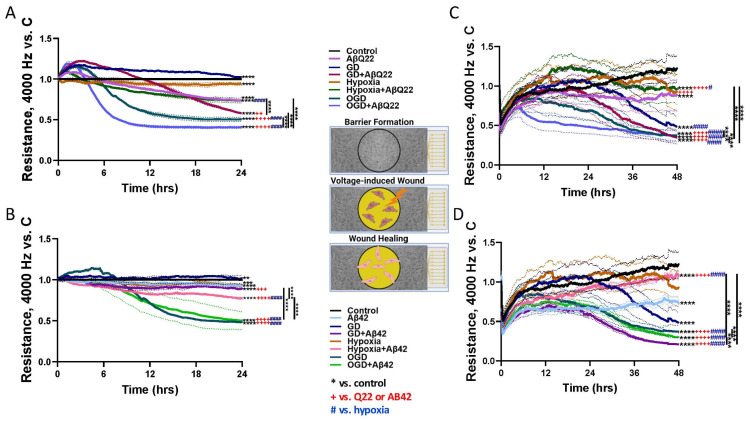 Figure 2
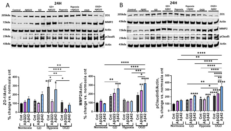 Figure 3
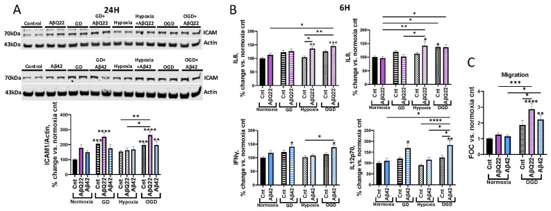 Figure 4
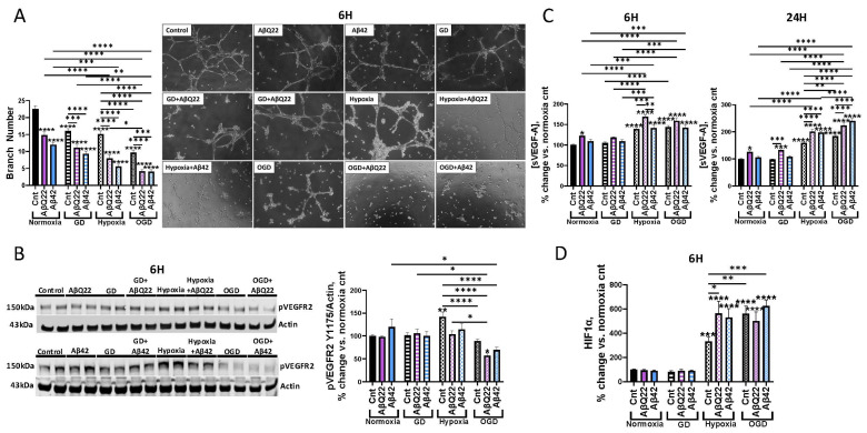 Figure 5
- —NIH
- —Alzheimer’s Disease Drug Discovery Research
- —Alzheimer’s Association
- —Pennsylvania Department of Heath Collaborative Research on Alzheimer’s Disease (PA Cure) Grant
- —Karen Toffler Charitable Trust Scholarship
- —NIH NHLBI T32 HL091804 Training Grant
Peer Reviews
No public reviews on file for this paper yet. If you reviewed it on a platform where reviews are public (OpenReview, ICLR, NeurIPS, ICML), you can paste yours below so the community can read it here.
Videos
No videos yet. Explain this paper in a talk, walkthrough, or lecture? Add one.
Taxonomy
TopicsNeurological Disease Mechanisms and Treatments · Barrier Structure and Function Studies · Alzheimer's disease research and treatments
1. Introduction
Alzheimer’s Disease (AD) is the most prevalent age-associated dementia and is currently affecting more than 6.9 million Americans, with case numbers rising exponentially and the annual death rate climbing [1]. The deposition of amyloid beta (Aβ) plaques, accumulation of hyperphosphorylated tau neurofibrillary tangles, chronic neuroinflammation and cerebrovascular dysfunction pathologically define AD, with these pathologies promoting neuronal death and ultimately cognitive decline [2]. A common phenomenon within the general aging population and particularly in 85–95% of AD patients is Cerebral Amyloid Angiopathy (CAA), characterized by Aβ deposition within and around the cerebral vasculature [2,3,4]. CAA’s consequences include focal ischemia and cerebral hypoperfusion (CH), cerebral blood flow (CBF) impairments, and blood–brain barrier (BBB) dysfunction, as well as cerebral hemorrhages and microinfarcts [2,5]. CAA deposits are mainly composed of the Aβ40 species, which tend to accumulate within the cerebral vessel walls, while plaques mostly containing Aβ42 are located within the cerebral parenchyma [5,6]. The vasculotropic familial Dutch mutant, Aβ40_E22Q_ (AβQ22), contains an amino acid 22 glutamic acid to glutamine substitution that fast-tracks the peptides’ oligomerization. Specifically, this Aβ mutant is closely associated with early-onset CAA, with disease progression beginning as early as 30–40 years of life and pathologies encompassing hemorrhages, strokes, and dementia [4,7]. AβQ22 is more aggregation-prone and exerts more aggressive effects on endothelial cells (ECs) compared to wild-type (WT) Aβ40 [4]; thus, utilizing AβQ22 within our in vitro experiments serves as a vital tool to investigate the specific effects of vascular Aβ.
It is now widely recognized that more than half of dementia cases are mixed pathology dementias, with AD and cerebrovascular pathology being the most frequent combination [8]. Additionally, mixed vascular and Alzheimer’s dementia is common within the general aging population, with a prevalence of 22% [9]. Mounting evidence over the past decades reveals that cerebrovascular dysfunction and CBF impairments, resulting in CH, are likely strong influencers of early AD pathogenesis, occurring prior to significant amyloid accumulation [10,11,12,13]. Recent studies have proposed that chronic CH is a driver of neurodegeneration by promoting cerebral oxygen and nutrient deficits as well as increased oxidative stress and neuroinflammation, resulting in neuronal death and accelerated cognitive decline [14,15]. Studies have revealed not only that AD patients demonstrate CH in distinct cortical areas [16,17,18], but within the general population, increased CH has also been predictive of exacerbated dementia risk and cognitive decline [19,20,21]. Taken together, it is clear there is a vital necessity to better understand the contributions of early cerebrovascular changes, such as CH, to the mechanisms responsible for the vascular contributions to cognitive impairment and dementia (VCID), as these mechanisms could be targeted for prevention and treatment options.
A majority of cardiovascular diseases and risk factors have been found to promote CH [22] and to increase dementia and AD risk [15]. The natural aging process itself is also known to promote CH, with studies demonstrating a 20% decrease in CBF by age 60 [22], resulting in decreases in oxygen and nutrient availability and potentiated neuronal death [22]. Notably, hypertension has been found to promote CH, with the proposed pathological mechanisms involving cerebral ischemia and hemorrhages, capillary rarefaction, endothelial dysfunction and death, BBB permeability and neuroinflammation [23]. Similarly, ischemic strokes are known to promote CH, as they specifically result from vessel occlusions caused by vascular complications (blood clots, stenosis, intracranial atherosclerotic plaques) [24]. Additionally, CH and Aβ have been shown to produce similar cerebrovascular pathologies. Despite this, whether CH and amyloidosis operate through common molecular mechanisms to promote cerebral endothelial dysfunction and whether they do so in an additive or synergistic manner remains unknown. Dissecting the molecular pathways activated by both Aβ and CH that result in endothelial pathology will be imperative for pinpointing novel molecular targets that could be utilized to treat the comorbid vascular effects of Aβ and CH within AD and dementia patients and the aging population.
Our lab was a pioneer in outlining the mechanisms by which Aβ, particularly AβQ22 and Aβ42, affect cerebral EC function. We have demonstrated that treatment of human cerebral microvascular ECs (HCMECs) with AβQ22 and Aβ42 results in increased mitochondria-mediated apoptosis [4,25,26]. Additionally, our studies have revealed that AβQ22 and Aβ42 treatments cause HCMEC barrier dysfunction, promoting trans-endothelial electrical resistance (TEER) loss and dysregulation of BBB-regulating proteins [26,27,28], and also that these Aβ species decrease HCMEC angiogenic and wound healing capabilities [26,28]. Likewise, previous literature reveals that oxygen glucose deprivation (OGD), an in vitro model of hypoperfusion, promotes similar cerebral EC dysfunction, specifically promoting apoptosis and necrosis [29], barrier permeability [30,31], and angiogenesis/wound healing impairments [32,33,34,35]. This study aims to determine whether partial OGD potentiates specific mechanisms of Aβ-induced HCMEC death, barrier dysfunction and angiogenic impairments. An additional goal of this study is to understand whether the EC dysfunction resulting from the combination of OGD and Aβ is Aβ-species specific. We hypothesize that OGD will exacerbate Aβ-induced HCMEC apoptosis, barrier impairments, and angiogenic deficits in an additive manner and through activation of common molecular pathways, thus fast-tracking cerebrovascular pathological progression and increasing dementia risk.
2. Materials and Methods
2.1. Cell Culture
HCMECs/D3, immortalized human cerebral microvascular ECs, were acquired from Babette Weksler (Cornell University) [4]. EBM-2 (Lonza, Basel, Switzerland) supplemented with growth factors (Hydrocortisone, hFGF-B, VEGF, R3-IGF-1, ascorbic acid, hEGF, GA-1000) and 5% fetal bovine serum (FBS) was used to grow the cells. Cells were maintained in a humidified cell culture incubator (37 °C) under a 5% CO_2_ atmosphere. The EVOS M5000 Imaging System (Thermo Fisher, Waltham, MA, USA) was utilized for cell visualization and imaging.
2.2. AβQ22 and Aβ42 Peptide
The Aβ42 peptide, the most aggregation-prone brain amyloid, and the genetic variant of the WT Aβ40 peptide containing the E22Q vasculotropic substitution (AβQ22) were utilized for cell treatments. Specifically, AβQ22 is the synthetic homolog of the amyloid subunit present in the vascular deposits in sporadic and familial Dutch-AD cases. Peptide synthesis was performed by Peptide 2.0 (Chantilly, VA, USA). Aβ42 and AβQ22 were dissolved to 1 mM in 1,1,1,3,3,3-hexafluoro-2-propanol (HFIP, Sigma, St. Louis, MO, USA) and incubated for 24 h to break down pre-existing β-sheet structures. After incubation, the peptide was lyophilized and then dissolved to a 10 mM concentration in DMSO, followed by the addition of dH_2_O to a 1 mM concentration. Aβ peptides were further diluted in culture medium (DMEM (Gibco, Waltham, MA, USA), 1% FBS and no growth factors) to the required experimental concentrations. To note, equivalent levels of DMSO alone did not cause any toxicity within these cells.
2.3. Oxygen and Glucose Deprivation Treatments
Glucose-free DMEM (Gibco, Waltham, MA, USA) was supplemented with 1 M glucose (Agilent, Santa Clara, CA, USA) to achieve final concentrations of 0.1 mg/mL, defined as glucose deprivation (GD), and 1 mg/mL, defined as normal glucose. Partial oxygen deprivation (hypoxia) was achieved by placing cells in a hypoxic chamber (Coy Laboratories, Grass Lake, MI, USA), maintained at 1% oxygen and 5% CO_2_. OGD was achieved by treating cells with GD medium and hypoxia (as above).
2.4. Cell Death ELISA
Apoptosis was evaluated by measuring fragmented nucleosome levels using the Cell Death Detection ELISA^Plus^ (Roche, Basel, Switzerland) according to the manufacturer’s instructions. HCMECs were seeded and, after 24 h, treated with 25 µM AβQ22 or 5 µM Aβ42 (concentrations that we know from our previous studies cause EC apoptosis) [25,28], GD, or a combination of Aβ and GD under conditions of normoxia, hypoxia, or hypoxia/reoxygenation (HR = 24 h hypoxia, followed by 24 h normoxia) for 48 h. Extranuclear DNA-histone fragmented complexes were measured as absorbance using the SpectraMax i3x Multi-Mode Microplate Reader (Molecular Devices, San Jose, CA, USA). Results are expressed as percent change versus untreated controls.
2.5. Lactate Dehydrogenase Assay
Necrosis was assessed by measuring lactate dehydrogenase (LDH) levels released within cell media utilizing an LDH assay kit (Roche, Basel, Switzerland) according to the manufacturer’s instructions. HCMECs were seeded for 24 h and subsequently treated with 25 µM AβQ22 or 5 µM Aβ42, GD, or a combination of both under conditions of normoxia, hypoxia, or HR for 48 h. Cell media were collected, and LDH levels were measured as absorbance (492 nm) using the SpectraMax i3x Microplate Reader (Molecular Devices, San Jose, CA, USA). Results are expressed as percent change versus untreated controls.
2.6. Cell Event Fluorescent Caspase-3/7 Assay
Cleaved/active caspase-3/7 expression was evaluated with the CellEvent^TM^ Caspase-3/7 Green Detection Reagent (Thermo Fisher, Waltham, MA, USA). Cells were seeded in a 96-well plate (10,000 cells/well) and treated for 24 h with 25 µM AβQ22 or 5 µM Aβ42, GD, or a combination of both under normoxia or hypoxia conditions. Post-treatment, cell media were removed, the fluorescent Caspase-3/7 Detection Reagent was added to each well (5 µM), and cells were incubated for 1 h at 37 °C. Cells were imaged with bright-field and GFP filters via the EVOS M5000 imaging system (4 randomized images/well, Thermo Fisher, Waltham, MA, USA). Percent of cleaved caspase-3/7-positive cells was determined by counting the total cell number in each picture, followed by the creation of a mask for the fluorescent signal to count the number of cells demonstrating active caspase-3/7 fluorescence [% Caspase-3/7 + Cells = (# of cells with cleaved caspase-3/7/total cell #) × 100].
2.7. ECIS Trans-Endothelial Electrical Resistance
The ECIS Zθ system (Applied Biophysics, Troy, NY, USA) was utilized to assess HCMEC barrier integrity. Experimental procedures were performed in 8-well ECIS (8WE10+) 40-electrode gold-plated arrays, which were pre-treated according to the manufacturer’s recommendations. HCMECs were seeded and monitored for 48 h until TEER reached a plateau at a frequency of 4000 Hz, which is indicative of complete barrier formation. The cell monolayers were then treated with 25 µM AβQ22 or 5 µM Aβ42, GD, or a combination of both under normoxia or hypoxia and followed for 24 h post-treatment. Barrier permeability was defined as a decrease in TEER at 4000 Hz versus untreated controls.
2.8. ECIS Wound Healing Assay
HCMEC wound healing capability was evaluated utilizing the corresponding ECIS Zθ assay. Experimental procedures were performed in 8-well ECIS (8W1E) gold-plated arrays containing one central electrode, and the arrays were pre-treated according to the manufacturer’s recommendations. HCMECs were seeded and monitored for 48 h until TEER reached a plateau at a 4000 Hz frequency (barrier formation). The cell monolayers were then treated with 25 µM AβQ22 or 5 µM Aβ42, GD, or a combination of both under normoxia or hypoxia conditions. One hour post-treatment, a wound was inflicted to the EC monolayer lasting 20 s (60,000 Hertz; amplitude = 5 V; wound current = 1400 µAmps) and wound healing was assessed for 48 h post-wound. Impairments in wound healing were quantified as decreased post-wound recovery TEER versus untreated controls.
2.9. Western Blot
Evaluation of ZO1 (61-7300, Invitrogen, Carlsbad, CA, USA), ICAM (MA5407, Invitrogen, Carlsbad, CA, USA), phosphorylated claudin-5 (ab172968, abcam, Cambridge, UK), MMP2 (ab86607, abcam, Cambridge, UK), phosphorylated VEGFR2 Y1175 (ab194806, abcam, Cambridge, UK), and VEGF-A (19003-1-AP, Proteintech, Rosemont, IL, USA) expression was performed using Western blot (WB) analysis after electrophoretic separation on 4–12% Bolt Bis-Tris SDS polyacrylamide gels. Anti-β actin (MAB1501, Millipore, Burlington, MA, USA) was utilized for normalization. Proteins were electrotransferred to nitrocellulose membranes (0.45 μm pore, Cytiva Life Sciences, Marlborough, MA, USA) at 110 V for 70 min using towbin buffer containing 20% (v/v) methanol. Intercept Blocking Buffer (Licor, Lincoln, NE, USA) was utilized to block membranes, and membranes were subsequently immunoreacted with the corresponding primary antibodies for each experiment, followed by incubation with the appropriate anti-rabbit or anti-mouse secondary antibodies (1/20,000, Licor, Lincoln, NE, USA). Membranes were developed utilizing the LICOR Odyssey CLx Immunoblot Imager (Lincoln, NE, USA), and blots were analyzed with LICOR Image Studio software version 5.5.
2.10. Proinflammatory Panel MSD
Proinflammatory cytokine expression was measured via the V-PLEX proinflammatory panel 1 kit from MesoScale Discovery (MSD, Rockville, MD, USA), which measures expression levels of 10 cytokines/sample (IL1β, IL2, IL4, IL6, IL8, IL10, IL12p70, IL13, IFNγ, TNFα). HCMECs were seeded for 24 h and then treated with 25 µM AβQ22 or 5 µM Aβ42, GD, or a combination of both under normoxia or hypoxia conditions for 6 h. Media were collected post-treatment and stored at ~80 °C until the MSD was conducted. The assay was performed in accordance with the manufacturer’s recommendations, and sample protein concentration was utilized for normalization.
2.11. U937 Human Monocytes
The human monocyte cell line U937 was obtained from ATCC (Manassas, VA, USA) and maintained in RPMI-1640 medium supplemented with 10% heat-inactivated pooled FBS. Cells were serum-starved for 24 h prior to extravasation assays.
2.12. Monocyte Trans-Endothelial Extravasation Assay
A monocyte extravasation assay was performed as previously described [36]. Briefly, HCMECs were plated on rat tail collagen type I-coated FluoroBlok^™^ HTS 96-well inserts with 8 µM pores (Corning, Corning, NY, USA) (2.5 × 10^4^ cells/insert). After confluent monolayer formation, HCMECs were treated with 25 µM AβQ22 or 5 µM Aβ42, OGD, or a combination of the two for 24 h. All conditions were also supplemented with 1× GlutaMAX. On extravasation challenge day, U937 monocytes were loaded with Calcein-AM at 5 µM/1 × 10^6^ cells for 45 min. Permeable supports with HCMEC monolayers were rinsed post-treatment and transferred to wells filled with phenol-free DMEM supplemented with 1 g/L glucose, 1× GlutaMAX, and 10% FBS. Calcein-AM-labeled U937 (40,000 cells/support) were added to the luminal side of the HCMECs and allowed to migrate and adhere to the abluminal side of the supports for 6 h at 37 °C. For fluorescence measurement, supports were transferred to clean 24-well plates filled with HBSS, and fluorescence was acquired with a Spectramax M5 fluorescence plate reader (Molecular Devices, San Jose, CA, USA). The data was calculated based on the standard curve derived from the fluorescent intensity of known numbers of labeled monocytes.
2.13. Angiogenesis Inhibition Assay
Angiogenic potential was evaluated with the Millicell μ-Angiogenesis Inhibition Assay (Millipore, Burlington, MA, USA) according to the manufacturer’s instructions. HCMECs were seeded in μ-angiogenesis slides containing ECMatrix gel solution and simultaneously treated with a sublethal concentration (1 µM) of AβQ22 or Aβ42, GD, or a combination of both under normoxia or hypoxia conditions [28]. Tube formation was assessed after 6 h by acquiring pictures with the EVOS M5000 imaging system (4 randomized images/well, Thermo Fisher, Waltham, MA, USA). Capillary branches meeting length criteria were counted within the images.
2.14. HIF1α ELISA
HIF1α expression was measured with a Total HIF1α Duoset ELISA (Bio-techne R&D Systems, Minneapolis, MN, USA), following the manufacturer’s recommendations. HCMECs were seeded for 24 h and then treated with 25 µM AβQ22 or 5 µM Aβ42, GD, or a combination of both under normoxia or hypoxia conditions for 6 h. Post-treatment, protein lysate was obtained and stored at ~80 °C until the ELISA was conducted. Sample protein concentration was utilized for normalization.
2.15. VEGF-A ELISA
Soluble VEGF-A release was measured via the VEGF-A Quantikine ELISA (Bio-techne R&D Systems, Minneapolis, MN, USA), following the manufacturer’s recommendations. HCMECs were seeded for 24 h and then treated with 25 µM AβQ22 or 5 µM Aβ42, GD, or a combination of both under normoxia or hypoxia conditions for 6 h and 24 h. Post-treatment, media were collected and stored at −80 °C until the ELISA was conducted. Sample protein concentration was utilized for normalization.
2.16. Statistical Analysis
All experimental graphs are representative of at least 3 independent experiments with 2 or more technical replicates. All data is represented as means ± SEM. Statistical significance for ECIS TEER and wound healing was evaluated by one-way ANOVA followed by Tukey’s post hoc test, and statistical significance for all other experiments was assessed by two-way ANOVA followed by Tukey’s post hoc test using GraphPad Prism 9. Statistically significant differences required a p-value ≤ 0.05.
3. Results
3.1. Exposure of HCMECs to AβQ22 or Aβ42 and GD, Under Varying Oxygen Conditions, Differentially Affects Apoptosis and Necrosis
It has been shown that AβQ22 and Aβ42 promote increased fragmented nucleosomes formation, indicative of elevated apoptosis, as well as increased LDH release, indicative of increased necrosis levels [4,26,37], with OGD (in vitro CH model) also being reported to activate similar cell death pathways within brain ECs [29]. Aβ accumulation and hypoperfusion are simultaneously occurring within AD and CAA brains [38,39], and both pathological factors can promote cerebrovascular cell death, but it is currently unknown whether the combined challenge of specific Aβ species and OGD will exacerbate cerebral EC death through common cell death mechanisms. To investigate this, we measured cleaved caspase-3, apoptosis, and necrosis levels in HCMECs treated with 25 µM AβQ22 or 5 µM Aβ42 (concentrations known to start activating apoptotic pathways [25,26,28]), GD, or a combination of both under varying oxygenation conditions. The rationale for treating HCMECs with lower concentrations of Aβ42 versus AβQ22 was based on our lab’s prior findings demonstrating that Aβ42’s toxic effects are more rapid and drastic versus Aβ40 and its variants, due to Aβ42 being extremely aggregation-prone and showing faster oligomerization kinetics [4,26]. Due to this, HCMECs were treated with a higher concentration of AβQ22 (25 µM) versus Aβ42 (5 µM) to take into consideration these aggregation and toxicity differences. CH conditions within this study were modeled by exposing HCMECs to OGD conditions, defined here as partial depletion of oxygen (hypoxia: 1% O_2_) and glucose (controls: 1 mg/mL vs. GD: 0.1 mg/mL).
Cleaved (activated) caspase-3/7, the executioner caspases within the apoptotic pathway, were measured using a fluorescent active caspase-3/7 assay following 24 h of treatment. HCMECs exposed to AβQ22 or Aβ42 and GD both demonstrated a potentiated increase in cleaved caspase-3/7 levels compared to control cells and the challenges alone, with Aβ42+GD more significantly increasing cleaved caspase-3/7 levels versus AβQ22+GD-exposed cells (Figure 1A). HCMECs treated with Aβ and/or GD under hypoxic conditions demonstrated a similar trend of potentiated cleaved caspase-3 levels, although hypoxia did not further exacerbate cleaved caspase-3 increases when added to GD (OGD) (Figure 1A). To confirm that caspase-3 activation resulted in the execution of the apoptotic pathway, we measured DNA fragmentation, indicative of the terminal stage of apoptosis, after the 48 h challenge. Treatment of HCMECs with GD significantly potentiated AβQ22-mediated increases in apoptosis versus controls and each challenge alone (Figure 1B).
However, we did not observe this increase with Aβ42+GD (Figure 1C), as Aβ42 alone already resulted in much higher apoptosis levels versus AβQ22. Instead, the Aβ42+GD challenge resulted in a significant increase in necrosis (Figure 1C). Interestingly, the hypoxia and GD combined challenge (OGD) resulted in a significant increase in Aβ42-induced necrosis while reducing the levels of apoptosis caused by this peptide. Since a common phenomenon in ischemic or hypoxic tissue environments is hypoxia-reperfusion injury, where blood flow restoration may cause additional damage, resulting in exacerbated EC death [40], an HR condition (24 h hypoxia, then 24 h normoxia) was also considered when measuring apoptosis and necrosis. HR itself significantly increased HCMEC apoptosis levels (Figure 1B,C), while hypoxia and HR did not further potentiate HCMEC apoptosis levels when in combination with Aβ or GD. Aβ42 and OGD exposure resulted in an additive increase in HCMEC necrosis levels (Figure 1C). HR alone also significantly increased HCMEC necrosis (Figure 1B,C), with ECs exposed to HR+AβQ22 or HR+AβQ22+GD demonstrating the most significant increase in necrosis versus controls (Figure 1B).
3.2. Treatment of HCMECs with AβQ22 and OGD Additively Decreases TEER and Wound Healing Capabilities
We have shown previously that AβQ22 and Aβ42 promote HCMEC barrier permeability [26,28]. Additionally, our recent study shows that AβQ22 impairs HCMEC wound healing [28]. OGD has also been shown to promote barrier dysfunction and permeability [30,31,41], as well as wound healing impairments [32,33] in cerebral ECs. To determine whether HCMECs exposed to either Aβ species and/or OGD demonstrate additive increases in endothelial barrier permeability and wound healing impairments, ECIS technology was utilized. Once a stable EC barrier was established, HCMECs were treated with AβQ22 (25 µM) or Aβ42 (5 µM), GD, or a combination of both under normoxia or hypoxia, and TEER was measured over time for 24 h. As expected, AβQ22 and Aβ42 significantly decreased HCMEC TEER versus controls (Figure 2A,B).
HCMECs exposed to AβQ22+OGD demonstrated an additive decrease in TEER compared to the treatments alone (Figure 2A). Similarly, when Aβ42 was given in combination with either GD, hypoxia, or OGD, HCMEC TEER was more significantly reduced compared to cells treated with Aβ42 alone (Figure 2B), but Aβ42 addition at this concentration did not have a significant additive effect on OGD-induced TEER decreases at later timepoints, possibly due to a floor effect where maximal TEER decreases were achieved, suggesting that the barrier impairment in Aβ42+OGD conditions is primarily driven by OGD rather than Aβ42 (Figure 2B). TEER data at 12 h and 24 h was represented as bar graphs in Supplemental Figure S5.
To assess wound repair ability, a wound healing assay was conducted utilizing a dedicated ECIS assay. Following barrier formation, HCMECs were treated with 25 µM AβQ22 or 5 µM Aβ42, GD, or a combination of both under normoxia or hypoxia conditions. At 1 h post-treatment, a strong electrical current was applied to the center of the EC barrier to inflict a circular wound. Recovery from the wound was monitored for 48 h and was indicated by post-wound TEER increases. As our lab has recently demonstrated, AβQ22-treated HCMECs demonstrated impaired wound healing versus controls (Figure 2C) [28]. Similarly, treatment of HCMECs with Aβ42 resulted in impaired wound healing versus controls (Figure 2D). Combined exposure of HCMECs to AβQ22+GD exacerbated wound healing impairments compared to cells treated with each challenge alone, while HCMECs treated with AβQ22+OGD demonstrated an additive decrease in wound healing levels (Figure 2C). Interestingly, Aβ42+OGD did not have an additive effect on HCMEC wound healing versus OGD alone, but HCMECs treated with Aβ42+GD, Aβ42+OGD, or OGD alone all demonstrated the most impaired wound healing capabilities (Figure 2D). Wound healing TEER data at 24 h and 48 h was represented as bar graphs in Supplemental Figure S6.
3.3. Exposure of HCMECs to AβQ22 or Aβ42 in Combination with OGD Exacerbates Changes in Expression of BBB-Modulating Proteins
Strong evidence from previous studies indicates that Aβ species can promote BBB permeability by disrupting the expression and phosphorylation state of BBB-regulating proteins, such as tight junctions (TJs) [42,43,44,45,46]. Exposure of brain ECs to OGD has previously been reported to disrupt the expression and regulation of barrier proteins as well [30,47,48]; therefore, we aimed to understand whether combined exposure of HCMECs to Aβ+OGD additively exacerbates expressional changes within BBB-related proteins and whether these changes are Aβ-species specific and correlate to the TEER decreases observed in Figure 2. To do so, we analyzed zona occludin 1 (ZO1), phosphorylated claudin-5 (pClaudin5), and MMP2 (matrix metalloproteinase 2) protein expression in HCMECs treated with 25 µM AβQ22 or 5 µM Aβ42, GD, or a combination of both under normoxia or hypoxia conditions for 24 h, a timepoint revealing drastic TEER decreases (Figure 2).
ZO1 is an anchor protein vital for organizing TJ proteins and connecting them to the actin cytoskeleton [49]. Hypoxia-treated HCMECs demonstrated a significant increase in ZO1 expression versus controls, possibly indicating a HIF-1-mediated compensatory mechanism to maintain barrier integrity under low oxygen [50] (Figure 3A,B). However, HCMECs treated with AβQ22 under hypoxia or OGD conditions demonstrated significant decreases in ZO1 expression compared to hypoxia controls, with an additive decrease in ZO1 expression being revealed by HCMECs treated with AβQ22+OGD (Figure 3A). Similarly, HCMECs treated with Aβ42+OGD demonstrated a decrease in ZO1 expression compared to Aβ42 alone or Aβ42+hypoxia, but the decrease did not reach the same significance as in AβQ22+OGD-exposed HCMECs (Figure 3B). Claudin-5 is a major TJ protein, and its phosphorylation has been directly linked to increased endothelial barrier permeability [51]. Interestingly, we revealed that OGD challenge increased pClaudin5 expression versus controls. Moreover, HCMECs treated with AβQ22+OGD or Aβ42+OGD demonstrated an additive increase in pClaudin5 expression compared to cells challenged with the peptides alone, with Aβ42+OGD more significantly increasing pClaudin5 levels than AβQ22+OGD (Figure 3A,B). MMP2 is an enzyme responsible for degrading the extracellular matrix and is a known player in BBB breakdown and cerebral hemorrhage in AD and CAA [52,53]. Our lab and others have demonstrated that exposure of HCMECs to Aβ species, particularly AβQ22, increases MMP2 expression and activity [27,28], with similar effects being reported for OGD-exposed HCMECs [54]. Following 24 h of treatment, HCMECs challenged with AβQ22 or Aβ42 under conditions of GD and OGD demonstrated potentiated increases in MMP2 protein expression, with HCMECs treated with Aβ42+OGD revealing the most significant increase in MMP2 levels (Figure 3A,B). We also measured the expression of these proteins following an acute treatment (6 h), a timepoint where TEER decreases began to be observed (Figure 2). Very similar trends to the 24 h timepoint were observed at this early timepoint (Supplemental Figure S1). Overall, the exacerbated ZO1 loss, claudin-5 phosphorylation, and MMP2 upregulation induced by exposure of HCMECs to Aβ+OGD indicate that Aβ and OGD act in an additive manner on the same protein mediators to potentiate the loss of blood–brain barrier (BBB) function.
3.4. AβQ22 or Aβ42 Combined with OGD Differentially Potentiates Increases in Cerebrovascular Inflammatory Modulators and Monocyte Trans-Endothelial Extravasation
Intercellular adhesion molecule 1 (ICAM1) is a key player in the extravasation of immune cells from the lumen of cerebral vessels into the brain parenchyma, where these immune cells release proinflammatory factors and drive neuroinflammation in AD and diseases associated with CH [55]. Additionally, proinflammatory cytokines, including IL-6, IL-8, and IFN-γ, have been shown to induce BBB permeability, promoting reduced TJ expression and improper TJ localization [56,57,58]. To understand whether combined exposure of HCMECs to Aβ species and OGD exacerbates the expression of proinflammatory mediators, we measured ICAM1 protein expression in HCMECs treated with 25 µM AβQ22 or 5 µM Aβ42, GD, or a combination of both for 24 h under normoxia or hypoxia conditions. While GD alone resulted in an increase in ICAM expression, AβQ22+GD, as well as AβQ22+OGD, induced potentiated increases in ICAM1 expression. This overexpression appeared to be mainly driven by GD and not hypoxia (Figure 4A). Different from AβQ22, the Aβ42 challenge did not cause additive effects on GD-mediated ICAM1 overexpression (Figure 4A).
Furthermore, we aimed to understand whether combined exposure of HCMECs to Aβ species and OGD additively promoted the secretion of proinflammatory cytokines. To do so, we utilized a multiplex cytokine array (MSD). HCMECs were treated with 25 µM AβQ22 or 5 µM Aβ42, GD, or a combination of both under normoxia or hypoxia conditions for 6 h, and the media were collected. The 6 h timepoint was selected to understand whether early changes in the HCMEC inflammatory state contribute to the decreases in TEER observed in Figure 2. AβQ22 and Aβ42 differentially affected the expression of certain proinflammatory cytokines. Treatment of HCMECs with AβQ22+OGD resulted in an additive increase in IL6 secretion, a potent inducer of BBB permeability, versus controls and cells exposed to each treatment alone (Figure 4B). HCMECs treated with AβQ22+OGD also demonstrated a significant increase in IL8 expression, another cytokine linked to decreased BBB integrity, compared to control cells, but this effect appeared to be similar also in cells challenged with hypoxia+AβQ22 and with OGD alone (Figure 4B). Conversely, treatment of HCMECs with Aβ42 and GD or OGD resulted in a comparable potentiation in IFN-γ secretion levels, another cytokine that is known to promote BBB breakdown (Figure 4B). Additionally, treatment of HCMECs with Aβ42+GD or Aβ42+OGD resulted in a significant increase in IL12p70, a pleotropic proinflammatory cytokine (Figure 4B). Other proinflammatory cytokines were measured and did not reveal significant differences between treatments (Supplemental Figure S2).
To understand whether the combined challenge with Aβ+OGD also impacted a functional measure of the microvascular inflammatory activation, a monocyte trans-endothelial extravasation assay was conducted. HCMECs were allowed to form monolayers and were subsequently treated with 25 µM AβQ22 or 5 µM Aβ42, OGD, or a combination of both for 24 h. Post-treatment, fluorescently labeled human monocytes were allowed to migrate across EC barriers for 6 h, and the number of extravasated monocytes was determined. Treatment of HCMECs with a combination of either AβQ22 or Aβ42 and OGD resulted in a significant potentiation of monocyte migration across EC monolayers, with HCMECs exposed to AβQ22+OGD demonstrating the most significant increase in monocyte migration versus controls and each treatment condition alone (Figure 4C).
Taken together, the increases in ICAM1 and proinflammatory cytokine expression resulting from HCMEC exposure to Aβ+OGD unveil specific molecular mechanisms through which Aβ+OGD work in concert to exacerbate EC activation, which results in increased monocyte migration across the HCMEC barrier, thus providing insights into the pathways promoting HCMEC barrier permeability and microvascular inflammatory activation in conditions with comorbid vascular amyloidosis and hypoperfusion.
3.5. Aβ and OGD Additively Decrease HCMECs’ Angiogenic Capabilities
Cerebral angiogenesis is an essential homeostatic process allowing ECs to form new vessels to maintain proper cerebral blood flow levels, particularly in conditions of poor perfusion or microvascular damage [59]. Our lab has demonstrated that challenging HCMECs with low doses of AβQ22 and Aβ42 impairs angiogenesis, specifically decreasing vessel branching [26]. Previous evidence reveals that OGD-exposed ECs demonstrate similar angiogenesis impairments, specifically decreased vessel sprouting, migration, and tube formation [34,35]; thus, we sought to elucidate whether exposure of HCMECs to Aβ in combination with OGD potentiates angiogenic impairment and whether both challenges operate through mutual molecular mechanisms to produce this dysfunction. HCMECs were treated with 1 µM AβQ22 or Aβ42 (the lowest dose capable of inducing angiogenesis inhibition in previous studies [26,28]), GD, or a combination of the two, and the ability to form vessels was monitored after 6 h under normoxia or hypoxia through an angiogenesis assay. The 1 µM concentration for Aβ was used specifically for this assay because it requires cells to be seeded and treated simultaneously, and higher concentrations of amyloid dilute differences between treatment groups and impair the ability of HCMECs to attach and have a chance of forming vessels. HCMECs demonstrated significantly decreased vessel branch numbers versus controls in response to all treatment conditions (Figure 5A), indicating decreased angiogenesis. Specifically, either peptide alone, as well as GD or hypoxia alone, significantly inhibited angiogenesis. However, HCMECs exposed to Aβ peptides+OGD demonstrated the most drastic inhibition of vessel branching versus cells exposed to the treatments alone (Figure 5A).
Since combined exposure of HCMECs to Aβ+OGD additively decreased angiogenic capabilities, we then aimed to determine whether we observed analogous changes in the protein expression of key angiogenic mediators. HCMECs were treated with Aβ peptides, GD, or a combination of both at the same 6 h timepoint under normoxia or hypoxia conditions. Protein expression of the phosphorylated vascular endothelial growth factor receptor 2 (VEGFR2) Y1175 (pVEGFR2), the VEGFR2 phosphorylated at tyrosine1175, a known phosphorylation site associated with angiogenic signaling activation, was evaluated. As expected, hypoxia-exposed HCMECs demonstrated a significant increase in pVEGFR2 expression versus controls (Figure 5B). Hypoxic conditions are known to stabilize the transcription factor HIF1α, which transcribes VEGF. VEGF subsequently binds to VEGFR2 to induce its phosphorylation and activation [60]. The hypoxia-induced increase in active pVEGFR2 was prevented when HCMECs were exposed to hypoxia+AβQ22 or Aβ42 (Figure 5B). HCMECs exposed to Aβ42+OGD demonstrated a decrease in pVEGFR2 compared to cells treated with Aβ42 alone, while cells challenged with AβQ22+OGD demonstrated the most significant decrease versus hypoxia and normoxia controls (Figure 5B). Accordingly, we found that HCMECs treated with AβQ22+OGD demonstrated a significant decrease in VEGF-A intracellular protein abundance versus hypoxia and normoxia controls (Supplemental Figure S3). Because VEGF-A is normally secreted by vascular ECs, we also measured soluble VEGF-A (sVEGF-A) levels within HCMEC-conditioned media after 6 h and 24 h. At both the short (6 h) and long (24 h) timepoints, a significant increase in sVEGF-A levels was observed when HCMECs were exposed to hypoxia, coinciding with previous studies (Figure 5C). Interestingly, AβQ22-exposed HCMECs demonstrated a significant increase in sVEGF-A release versus controls at 6 h and 24 h (Figure 5C). Specifically, at 6 h, HCMECs treated with AβQ22 under hypoxia conditions revealed the highest sVEGF-A level versus normoxia and hypoxia controls, while Aβ42 treatment had no effect on sVEGF-A expression (Figure 5C). At the 24 h timepoint, an additive increase in sVEGF-A level was observed when cells were exposed to Aβ42+OGD, and the same additive increase was observed when HCMECs were treated with AβQ22+OGD (Figure 5C). Overall, these results suggest that an increased release of sVEGF-A from the cells may be a compensatory mechanism in response to the hypoxia/OGD. However, this does not result in a functional VEGFR2 activation, particularly in the combination treatment with Aβ and OGD.
HIF1α is the main regulator of the hypoxic response and the transcription factor that promotes pro-angiogenic gene expression, such as VEGF. Therefore, we also measured HIF1α protein expression in HCMECs treated with AβQ22 or Aβ42, GD, or a combination under normoxia or hypoxia conditions for 6 h. The 6 h timepoint was chosen due to previous evidence demonstrating that 6 h of hypoxia results in peak HIF1α expression in vascular ECs [61]. As anticipated, exposure of HCMECs to hypoxia alone resulted in a significant increase in HIF1α expression (Figure 5D). Treatment of HCMECs with AβQ22 under hypoxic conditions significantly potentiated HIF1α increases, but no further increase was observed from AβQ22+OGD treatment (Figure 5D). HCMECs treated with Aβ42+OGD demonstrated the most significant upregulation in HIF1α protein expression versus normoxia and hypoxia controls, which appeared to be mainly driven by the OGD treatment. Taken together, these data suggest that Aβ potentiates the hypoxic/OGD response, exacerbating HIF1α upregulation as well as sVEGF-A secretion, but these pro-angiogenic factors accumulate without promoting angiogenic signaling, since we observe an additive decrease in pVEGFR2 expression and vessel branching after Aβ+OGD challenge.
4. Discussion
AD is a multifactorial syndrome not only driven by Aβ and tau accumulation. It is now well accepted that the vascular contributions to cognitive impairment and dementia (VCID), and particularly vascular disruptions resulting in CH, are key drivers of early AD pathogenesis, even before considerable Aβ deposition [10,11,12,13,62,63,64]. Indeed, since vascular and amyloid pathologies constitute the most frequent combination of mixed etiology dementias, Aβ accumulation can often occur within an already hypoperfused cerebral environment, where EC death, barrier dysfunction, and angiogenic impairment may be common consequences of the combined exposure to both Aβ and OGD. This study sought to determine whether exposure of HCMECs to Aβ and OGD results in additive increases in common molecular mediators of apoptotic and necrotic cell death mechanisms, barrier permeability, and angiogenesis failure. Additionally, we aimed to understand whether two different Aβ species that are known to be pro-apoptotic for cerebral ECs and to promote BBB permeability (AβQ22 and Aβ42 [26,28,65]) promote similar or differential mechanisms of HCMEC dysfunction in hypoxia, GD, and OGD conditions.
We began by investigating how the combined exposure of HCMECs to Aβ+OGD influences the activation of cell death mechanisms. Our lab has previously demonstrated that both AβQ22 and Aβ42 increase HCMEC apoptosis and, later, necrosis levels [4,25,26,65,66]. Hypoxia has also been linked to increased apoptosis and necrosis [67], and OGD in vitro has been found to specifically increase microvascular EC death through the same ischemia-mediated cell death pathways [29]. This study demonstrates that exposure of HCMECs to AβQ22 or Aβ42 in combination with GD significantly potentiates caspase-3 activation. Similarly, we observed a potentiated increase in apoptosis levels in HCMECs exposed to AβQ22+GD, while an additive increase in necrosis levels was observed in HCEMCs exposed to Aβ42+OGD, particularly after HR injury. Overall, these results suggest that lack of glucose in the presence of AβQ22 pushes HCMECs towards potentiated apoptosis, while reduction and variability in oxygen levels, such as during HR, drives AβQ22-exposed HCMECs towards necrosis. Both AβQ22- and Aβ42-exposed HCMECs under GD conditions demonstrate potentiated increases in cleaved caspase-3, with this trend being significantly more evident with Aβ42 treatment. This strong caspase-3 activation at 24 h in Aβ42-exposed cells may accelerate the terminal stages of apoptosis, shifting HCMEC fate to secondary necrosis faster, which was particularly evident in HCMECs treated with Aβ42+OGD. It is also possible that not all HCMECs demonstrating active caspase-3 will undergo apoptosis. Indeed, when measuring DNA fragmentation, one of the last steps of the apoptotic cascade, at 48 h, it appears that not all groups that were caspase-3-positive at 24 h reached this step. Particularly, the observed decrease in apoptosis and shift to necrosis evident in hypoxia-exposed cells suggests a switch in cell death mechanism, as it is possible that cells with active caspase-3 ultimately undergo secondary necrosis instead of apoptosis, for example, through caspase-3-mediated cleavage of DFNA5, which induces membrane permeabilization [68]. Additionally, ATP is required for apoptosis execution, so the lack of oxygen, which is known to trigger decreased mitochondrial respiration and ATP production, could be an underlying reason for this necrotic shift, but further studies are warranted to understand the underlying molecular mechanisms at play. These results confirm that manipulation of oxygen levels, particularly during HR, is sufficient to potentiate necrotic cell death in cerebral ECs, even in the absence of Aβ peptides. In line with these findings, it is known that in ischemic stroke, where oxygen delivery is disrupted, necrosis is the major cell death mechanism within the ischemic core, and ischemia/reperfusion injury is highly associated with necrosis, with this tissue necrosis often resulting in substantial activation of neuroinflammatory processes [69,70,71].
Cerebral ECs’ ability to preserve proper barrier integrity is crucial for maintenance of healthy BBB structure and function. Increased BBB permeability is a hallmark of AD pathology, with a collection of studies reporting leakage of blood proteins as well as loss of endothelial, basement membrane, and TJ proteins within post-mortem AD brains [72]. Prior studies from our lab have demonstrated that Aβ species (AβQ22/Aβ42) are capable of decreasing TEER, an indicator of EC barrier permeability [26,28]. Similar to AD, ischemic strokes are also known to cause substantial BBB breakdown, resulting from dysregulation of TJs and upregulation of proinflammatory mediators and MMP activity [73]. Through this study, we provide novel evidence that combined exposure of HCMECs, specifically to AβQ22+OGD, results in a continued additive decrease in TEER, reflecting the most permeable HCMEC barrier. This additive effect also appears at early (before 12 h) but not late timepoints for Aβ42+OGD, suggesting that the combined treatment may reach maximal toxicity early after challenge.
To dissect the molecular players involved in promoting the drastic EC barrier permeability displayed by Aβ+OGD-challenged HCMECs, we measured the expression of proteins that mediate EC barrier integrity. We found that ZO1, a BBB anchor protein that links TJs and cytoskeletal proteins, was significantly upregulated in response to hypoxia. Similarly, a recent study utilizing a multicellular cerebral organoid found a significant increase in vascular ZO1 expression following hypoxia exposure [50]. This hypoxia-induced ZO1 upregulation could reflect a possible compensatory mechanism employed by HCMECs to contrast barrier damage upon oxygen deprivation, but the structure and localization of this upregulated ZO1 would need to be explored to understand if this modulation is functional. Despite this, when HCMECs are challenged with AβQ22 or Aβ42 together with OGD, ZO1 expression is significantly decreased versus hypoxia controls, with specifically AβQ22+OGD-challenged cells presenting decreased ZO1 expression versus untreated controls (Supplemental Figure S4). If ZO1 upregulation is a compensatory mechanism for cerebral EC barrier maintenance under hypoxia, and this upregulation is blocked when the additional Aβ challenge is introduced, this could explain why HCMECs exposed to hypoxia alone maintain TEER in close proximity to control cells, while HCMECs exposed to hypoxia/OGD + Aβ demonstrate significantly reduced TEER. Additionally, we found that HCMECs exposed to Aβ+OGD demonstrated the most significant increase in pClaudin5 expression. Claudin-5 phosphorylation is a post-translational modification associated with TJ instability and decreased EC barrier integrity, providing additional insight into possible molecular players involved in Aβ+OGD-induced TEER loss [51]. Interestingly, we also found that MMP2 expression was most significantly increased in HCMECs exposed to Aβ+OGD. MMP2, a matrix metalloproteinase responsible for extracellular matrix breakdown, plays a major role in BBB dysfunction in AD, and several Aβ species have been found to increase MMP2 expression and activity within cerebral ECs [27]. Moreover, activation of MMPs whose expression is HIF1α-dependent, including MMP2, is a characteristic molecular event within ischemic strokes [73]. Overall, the increase in MMP2 and pClaudin5 expression and decrease in ZO1 expression resulting from combined exposure of HCMECs to Aβ+OGD reveals BBB modulators that contribute to Aβ+OGD’s potentiated TEER loss.
It is known that the AD brain presents a chronic neuroinflammatory state, which promotes neurodegeneration and potentiates Aβ-associated pathology [74]. CH also triggers substantial neuroinflammatory events in efforts to restore the injured cerebral area, but, in turn, this also results in BBB breakdown, neurodegeneration, and cognitive impairment [75]. Here we have demonstrated that combined exposure of HCMECs to specifically AβQ22 and GD or OGD resulted in the potentiated expression of ICAM1, an immune cell adhesion molecule highly involved in promoting immune cell extravasation across the BBB. We also found differing inflammatory cytokines whose expression was affected upon AβQ22 or Aβ42 exposure in conditions of hypoxia or OGD. Both IL6 and IL8 secretion were significantly increased by HCMECs exposed to AβQ22+ hypoxia or +OGD. Interestingly, IL8 release was also increased by HCMECs exposed to OGD alone. Both IL6 and IL8 have been shown to directly cause cerebrovascular damage and BBB breakdown [56,57]. Since AβQ22, the Dutch mutant, is strongly associated to CAA, cerebrovascular dysfunction, strokes and hemorrhages in the human familial disease, its greater effect found here on potentiating vascular factors that promote inflammation and increasing secretion of cytokines associated with BBB permeability versus Aβ42, a parenchymal species, is highly correlated to the effects observed in the human pathology and can explain its exacerbated vascular toxicity.
Conversely, HCMECs treated with Aβ42 and GD or OGD demonstrated potentiated increases in IFN-γ and IL12p70, both involved in promoting proinflammatory processes and regulating immune homeostasis. IL12p70, the biologically active heterodimeric form of IL12, mainly functions to promote IFN-γ production, and IFN-γ, in return, promotes more IL12 subunit production to continue the feedback loop, which is validated by both cytokines displaying similar trends within the same treatment condition [76]. IFN-γ has also been found to disrupt cerebral EC barriers, decreasing TEER and promoting trans-endothelial immune cell migration [58]. It is important to note that within the AD and CAA brain, Aβ40 and Aβ42 aggregated species coexist simultaneously on the vessel wall, thus they may concurrently promote the secretion of these differing cytokines, which may all work together to promote similar cerebrovascular damage and BBB dysfunction. Overall, our data underscores that exposure of HCMECs to Aβ+OGD potentiates a proinflammatory activation state within cerebral ECs and a leaky BBB. This was further confirmed by the observed increase in monocyte trans-endothelial migration, demonstrating that the inflammatory response is functional, with Aβ+OGD (particularly AβQ22+OGD) inducing exacerbated monocyte migration across the EC barrier. Additional validation of these inflammatory changes in the vasculature of animal models of amyloidosis and comorbid hypoperfusion will be important to strengthen the physiological relevance of these findings.
Although the concentrations of Aβ used in this study could be considered high, in patients with CAA, severe amyloid deposition covers the vessel walls for years [77], often replacing smooth muscle cells in arterioles. Therefore, a very high concentration of amyloid is in direct contact with endothelial cells, reasonably 100×–1000× higher compared to average brain parenchymal levels. Therefore, the concentrations used within the study can be pathologically relevant, especially considering the recent use of anti-amyloid antibodies for AD treatment, which are known to bring even more amyloid to the vessels, further increasing CAA and the concentration of Aβ that endothelial cells are exposed to.
Angiogenesis is another important homeostatic mechanism that is disrupted within AD. When CBF is reduced, like in AD or ischemic stroke, formation of new cerebral vessels is vital to restore proper brain perfusion. Increasing evidence demonstrates that aberrant VEGF/VEGFR2 signaling dysregulation may contribute to AD pathogenesis [78]. Our lab has demonstrated that Aβ species (AβQ22/Aβ42) potently inhibit HCMEC angiogenic capabilities [26,28]. We observed that OGD blocked practically all wound healing ability of cerebral ECs, with AβQ22+OGD showing even more dramatic effects. HCMECs treated with both AβQ22 or Aβ42 and OGD also demonstrated the most impaired angiogenesis, as evidenced by the highest reduction in vessel branch number versus controls.
We showed that the reduction in phosphorylated (active) VEGFR2, pVEGFR2 Y1175, appears to be the molecular mediator that contributes to this angiogenic failure. When bound and auto-phosphorylated at tyrosine1175, pVEGFR2 activates downstream pro-angiogenic signaling to promote EC proliferation, migration, and new vessel formation [79]. We found a significant decrease in pVEGFR2 Y1175 expression in HCMECs treated with Aβ+OGD, with specifically AβQ22 revealing a significant decrease from controls as well as hypoxia-treated cells (which had, as expected, elevated VEGF and pVEGFR2 levels [60]). We do acknowledge that this observed decrease in pVEGFR2 expression could be influenced by an overall decrease in VEGFR2 levels, but regardless, a decrease in pVEGFR2 and VEGFR2 will both have the same outcome, impaired angiogenesis.
VEGF-A is the main ligand for VEGFR2, and HIF1α, a transcription factor stabilized under hypoxia, promotes transcription of pro-angiogenic genes, such as VEGF [80]. Interestingly, HCMECs exposed to AβQ22 or Aβ42 and OGD exhibited an exacerbated increase in sVEGF-A release, while the intracellular VEGF protein level was decreased, possibly due to the ECs more quickly and robustly releasing this growth factor into the media in an attempt to stimulate angiogenesis activation to combat OGD conditions. This study is also the first to reveal that exposure of HCMECs to Aβ in a hypoxic environment potentiates HIF1α upregulation versus hypoxia alone. Overall, we can then hypothesize that exposure of cerebral ECs to Aβ within a hypoxic or ischemic environment exacerbates HIF1α overexpression, leading to increased transcription and overexpression of VEGF-A, which is released and should bind and activate VEGFR2. However, the observed decrease in pVEGFR2 in Aβ+OGD-treated cells suggests that the VEGF-A overexpression is maladaptive, with VEGF-A not binding to and activating VEGFR2, thus leading to angiogenic failure. In line with these findings, VEGF has also been reported to be overexpressed within certain regions of the AD human brain [18,81], and increased in human AD plasma [82], yet AD brains exhibit decreased microvessel density and a chronic state of CH and CBF decrease [78].
The results of this study demonstrated strong exacerbated endothelial cell dysfunction resulting from combined Aβ+OGD treatment, and while most effects were additive, we did see some synergistic increases in endothelial dysfunction. Particularly, we observed synergistic increases in cleaved caspase-3/7 due to hypoxia/OGD+Aβ42. We observed synergistic decreases in TEER and wound healing due to OGD+AβQ22 and a synergistic increase in migration due to OGD+AβQ22. Also, we observed synergistic decreases in angiogenesis branching and pVEGFR2 levels. Although HCMECs exposed to AβQ22 and Aβ42 all demonstrated multiple mechanisms of EC impairment, this study also revealed subsets of EC dysfunction and specific molecular mediators that were differentially affected by specific Aβ species. HCMECs exposed to AβQ22 and varying glucose/oxygen conditions more strongly displayed increases in apoptosis, ICAM expression, and monocyte migration and decreases in TEER, ZO1 expression, and wound healing capabilities. In contrast, HCMECs exposed to Aβ42 and varying glucose/oxygen conditions more potently revealed exacerbated increases in necrosis, pClaudin5, and MMP2 levels. Additionally, AβQ22 and Aβ42 promoted increases in different cytokines, although all were linked to EC barrier dysfunction. Overall, both AβQ22 and Aβ42 potentiated activation of cerebral EC death pathways as well as angiogenic impairments and EC barrier dysfunction, but AβQ22 appeared to exacerbate factors more strongly reflective of EC apoptosis, activation and barrier permeability, particularly in hypoperfusion conditions, directly correlating with functional increases in barrier permeability and inflammation. Interestingly, we have conducted other studies exploring tau’s effects, and results from these experiments show that aggregated tau also has deleterious effects on HCMECs [83], which in part differ from the effects produced by amyloid and hypoperfusion. Due to this, we believe that the results observed in this study are specific to beta amyloid, but other, in part related, negative effects on the cerebral vasculature could also be induced by other proteinopathies, which will require further investigation.
Another interesting distinction that arose from this study was that different cerebral EC dysfunction mechanisms were produced in the face of GD versus hypoxia. GD had a greater influence on caspase-3 activation and apoptosis, while a reduction in oxygen levels (hypoxia/HR) pushed HCMECs towards a necrotic fate. Interestingly, GD and hypoxia individually had only moderate effects on barrier integrity and wound healing abilities, but OGD caused dramatic defects in barrier and wound healing properties. We also showed that GD particularly exacerbates increases in MMP2 and ICAM expression. As previously discussed, we observed a significant increase in ZO1 expression in HCMECs exposed to hypoxia alone. Recent evidence suggests that brain microvascular ECs can form mature EC monolayers under hypoxic conditions, and evidence of TJ protein upregulation, such as ZO1, under hypoxic conditions has been reported [84,85]. Additionally, the HIF1α signaling pathway, which is upregulated during hypoxia, has been linked to elevated TJ protein expression [84]. As expected, under hypoxic conditions, HCMECs also displayed a notable HIF1α-mediated increase in pro-angiogenic factors (sVEGF-A and pVEGFR2 Y1175), which is in line with the characteristic hypoxic response [60]. OGD potentiated the effects of hypoxia on decreasing TEER, wound healing, and vessel branching and increasing pClaudin5, sVEGF-A, and HIF1α expression. However, OGD mitigated the hypoxia-induced increase in ZO1 and pVEGFR2, resulting in even more impaired endothelial barrier function and angiogenesis.
One limitation of this study is that OGD in vitro does not directly mimic how CH operates in a living system, where cerebral ECs are exposed to other factors (changes in flow, shear stress, reperfusion following occlusion release) and other cells of the neurovascular unit, which would all also contribute to vessel damage and cerebral EC dysfunction in different ways. Despite this, this study’s experimental setting allowed us to mimic the specific endothelial effects of a nutrient/oxygen-deficient environment that would be expected within hypoperfused brain areas and allowed us to dissect whether deprivation of either oxygen, glucose, or their combination produced particular patterns of cerebral EC dysfunction and elucidate their molecular mediators. Testing the effects of Aβ40-WT would also be valuable. However, our previous studies have shown that AβQ22 induces toxic mechanisms extremely similar to Aβ40-WT in HCMECs, but in an accelerated manner due to its faster aggregation [4,25,26] into oligomers and protofibrils. Future studies should also conduct similar experiments on microfluidic chips capable of maintaining flow in real time or with disturbed flow. The use of microfluidic chips would also allow for a more complete multicellular BBB model containing additional cell types, such as pericytes and astrocytes. Additionally, future studies in progress in our lab and others are aiming to confirm these results in in vivo models, such as a combined AD model with partial cerebral artery occlusions or cardiovascular risk factors, to clarify how diseases that induce chronic CH impact the severity of AD pathology and cognitive dysfunction.
5. Conclusions
CBF impairment resulting in chronic CH is one of the earliest and most long-lasting pathological manifestations of AD and vascular dementia. Early in disease progression, hypoperfused cortical regions begin to display increased neuroinflammation, cell death, and BBB dysfunction, an environment perfectly primed to allow for poor clearance and increased Aβ accumulation [64,86], thus exacerbating Aβ detrimental effects. Within this study, we have demonstrated that depriving HCMECs of oxygen and/or glucose potentiates Aβ-induced cerebral EC dysfunction, specifically promoting increased apoptosis and necrosis, barrier instability and dysregulation of BBB proteins, inflammatory activation, and angiogenesis and wound healing failure. This study also reveals important distinctions between the effects of two Aβ species that are known to impair brain EC function, and between GD and hypoxia. This work reveals specific molecular changes responsible for the detrimental effects of Aβ on HCMECs within an environment modeling CH and directly demonstrates that OGD renders HCEMCs more vulnerable to Aβ effects. As CBF disruptions and CH appear early during the progression of AD, this likely weakens the defenses of ECs lining cerebral vessels, resulting in potentiated detrimental effects of Aβ, worsened vascular dysfunction, loss of microvessels and additional CH, thus instigating a vicious cycle. Monitoring for cerebral perfusion abnormalities, especially in midlife individuals with higher dementia risk, could be a useful tool to control the risk of AD and dementia pathology or to possibly prevent or reduce disease severity and progression. Additionally, this work clarifies specific molecular mechanisms that are mutually activated by Aβ and OGD, revealing novel targets for the development of treatments, as well as possible early biomarkers for AD, CAA, vascular dementias, and particularly mixed vascular/AD dementias.
The reference list from the paper itself. Each links out to its DOI / PubMed record.
- 12024 Alzheimer’s disease facts and figures Alzheimer’s Dement.2024203708382110.1002/alz.1380938689398 PMC 11095490 · doi ↗ · pubmed ↗
- 2De Ture M.A. Dickson D.W. The neuropathological diagnosis of Alzheimer’s disease Mol. Neurodegener.2019143210.1186/s 13024-019-0333-531375134 PMC 6679484 · doi ↗ · pubmed ↗
- 3Cortes-Canteli M. Iadecola C. Alzheimer’s Disease and Vascular Aging: JACC Focus Seminar J. Am. Coll. Cardiol.20207594295110.1016/j.jacc.2019.10.06232130930 PMC 8046164 · doi ↗ · pubmed ↗
- 4Fossati S. Cam J. Meyerson J. Mezhericher E. Romero I.A. Couraud P.O. Weksler B.B. Ghiso J. Rostagno A. Differential activation of mitochondrial apoptotic pathways by vasculotropic amyloid-β variants in cells composing the cerebral vessel walls FASEB J.20102422924110.1096/fj.09-13958419770225 PMC 2797039 · doi ↗ · pubmed ↗
- 5Greenberg S.M. Bacskai B.J. Hernandez-Guillamon M. Pruzin J. Sperling R. van Veluw S.J. Cerebral amyloid angiopathy and Alzheimer disease—One peptide, two pathways Nat. Rev. Neurol.202016304210.1038/s 41582-019-0281-231827267 PMC 7268202 · doi ↗ · pubmed ↗
- 6Gatti L. Tinelli F. Scelzo E. Arioli F. Di Fede G. Obici L. Pantoni L. Giaccone G. Caroppo P. Parati E.A. Understanding the Pathophysiology of Cerebral Amyloid Angiopathy Int. J. Mol. Sci.202021343510.3390/ijms 2110343532414028 PMC 7279405 · doi ↗ · pubmed ↗
- 7Levy E. Carman M.D. Fernandez-Madrid I.J. Power M.D. Lieberburg I. van Duinen S.G. Bots G.T. Luyendijk W. Frangione B. Mutation of the Alzheimer’s disease amyloid gene in hereditary cerebral hemorrhage, Dutch type Science 19902481124112610.1126/science.21115842111584 · doi ↗ · pubmed ↗
- 82020 Alzheimer’s disease facts and figures Alzheimer’s Dement.2020 Online ahead of print 10.1002/alz.1206832157811 · doi ↗ · pubmed ↗