Deep Learning–Based Choroidal Boundary Detection in Geographic Atrophy Using Spectral-Domain Optical Coherence Tomography
Elham Rahmanipour, Nasiq Hasan, Adarsh Gadari, James Whitley, Soumya Sharma, Shreyaa Lall, Cristian de los Santos, Elham Sadeghi, Sandeep Chandra Bollepalli, Kiran Kumar Vupparaboina, Mario J. Savaria, Jay Chhablani

TL;DR
A deep learning model helps detect choroidal boundaries in eyes with geographic atrophy using OCT scans, significantly reducing manual work but requiring human verification for accuracy.
Contribution
A deep learning model for choroidal boundary detection in GA is evaluated, showing high efficiency and accuracy with AI-assisted manual verification.
Findings
The model achieved 94.8% accuracy for inner choroidal boundary detection with high precision.
Outer choroidal boundary detection had higher error rates, but 94.2% were acceptable with minor deviations.
AI-assisted verification reduced processing time by 90% compared to manual segmentation alone.
Abstract
Background/Objectives: To evaluate the challenges and limitations of a deep learning model for automated choroidal boundary detection in eyes with geographic atrophy (GA) using spectral-domain OCT (SD-OCT), and to assess the workflow efficiency of an AI-assisted manual verification approach. Methods: In this retrospective study, total 5723 scans (Heidelberg Spectralis) with GA were analyzed. A previously validated tool (NMI ChoroidAI) was used to segment the choroidal inner (CIB) and outer (COB) boundaries. We compared the “AI-assisted” workflow (automated segmentation followed by manual verification) against “manual segmentation only” in terms of accuracy and time consumption. Slice-wise boundary errors were graded as 0 (accurate), 1 (≤33% deviation), 2 (33–66% deviation), or 3 (>66% deviation). Outcomes included error rates and weighted F1 score (and precision where applicable). Total…
Genes, proteins, chemicals, diseases, species, mutations and cell lines named across the full text — each resolved to its canonical identifier and authoritative record.
Click any figure to enlarge with its caption.
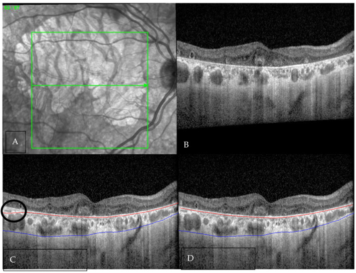 Figure 1
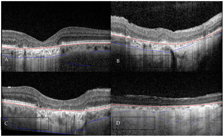 Figure 2
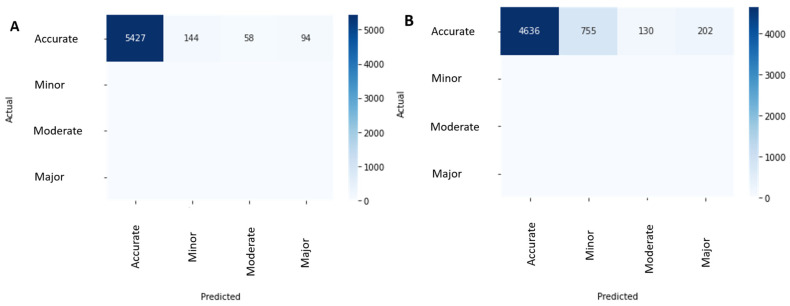 Figure 3
Peer Reviews
No public reviews on file for this paper yet. If you reviewed it on a platform where reviews are public (OpenReview, ICLR, NeurIPS, ICML), you can paste yours below so the community can read it here.
Videos
No videos yet. Explain this paper in a talk, walkthrough, or lecture? Add one.
Taxonomy
TopicsRetinal Diseases and Treatments · Glaucoma and retinal disorders · Retinal Imaging and Analysis