From Hematoxylin and Eosin to Masson’s Trichrome: A Comprehensive Framework for Virtual Stain Transformation in Chronic Liver Disease Diagnosis
Hossam Magdy Balaha, Khadiga M. Ali, Ali Mahmoud, Ahmed Aboudessouki, Mohamed T. Azam, Guruprasad A. Giridharan, Dibson Gondim, Ayman El-Baz

TL;DR
This paper introduces a new method for converting H&E-stained liver images to virtual Masson’s Trichrome stains, improving fibrosis diagnosis without extra tissue use.
Contribution
A transformer-based GAN with multi-stage alignment and fusion achieves high-quality virtual staining for liver fibrosis assessment.
Findings
The fused approach achieved state-of-the-art metrics like MI = 0.9815 and SSIM = 0.7474.
Statistical analysis showed enhanced stability with narrower interquartile ranges and fewer outliers.
Collagen morphology was preserved, supporting accurate fibrosis staging.
Abstract
Background/Objectives: Virtual histological staining offers a rapid, cost-effective alternative to physical reprocessing but faces challenges related to spatial misalignment and staining heterogeneity between Hematoxylin and Eosin (H&E) and Masson’s Trichrome (MT) domains. This study develops a robust framework for H&E-to-MT virtual staining to enable accurate fibrosis assessment without additional tissue consumption. Methods: We propose a transformer-based generative adversarial network (TbGAN) supported by a multi-stage alignment pipeline (SIFT (scale-invariant feature transform) coarse alignment, ORB/homography patch registration, and B-spline free-form deformation) and a weighted fusion mechanism combining four configuration outputs (O/10/3, O/3/10, R/10/3, and R/3/10). The framework was validated on 27 whole-slide images (>100,000 aligned patches) through 24 independent…
Genes, proteins, chemicals, diseases, species, mutations and cell lines named across the full text — each resolved to its canonical identifier and authoritative record.
Click any figure to enlarge with its caption.
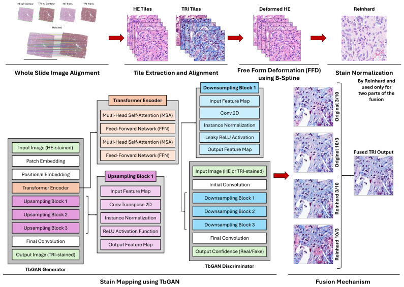 Figure 1
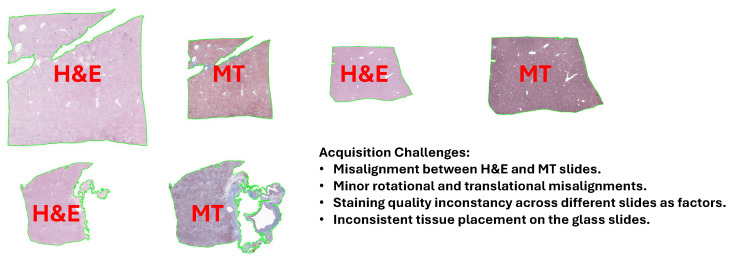 Figure 2
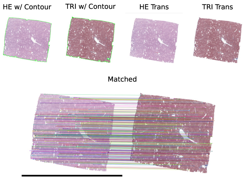 Figure 3
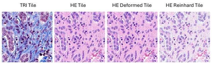 Figure 4
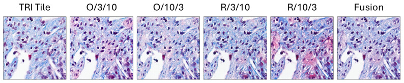 Figure 5
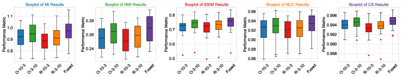 Figure 6
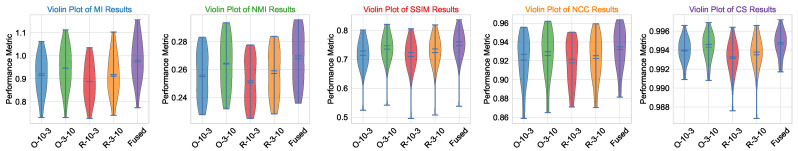 Figure 7
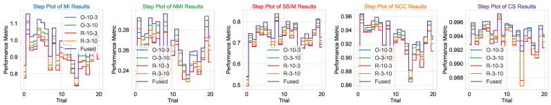 Figure 8
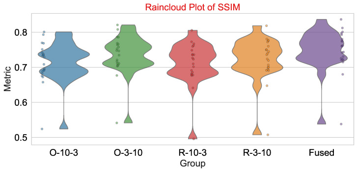 Figure 9
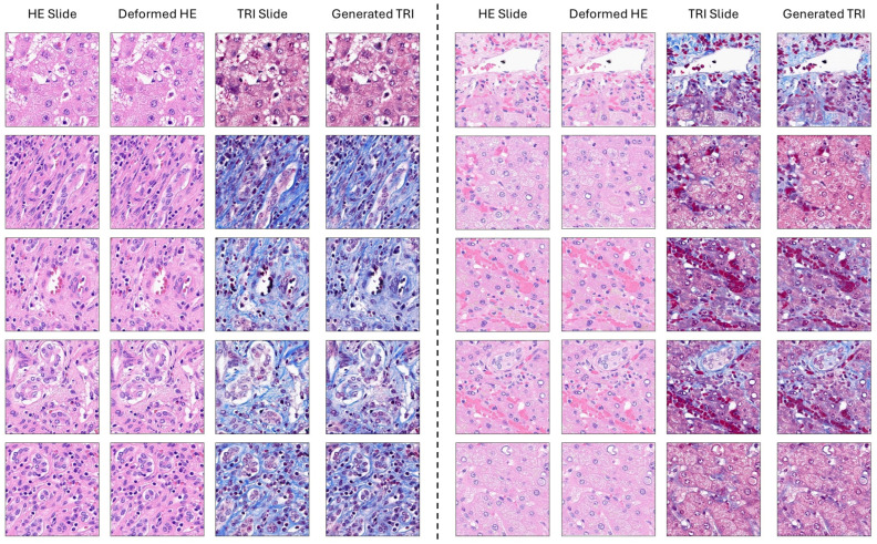 Figure 10
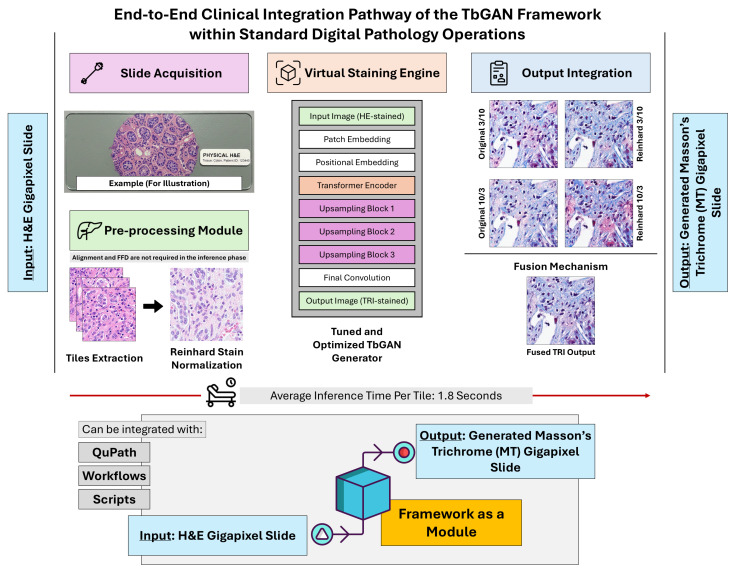 Figure 11
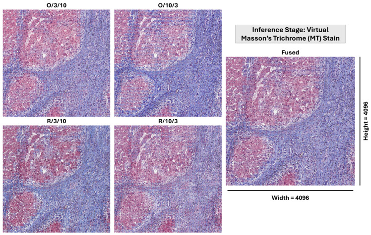 Figure 12
Peer Reviews
No public reviews on file for this paper yet. If you reviewed it on a platform where reviews are public (OpenReview, ICLR, NeurIPS, ICML), you can paste yours below so the community can read it here.
Videos
No videos yet. Explain this paper in a talk, walkthrough, or lecture? Add one.
Taxonomy
TopicsAI in cancer detection · Cell Image Analysis Techniques · Medical Image Segmentation Techniques