Comparison of measurement of the apical anatomy of maxillary central incisors using cone-beam computed tomography and conventional intraoral radiographs
Josephine Solgaard Henriksen, Eva Lauridsen, Simon Storgård Jensen, Tron Andre Darvann, Shumei Murakami, Tomomi Tsujimoto, Yuka Uchimoto, Nuno Vibe Hermann

TL;DR
This study compares how 2D X-rays and 3D CT scans measure the size of tooth root openings in maxillary incisors, finding that the opening shrinks with age and that 3D scans give different results than 2D images.
Contribution
The study introduces a novel comparison of apical foramen measurements using 2D radiographs and 3D CBCT, revealing age-related dimensional changes and measurement discrepancies.
Findings
The area of the apical foramen measured in 3D using CBCT decreased at -0.019 mm² per month.
The diameter of the foramen decreased with age in both mesiodistal and faciolingual directions.
The most common shape of the apical constriction was round, with a mean shape index of 0.992 ± 0.244.
Abstract
The aim of this study was to investigate the dimension of the apical foramen in maxillary incisors in relation to age as observed in conventional intra-oral periapical radiographs and Cone-Beam CT (CBCT). We hypothesized a discrepancy in apical foramen size between 2D radiographs and 3D CBCT, and that the foramen narrows with age. Seventy patients with 87 anterior maxillary teeth were included. The shape, size and the area of the apical foramen were measured corresponding to the narrowest part of the canal system (the apical constrictor/minor foramen) on intraoral conventional X-rays and CBCT scans taken in close succession. A comparison between the two imaging methods was performed and related to the age of the patient, with statistical analyses including t-test, regression, and Bland-Altman plots. In addition, the area of the foramen was measured, and a shape index was calculated…
Genes, proteins, chemicals, diseases, species, mutations and cell lines named across the full text — each resolved to its canonical identifier and authoritative record.
Click any figure to enlarge with its caption.
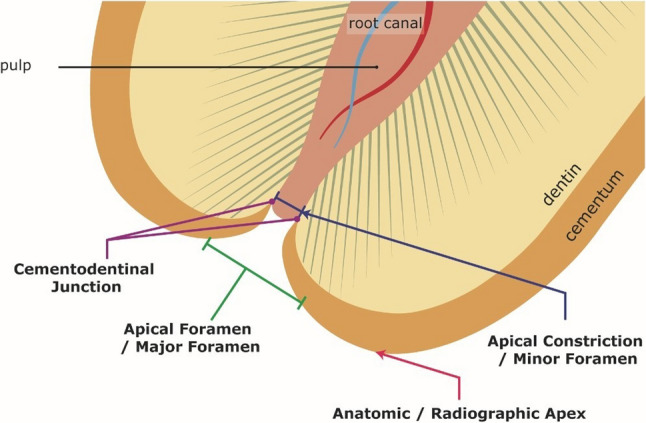 Figure 1
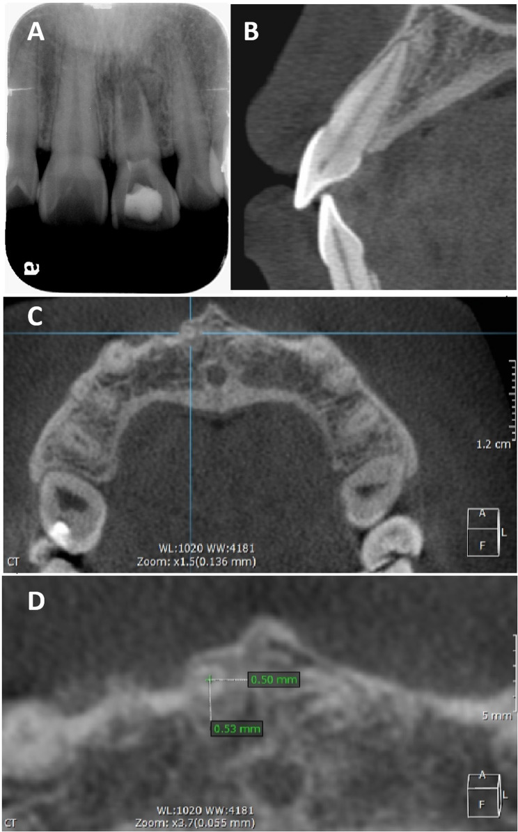 Figure 2
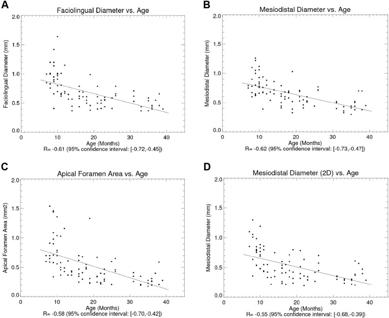 Figure 3
 Figure 4
Peer Reviews
No public reviews on file for this paper yet. If you reviewed it on a platform where reviews are public (OpenReview, ICLR, NeurIPS, ICML), you can paste yours below so the community can read it here.
Videos
No videos yet. Explain this paper in a talk, walkthrough, or lecture? Add one.
Taxonomy
TopicsEndodontics and Root Canal Treatments · Dental Radiography and Imaging · Orthodontics and Dentofacial Orthopedics