Tumor morphology on CT radiomics is largely driven by the local anatomical environment, not the primary tumor type
Sajjad Rostami, Corentin Guérendel, Marleen Soliman, Hannah W. Stutterheim, Olga Maxouri, Diana Ivonne Rodríguez Sánchez, Stephan Ursprung, Nino Boveradze, George Agrotis, Kalina Chupetlovska, Francesca Castagnoli, Federica Landolfi, Eun Kyoung Hong, Andrea Delli Pizzi

TL;DR
This study finds that tumor appearance on CT scans is more influenced by the surrounding anatomy than the tumor's origin, suggesting radiomic biomarkers should account for local tissue context.
Contribution
The study quantifies that tumor morphology on CT is predominantly shaped by the anatomical environment rather than the primary tumor lineage.
Findings
Tumor morphology clusters align more strongly with anatomical environment (ARI = 0.37) than with primary tumor type (ARI = 0.04).
In solid organ metastases, the anatomical environment's influence is even stronger (ARI = 0.60).
Intensity and texture features drive the association with anatomical environment more than shape features.
Abstract
Radiogenomics promises noninvasive tumor profiling; however, the extent to which imaging morphology reflects tumor lineage versus host-organ milieu remains unclear. This study aimed to quantify the relative influence of tumor type and anatomical environment on contrast-enhanced computed tomography (CT) radiomic phenotypes. A discovery cohort of 1,598 patients (10,485 lesions) and an external validation cohort of 2,440 patients (6,597 lesions) underwent portal-venous-phase CT. After manual segmentation, lesion-level radiomic features were standardized and embedded using t-distributed stochastic neighbor embedding. Bayesian-optimized agglomerative clustering defined morphology-based groups. Concordance with the primary tumor site (lineage) and anatomical environment was quantified using bootstrapped adjusted Rand indices (ARI); the silhouette score assessed clustering quality.…
Genes, proteins, chemicals, diseases, species, mutations and cell lines named across the full text — each resolved to its canonical identifier and authoritative record.
Click any figure to enlarge with its caption.
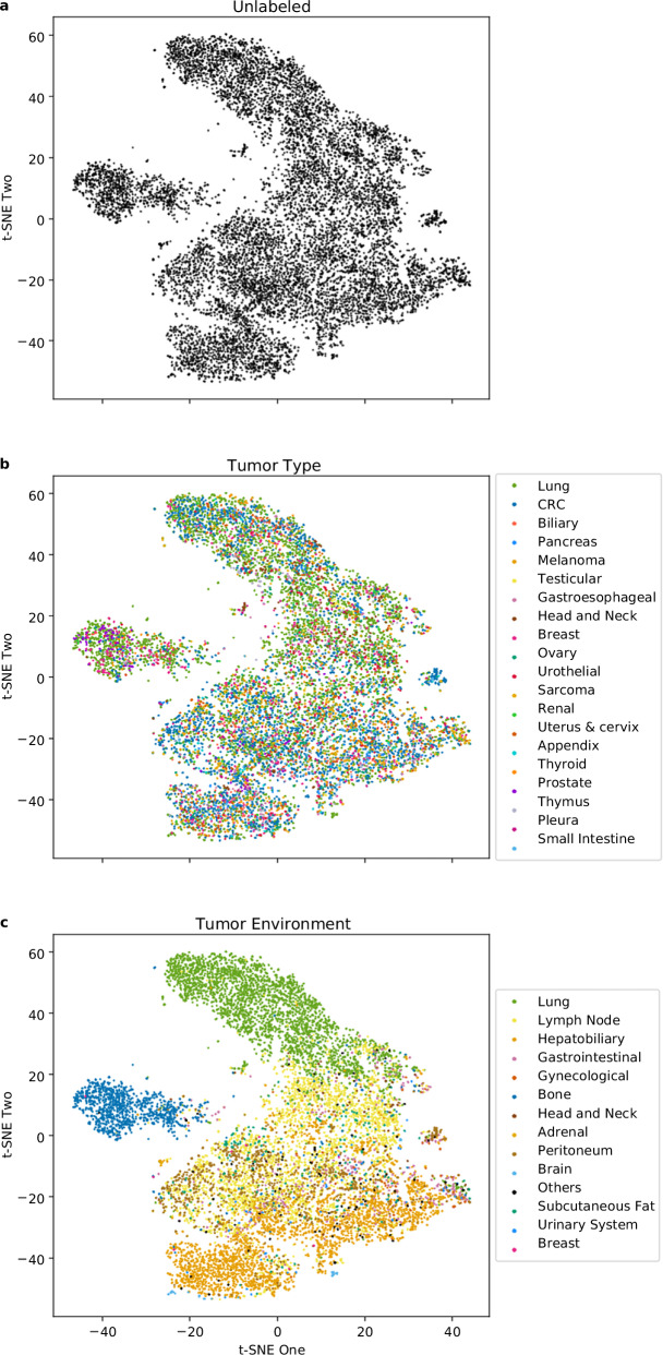 Figure 1
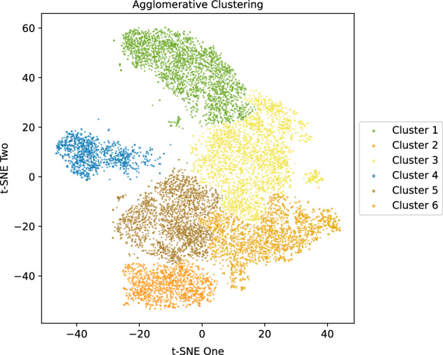 Figure 2
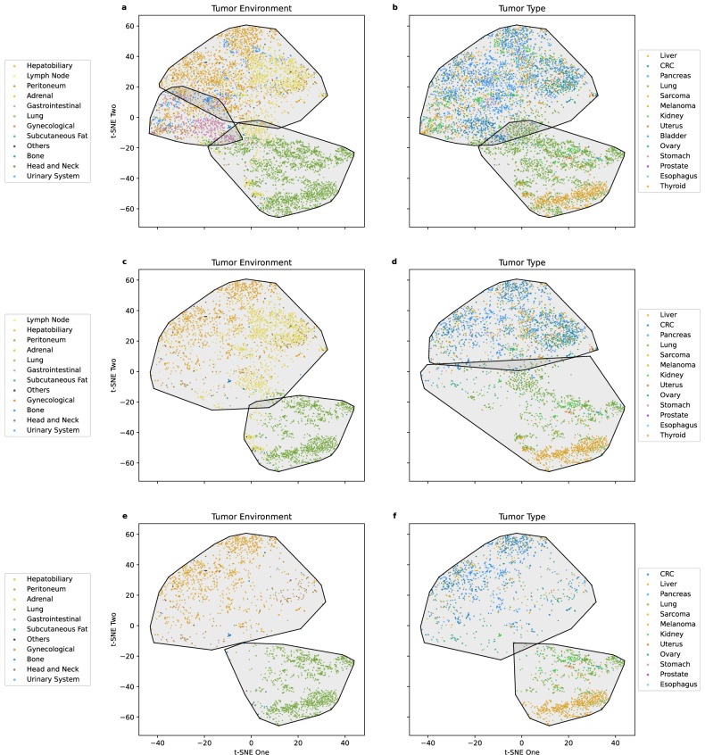 Figure 3
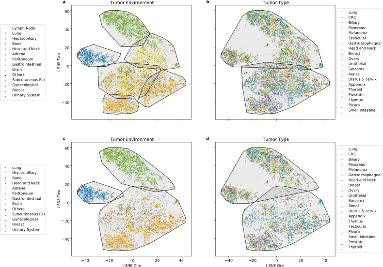 Figure 4
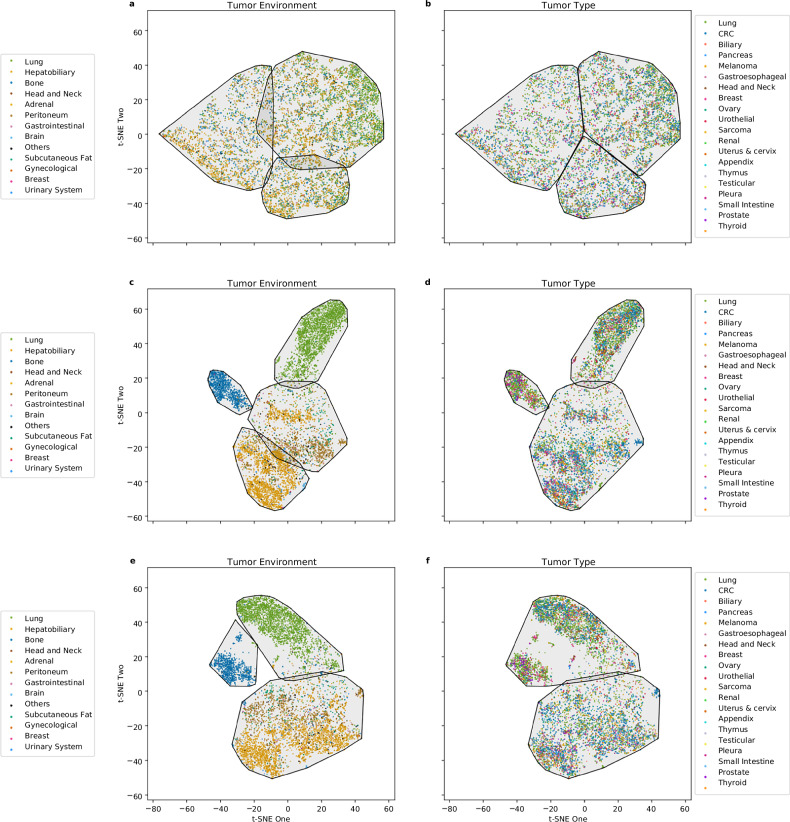 Figure 5
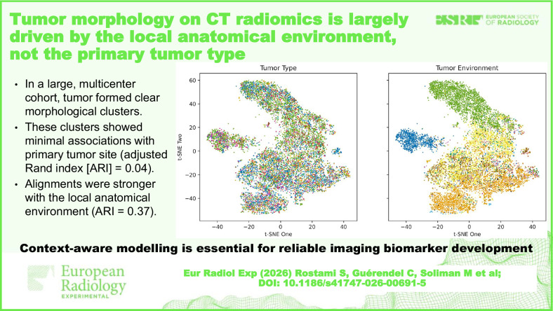 Figure 6
Peer Reviews
No public reviews on file for this paper yet. If you reviewed it on a platform where reviews are public (OpenReview, ICLR, NeurIPS, ICML), you can paste yours below so the community can read it here.
Videos
No videos yet. Explain this paper in a talk, walkthrough, or lecture? Add one.
Taxonomy
TopicsRadiomics and Machine Learning in Medical Imaging · MRI in cancer diagnosis · Advanced X-ray and CT Imaging