Decreased T2-signal intensities indicate positive response to front-line radiotherapy in pediatric low-grade gliomas
Simon Weiner, Monika Warmuth-Metz, Daniela Kandels, Beate Timmermann, Rolf-Dieter Kortmann, Stefan Dietzsch, Torsten Pietsch, Brigitte Bison, Mirko Pham, Astrid Katharina Gnekow, Annika Quenzer

TL;DR
This study shows that a decrease in T2-signal intensity on MRI scans can indicate a positive response to radiotherapy in children with low-grade gliomas.
Contribution
The study identifies decreased T2-signal intensity as a novel imaging marker for assessing treatment response in pediatric low-grade gliomas.
Findings
A significant decrease in T2-signal intensity was observed 24 months after radiotherapy.
T2-signal intensity reduction correlated with tumor volume reduction and treatment response.
Pseudoprogression cases showed stable T2-signal intensity despite other signs of progression.
Abstract
To evaluate MRI changes in T2-weighted imaging (T2WI) signal intensity (T2SI) as a potential imaging marker for assessing response to radiotherapy (RT) in pediatric low-grade glioma (pLGG). This retrospective study analyzed imaging data of 56 pLGG patients (mean age, 12.4 ± 3.5 years; 33/56 [58.9%] male) treated with photon-based or proton-based RT within the SIOP-LGG 2004 study and registry. Tumor signal characteristics on T2WI were qualitatively and quantitatively assessed at baseline and up to 24 months post-RT. Tumor volumes were calculated, and correlations between ∆T2SI and volumetric changes were examined. Statistical tests included inferential tests, correlation analysis, and linear regression. At baseline, 87.5% tumors were rated as hyperintense, while none was rated hypointense. The mean ratio between T2SI of the tumors compared to the cerebral cortex was 1.70. A significant…
Genes, proteins, chemicals, diseases, species, mutations and cell lines named across the full text — each resolved to its canonical identifier and authoritative record.
Click any figure to enlarge with its caption.
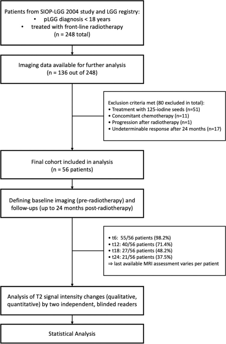 Figure 1
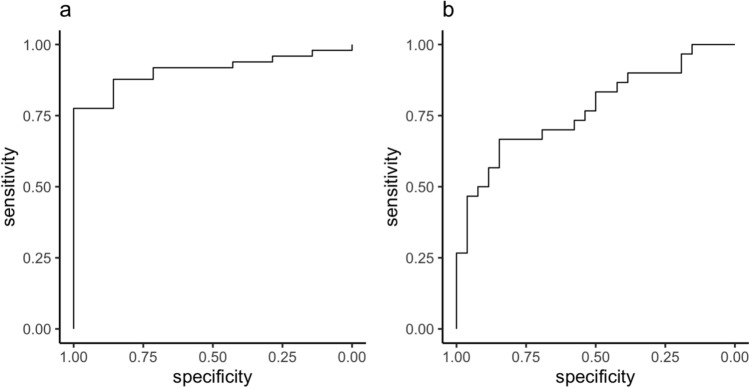 Figure 2
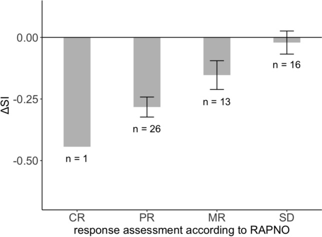 Figure 3
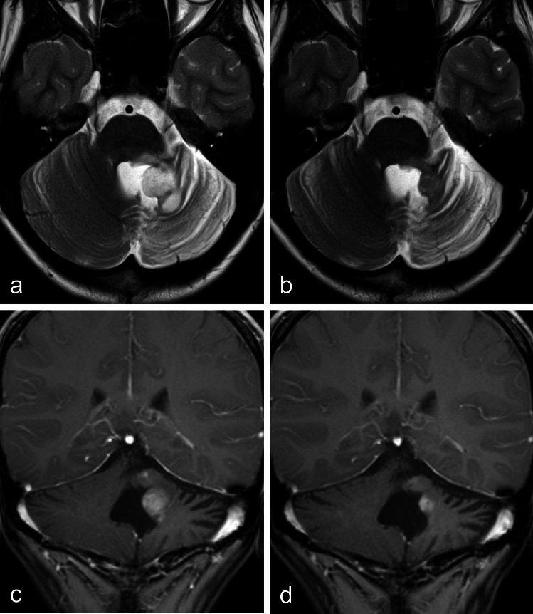 Figure 4
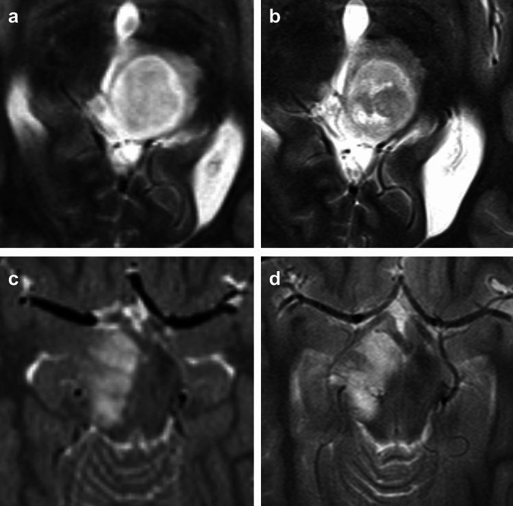 Figure 5
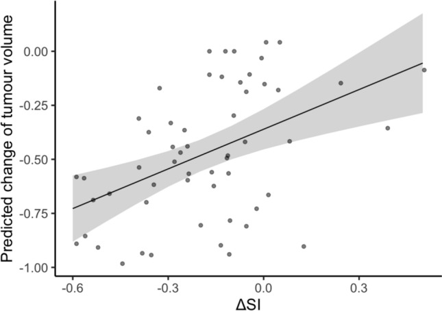 Figure 6
Peer Reviews
No public reviews on file for this paper yet. If you reviewed it on a platform where reviews are public (OpenReview, ICLR, NeurIPS, ICML), you can paste yours below so the community can read it here.
Videos
No videos yet. Explain this paper in a talk, walkthrough, or lecture? Add one.
Taxonomy
TopicsGlioma Diagnosis and Treatment · Advanced MRI Techniques and Applications · Meningioma and schwannoma management