Image segmentation of cervical grainy sandy patches lesions associated with female genital schistosomiasis using deep convolutional neural network with U-NET architecture
Karl Emil Jøker, Peter Christian Derek Leutscher, Kristine Brøndbjerg Øby, Karoline Jøker, Bodo Sahondra Randrianasolo, Maciej Plocharski, Louise Thomsen Schmidt Arenholt, Krystyna Cwiklinski, Krystyna Cwiklinski, Krystyna Cwiklinski

TL;DR
This study uses AI to detect cervical lesions caused by a common but neglected disease in African women, aiming to improve early diagnosis in underserved areas.
Contribution
A U-Net-based deep learning model is proposed for segmenting grainy sandy patches in cervical images of female genital schistosomiasis.
Findings
The model achieved a DICE score of 0.61, accuracy of 0.81, sensitivity of 0.84, and specificity of 0.81 in lesion segmentation.
Image quality and weak annotations were identified as factors affecting model performance.
The approach shows potential for integration into mobile diagnostic tools for FGS in resource-limited settings.
Abstract
Female genital schistosomiasis (FGS) is a neglected but highly prevalent disease in sub-Saharan Africa, caused by Schistosoma haematobium egg-induced inflammation in the pelvic region. FGS is characterized by four mucosal lesion types in the lower female genital tract: grainy sandy patches (GSP), homogeneous yellow sandy patches, abnormal blood vessels, and rubbery papules. This study focuses on the segmentation of cervical GSP lesions using a deep-learning convolutional neural network. A total of 583 cervical images from women in a S. haematobium endemic region of Madagascar, all exhibiting FGS-associated lesions, particularly GSP lesions, were used for this study. Weak annotations (non-pixel-wise) were generated using QubiFier software. A U-Net model with a focal loss function, and an Adam optimizer was trained to segment GSP lesions. A 5-fold cross validation was performed, thus…
Genes, proteins, chemicals, diseases, species, mutations and cell lines named across the full text — each resolved to its canonical identifier and authoritative record.
Click any figure to enlarge with its caption.
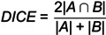 Figure 1
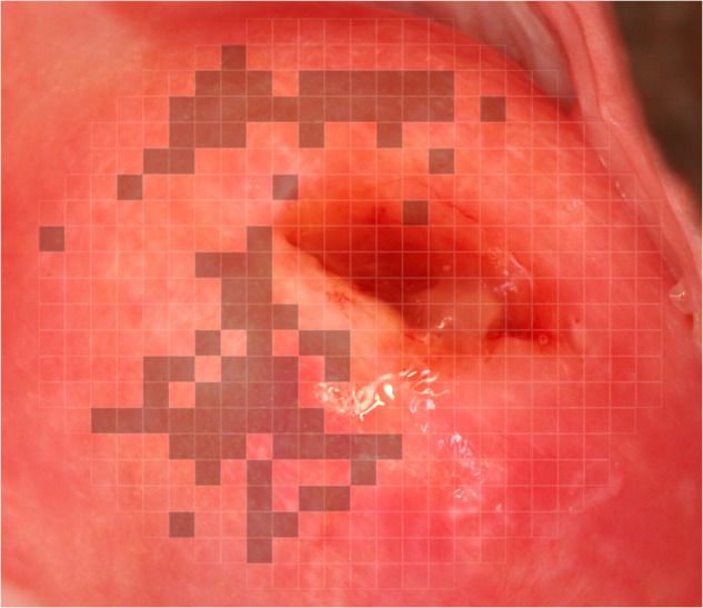 Figure 2
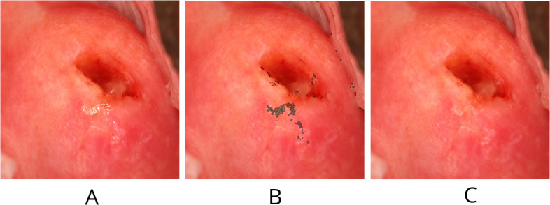 Figure 3
 Figure 4
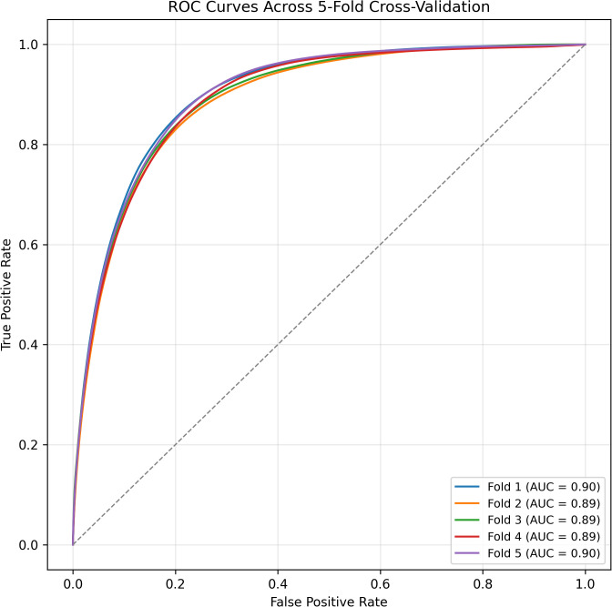 Figure 5
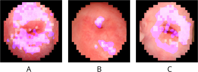 Figure 6
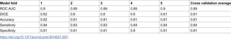 Figure 7
Peer Reviews
No public reviews on file for this paper yet. If you reviewed it on a platform where reviews are public (OpenReview, ICLR, NeurIPS, ICML), you can paste yours below so the community can read it here.
Videos
No videos yet. Explain this paper in a talk, walkthrough, or lecture? Add one.
Taxonomy
TopicsAI in cancer detection · Artificial Intelligence in Healthcare and Education · Parasites and Host Interactions