Surface Electromyography as a Method for Characterizing Mammogram Discomfort: Cross-Sectional Questionnaire Study of Procedural Stress
Krystyna Gielo-Perczak, Riley McNaboe, Hugo Posada-Quintero

TL;DR
This study uses surface electromyography to objectively measure muscle activity and stress during mammograms, revealing which muscles are most affected and how discomfort is experienced.
Contribution
The study introduces a novel sEMG-based method to objectively quantify mammogram-related stress and muscle activity.
Findings
The deltoid showed the highest muscle activation during mammogram simulations, with up to 436% increase in activity.
Muscle activations were ipsilaterally correlated, with significant differences when the breast was compressed on the same side as the measured muscle.
Patient-reported discomfort primarily localized to the shoulder and neck, aligning with the physiological data.
Abstract
Mammograms are the most readily used procedures for early breast cancer detection but are notorious for the discomfort they induce in patients. This physiological strain has been validated by many questionnaire-based investigations, some of which indicate that it may discourage and deter women from potentially lifesaving health care. While informative, these subjective measures are highly variable and do not provide an objective perspective regarding the coordinated physiological and ergonomic response required for the procedure. A multimuscle surface electromyography (sEMG) methodology is proposed to better understand the machine-patient dynamics and potentially develop objective measures of mammogram-related pain and stress. Seven different muscle pairs on the neck, shoulders, and torso were identified as being critical in postural positioning during the industry-standard mammogram…
Genes, proteins, chemicals, diseases, species, mutations and cell lines named across the full text — each resolved to its canonical identifier and authoritative record.
Click any figure to enlarge with its caption.
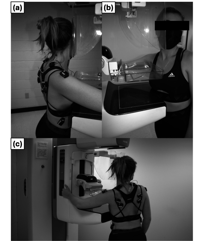 Figure 1
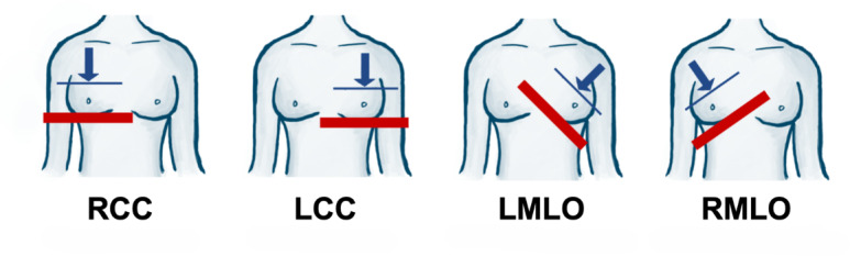 Figure 2
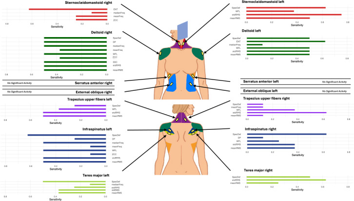 Figure 3
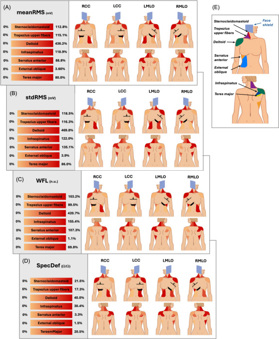 Figure 4
Peer Reviews
No public reviews on file for this paper yet. If you reviewed it on a platform where reviews are public (OpenReview, ICLR, NeurIPS, ICML), you can paste yours below so the community can read it here.
Videos
No videos yet. Explain this paper in a talk, walkthrough, or lecture? Add one.
Taxonomy
TopicsDigital Radiography and Breast Imaging · Breast Implant and Reconstruction · Optical Imaging and Spectroscopy Techniques