Seroprevalence of rift valley fever virus and associated risk factors in small ruminants at human-livestock-wildlife interface within Uganda’s conservation areas
Phiona Katushabe, Dennison Kizito, Charity Angella Nassuna, Joseph Kikwabanga Mutyaba, Swaib A. Lule, Gladys Kiggundu Nakanjako, Nackson Babi, Wilber Ssembajjwe, Tonny Kayizi, Milton Bahati, Martin Esau, Stella Atim, Brian Kivumbi, Teddy Nakayiki Muwawu, Deogratius Nteza

TL;DR
This study found a high rate of past exposure to Rift Valley Fever Virus in small ruminants at the human-livestock-wildlife interface in Uganda's conservation areas.
Contribution
The study provides new insights into RVFV seroprevalence and risk factors among small ruminants in conservation areas with high human-livestock-wildlife interactions.
Findings
Overall RVFV seropositivity in small ruminants was 41.1%, with higher rates in goats (42.4%) compared to sheep (34.5%).
Exotic breeds and females had higher seroprevalence rates at 55.5% and 41.8%, respectively.
Older animals had increased risk of RVFV exposure, while local breeds had reduced exposure risk.
Abstract
Following the first laboratory confirmed human Rift Valley Fever Virus (RVFV) outbreak in 48 years, Uganda has continued to detect sporadic outbreaks, particularly within the cattle corridor since 2016. Although wildlife potentially harbors RVFV strains, livestock exposure to RVFV at human-livestock-wildlife interfaces remains underexplored in major Ugandan conservation areas. A cross-sectional quantitative study was conducted between August, 2022 and March, 2023, at Satellite Research Sites (SRS), located in Bwindi-Mgahinga, Lake Mburo, Queen Elizabeth, Murchison Falls, and Pian Upe conservation areas in Uganda, which were selected for their high human-livestock-wildlife interactions. Using a two-stage sampling design, small ruminants were sampled from randomly selected herds within villages adjacent to the national parks. Blood samples were collected, and analysed with a validated…
Genes, proteins, chemicals, diseases, species, mutations and cell lines named across the full text — each resolved to its canonical identifier and authoritative record.
Click any figure to enlarge with its caption.
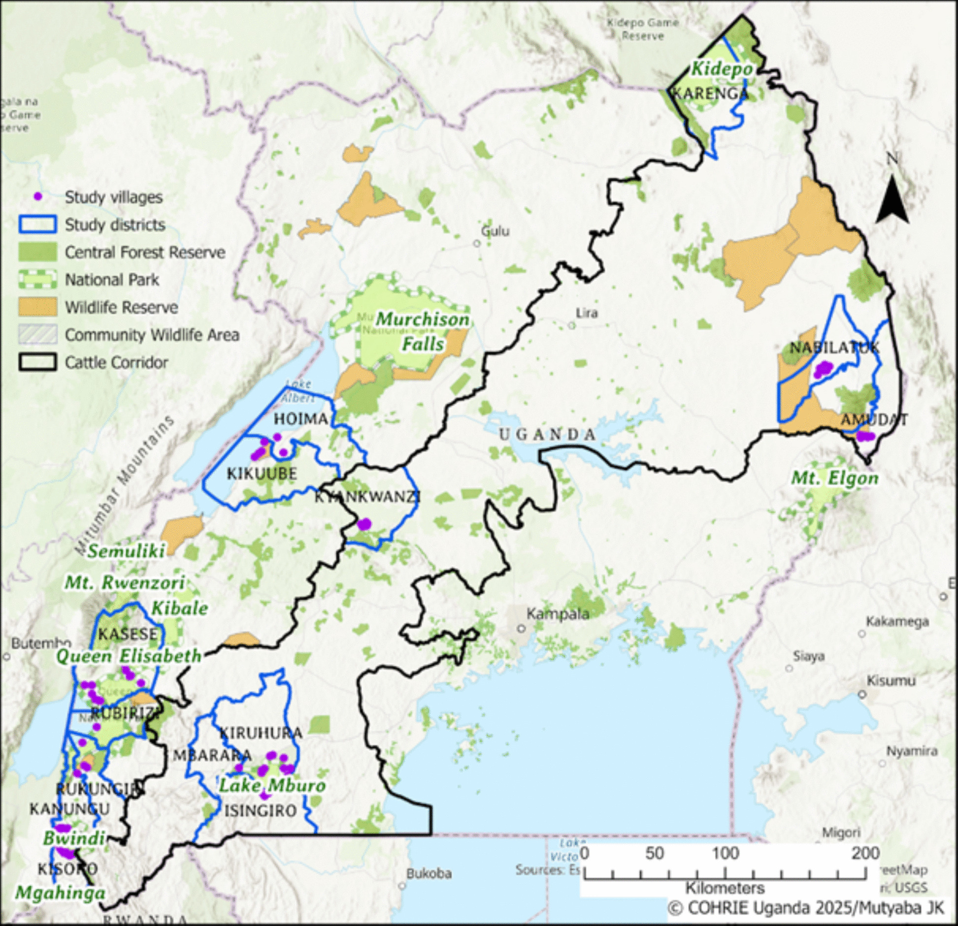 Figure 1
Peer Reviews
No public reviews on file for this paper yet. If you reviewed it on a platform where reviews are public (OpenReview, ICLR, NeurIPS, ICML), you can paste yours below so the community can read it here.
Videos
No videos yet. Explain this paper in a talk, walkthrough, or lecture? Add one.
Taxonomy
TopicsViral Infections and Vectors · Viral Infections and Outbreaks Research · Vector-Borne Animal Diseases