Cell-intrinsic vulnerability and immune activation cooperate to drive degeneration in a mitochondrial complex I deficiency model of optic neuropathy
Daniela Santamaría-Muñoz, Raenier V. Reyes, Miranda R. Krueger, Andrea García-Llorca, Brennan Marsh-Armstrong, Xin Duan, Yang Hu, Derek S. Welsbie, Nicholas Marsh-Armstrong, Elisenda Sanz, Albert Quintana, Sergi Simó, Anna La Torre

TL;DR
This study shows that mitochondrial dysfunction in retinal cells leads to optic neuropathy through a combination of cell-intrinsic vulnerability and immune system activation.
Contribution
The study introduces a conditional transgenic model to distinguish intrinsic and extrinsic mechanisms in optic neuropathy.
Findings
Deleting Ndufs4 in retinal cells causes vision loss and immune activation before RGC death.
Myeloid cell depletion preserves RGCs, showing inflammation's active role in degeneration.
Degeneration spreads beyond mutant regions, indicating non-cell autonomous propagation.
Abstract
Mitochondrial dysfunction is a central hallmark of many optic neuropathies, yet the mechanisms linking intrinsic metabolic stress to retinal ganglion cell (RGC) degeneration remain unclear. To bridge this gap, we developed conditional transgenic models targeting the mitochondrial complex I subunit Ndufs4 in the retina. Broad deletion of Ndufs4 in the retina resulted in vision loss, progressive RGC degeneration, and pronounced immune activation before overt RGC death. Strikingly, depletion of myeloid cells significantly preserved RGCs, demonstrating that inflammation is not simply a downstream consequence but a participant in the degeneration process. To further distinguish between intrinsic and extrinsic mechanisms, we generated a mosaic model in which only subsets of retinal cells lacked Ndufs4. In this paradigm, the degeneration first appeared selectively in mutant regions, suggesting…
Genes, proteins, chemicals, diseases, species, mutations and cell lines named across the full text — each resolved to its canonical identifier and authoritative record.
Click any figure to enlarge with its caption.
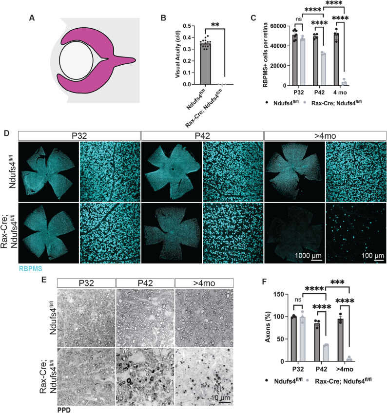 Figure 1
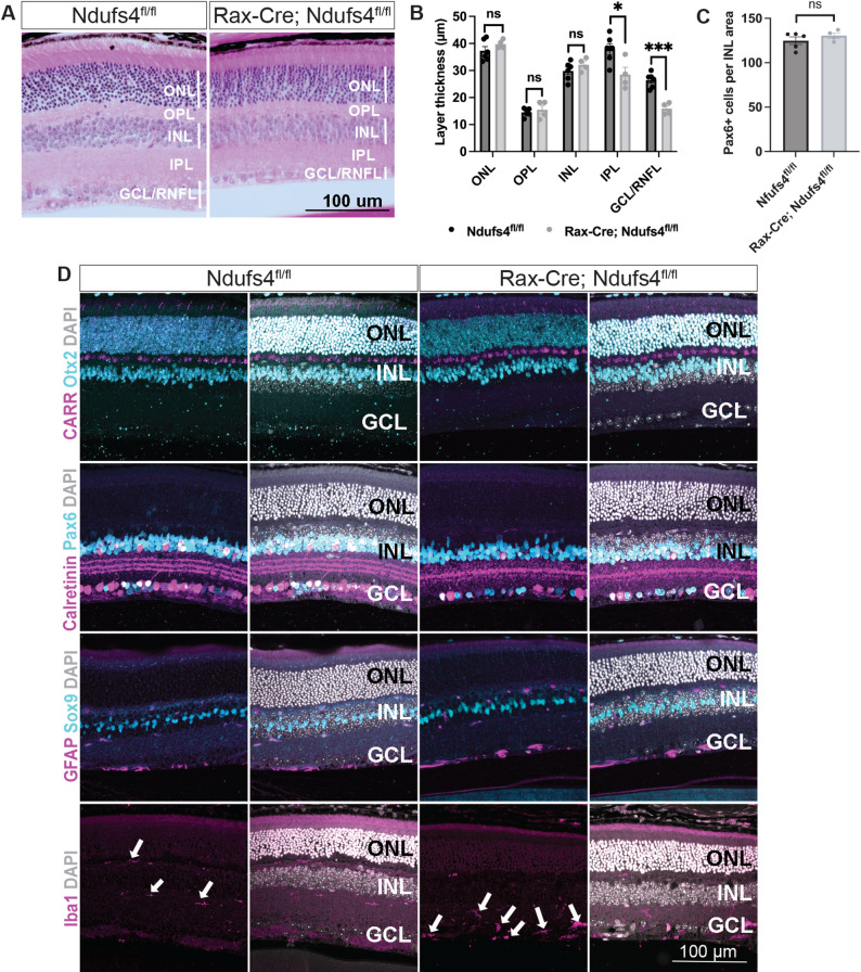 Figure 2
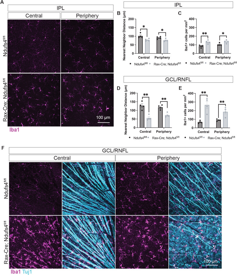 Figure 3
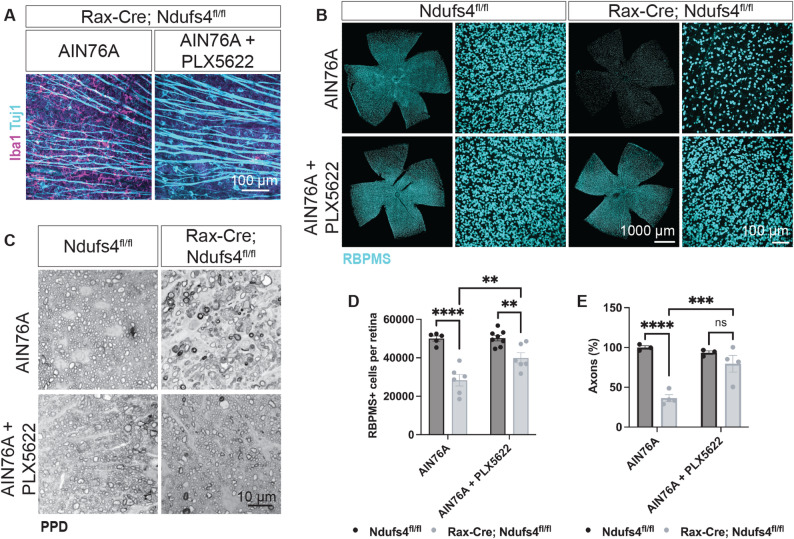 Figure 4
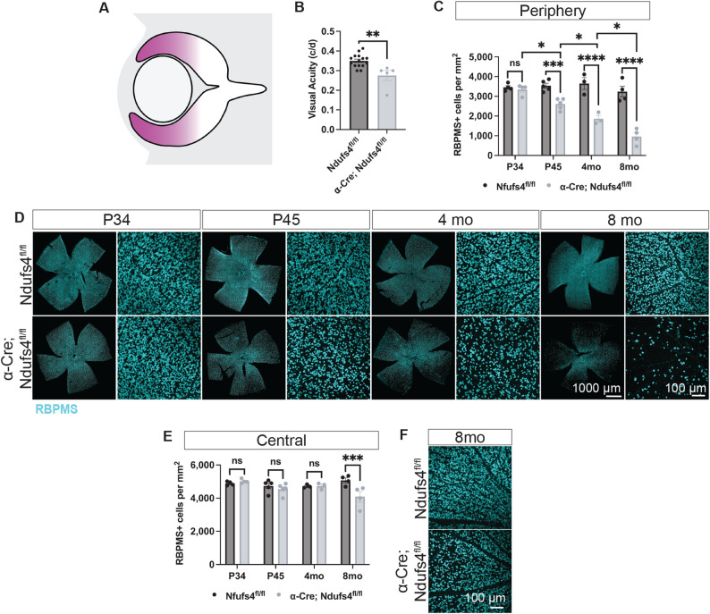 Figure 5
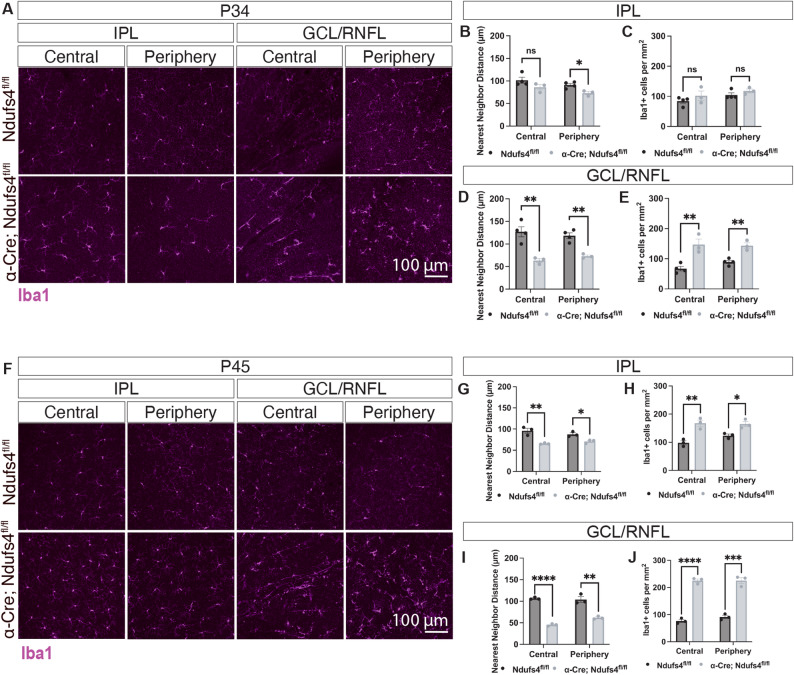 Figure 6
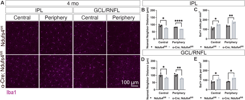 Figure 7
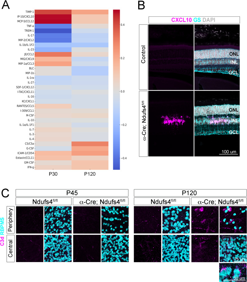 Figure 8
Peer Reviews
No public reviews on file for this paper yet. If you reviewed it on a platform where reviews are public (OpenReview, ICLR, NeurIPS, ICML), you can paste yours below so the community can read it here.
Videos
No videos yet. Explain this paper in a talk, walkthrough, or lecture? Add one.
Taxonomy
TopicsMitochondrial Function and Pathology · Multiple Sclerosis Research Studies · Ocular Diseases and Behçet’s Syndrome