Clinical Validation of Imaging Biomarkers in Mycosis Fungoides
Selinde S. Wind, Elise S. M. Beljaards, Rianne Rijneveld, Lisa Bruijnincx, Tessa Niemeyer‐van der Kolk, Manon A. A. Jansen, Yalcin Yavuz, Marieke de Kam, Jacobus Burggraaf, Naomi Klarenbeek, Jacobus Bosch, Koen D. Quint, Maarten H. Vermeer, Robert Rissmann, Rob Vreeken

TL;DR
This study evaluates non-invasive imaging techniques to objectively monitor skin disease severity in mycosis fungoides, a type of skin cancer.
Contribution
The study introduces and validates new imaging biomarkers that can reliably track disease progression and treatment response in mycosis fungoides.
Findings
MSI detected significant differences in skin parameters like erythema and pigmentation between healthy and diseased skin.
Four imaging biomarkers met all clinical validation criteria, including strong reliability and treatment responsiveness.
The new biomarkers showed moderate agreement with the traditional CAILS score and were well accepted by patients.
Abstract
The composite index lesion severity (CAILS) score is used to monitor disease and therapeutic response in mycosis fungoides (MF), but is limited by interobserver variability and low sensitivity. Emerging imaging techniques, such as multispectral imaging (MSI), colourimetry and laser speckle contrast imaging (LSCI), offer objective alternatives for quantifying CAILS parameters. The aim of this study was to evaluate non‐invasive imaging modalities for objective and reliable quantification of disease extent in MF. Sixty‐six participants were enrolled in two prospective studies: a cross‐sectional discovery cohort to assess baseline characteristics of 35 MF patients (IA–IVB) and 10 healthy controls using CAILS and MSI, and a longitudinal confirmation cohort including 21 early‐stage MF patients (IA–IIA) treated with chlormethine gel 0.016% for 16 weeks, in whom lesional and non‐lesional skin…
Genes, proteins, chemicals, diseases, species, mutations and cell lines named across the full text — each resolved to its canonical identifier and authoritative record.
Click any figure to enlarge with its caption.
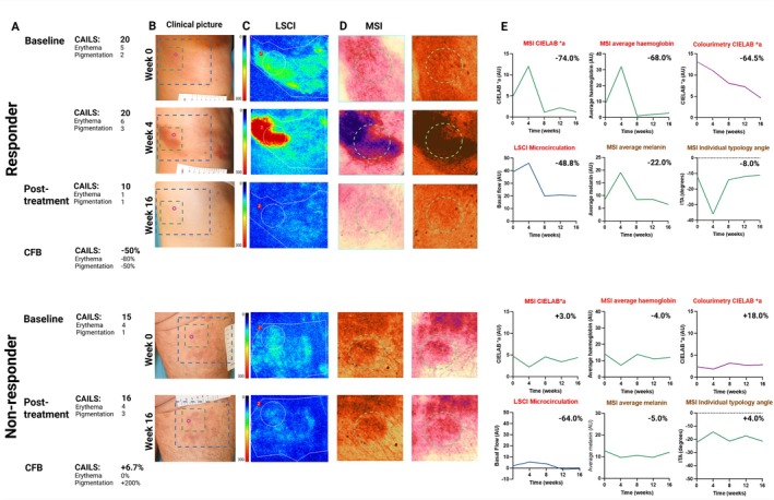 Figure 1
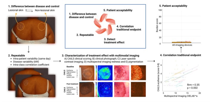 Figure 2
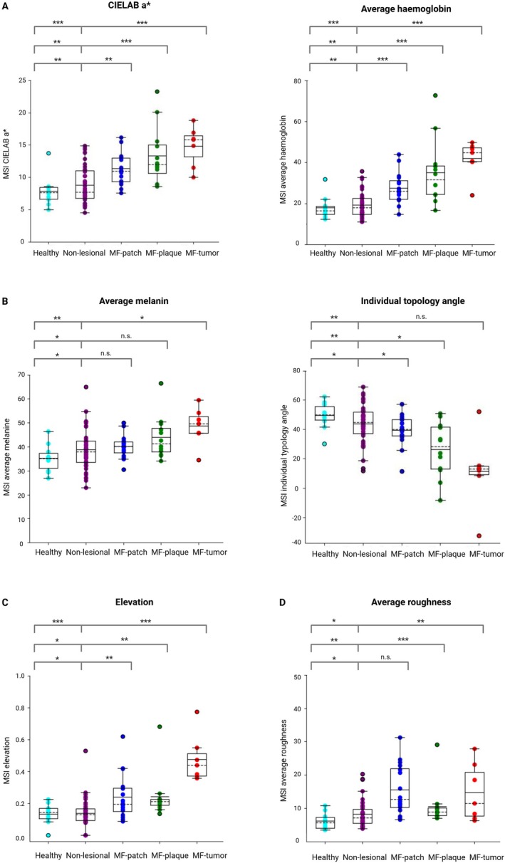 Figure 3
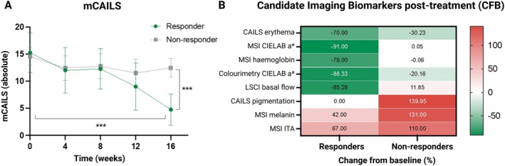 Figure 4
Peer Reviews
No public reviews on file for this paper yet. If you reviewed it on a platform where reviews are public (OpenReview, ICLR, NeurIPS, ICML), you can paste yours below so the community can read it here.
Videos
No videos yet. Explain this paper in a talk, walkthrough, or lecture? Add one.
Taxonomy
TopicsCutaneous lymphoproliferative disorders research · Retinoids in leukemia and cellular processes · Cutaneous Melanoma Detection and Management